Superior, Anterior & posterior mediastinum
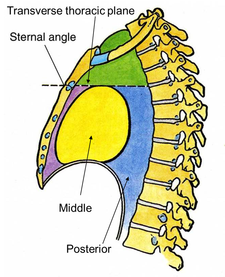
subdivisions of mediastinum
The superior mediastinum is located superior to the transverse thoracic plane passing through the sternal angle and the T4/5 IV disc.
Inferior part is further divided into three parts:
Anterior, Middle, Posterior
anterior mediastinum
The anterior mediastinum contains the inferior portion of the thymus gland in infants & children.
Lymph nodes
Sternopericardial ligaments
It is anterior to the pericardium and posterior to the body of the sternum
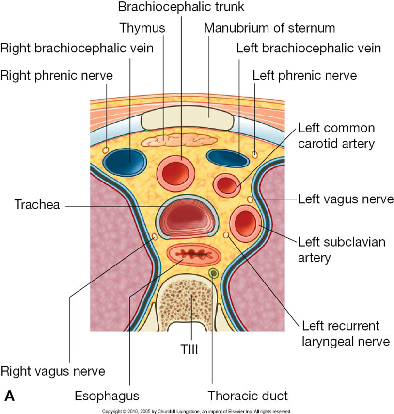
contents of superior mediastinum
from anterior to posterior
Thymus
Great vessels
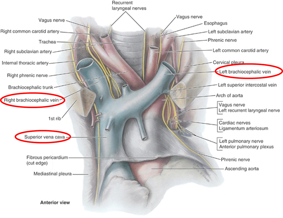
Brachiocephalic veins and SVC
Arch of aorta and roots of:
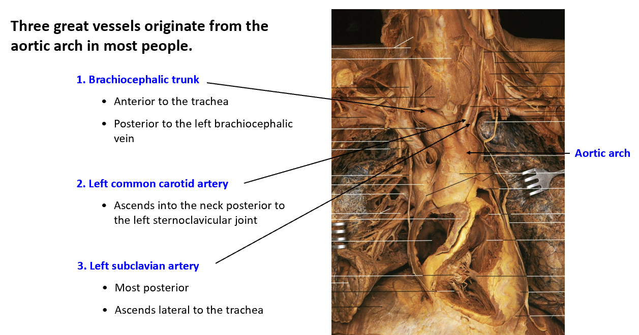
Brachiocephalic trunk
Left common carotid artery
Left subclavian artery
Phrenic nerves and vagus nerves
Cardiac nerve plexus
Left recurrent laryngeal nerve
Trachea and esophagus with nerves
Thoracic duct and lymphatic trunks
thymus
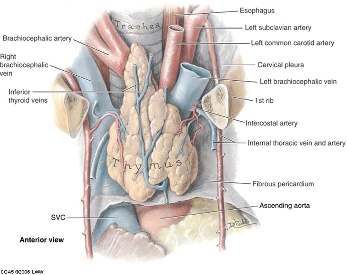
thymus occupies the most anterior part of the superior mediastinum, just posterior to the sternum.
After puberty, it gradually degrades and is replaced by fat.
Blood supply is from branches of the internal thoracic.
veins of the superior mediastinum
The brachiocephalic veins are formed by the union of the subclavian and internal jugular veins bilaterally.
They unite at the 1st costal cartilage to form the superior vena cava.
Left brachiocephalic vein is twice as long as the right, passes anterior to the arch of the aorta and great vessels.
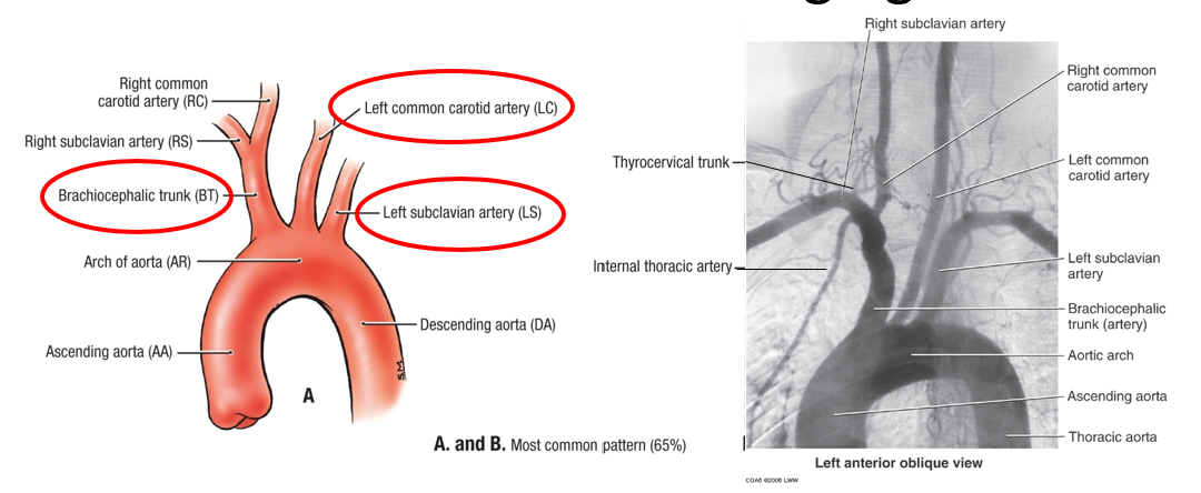
aortic arch branches & angiogram
Aortic arch begins posterior to the 2nd right sternocostal joint (level of sternal angle). It arches superiorly, posteriorly to the left, and then inferiorly.
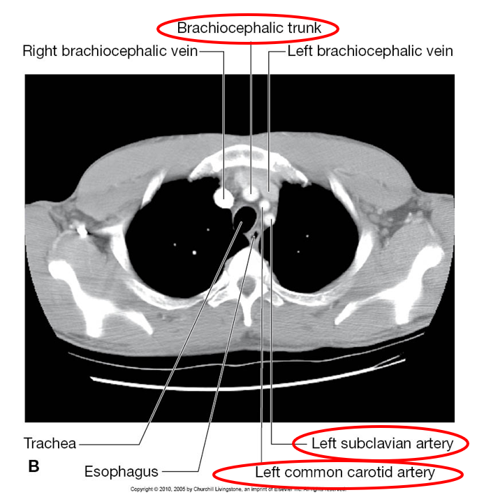
In most (65%) individuals, the typical branches arising from the aortic arch are the brachiocephalic trunk, the left common carotid and left subclavian arteries.
great vessels
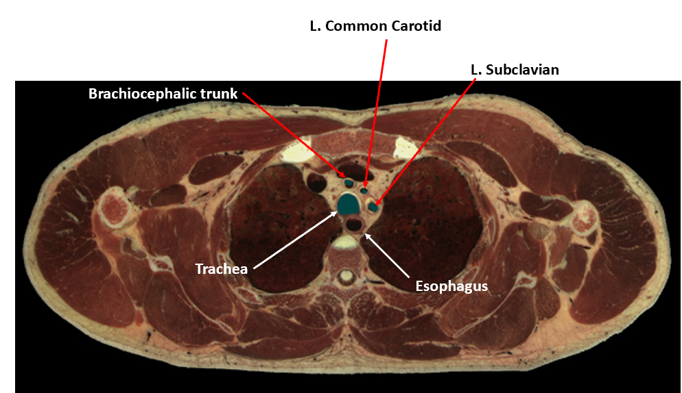
CT scan of thorax: superior mediastinum
superior mediastinum nerves
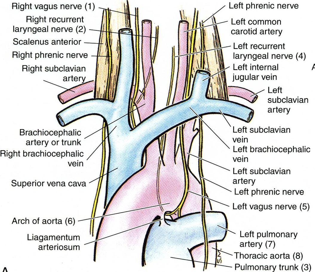
The phrenic nerves lie on the anterior surface of the anterior scalene muscles as they pass inferiorly into the thorax.
vagus nerves, and sympathetic nerves, form the carotid, pulmonary, and esophageal plexuses.
recurrent laryngeal nerves
Left recurrent laryngeal nerve arises from the vagus nerve and wraps underneath the aortic arch just lateral to the ligamentum arteriosum, ascends to the larynx between the trachea and esophagus.
Clinical note: aortic aneurysm, bronchogenic or esophageal carcinoma may compress the left recurrent laryngeal nerve causing a hoarse voice.
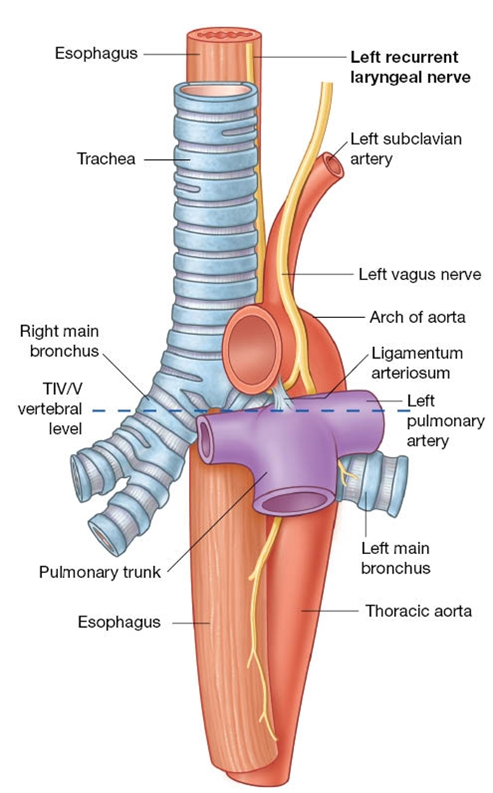
Left recurrent laryngeal is in the superior mediastinum.
right recurrent laryngeal nerve arises from the right vagus nerve and wraps underneath the right subclavian artery before ascending to the larynx.
It does not descend to the superior mediastinum
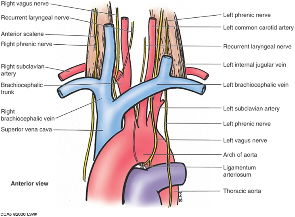
a deep dissection of superior mediastinum
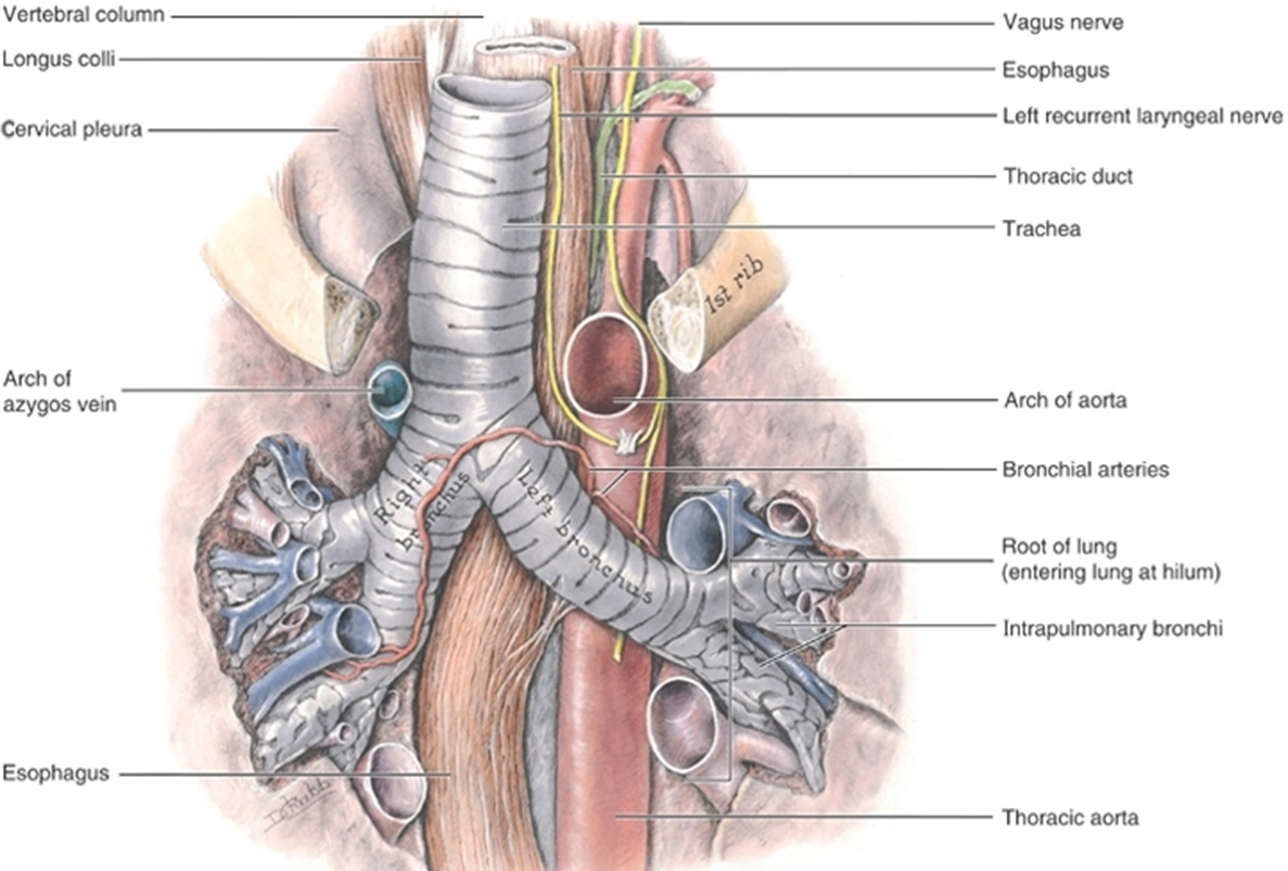
contents of posterior mediastinum
Thoracic aorta
Thoracic duct
Posterior mediastinal lymph nodes (tracheobronchial)
Azygos and hemiazygos veins
Esophagus and plexus
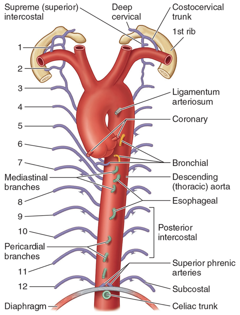
anterior view of thoracic aorta
Thoracic aorta begins at lower border of T4 body on left side. Approaching median plane as it descends to T12 vertebrae and passes through aortic hiatus.
Note the mediastinal, bronchial and esophageal, and paired branches.
Numbers 1-12 identify the paired posterior intercostal arteries and paired subcostal arteries.
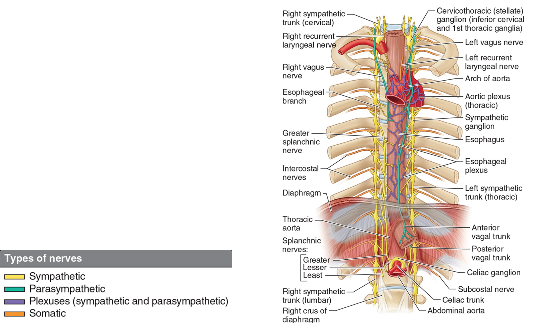
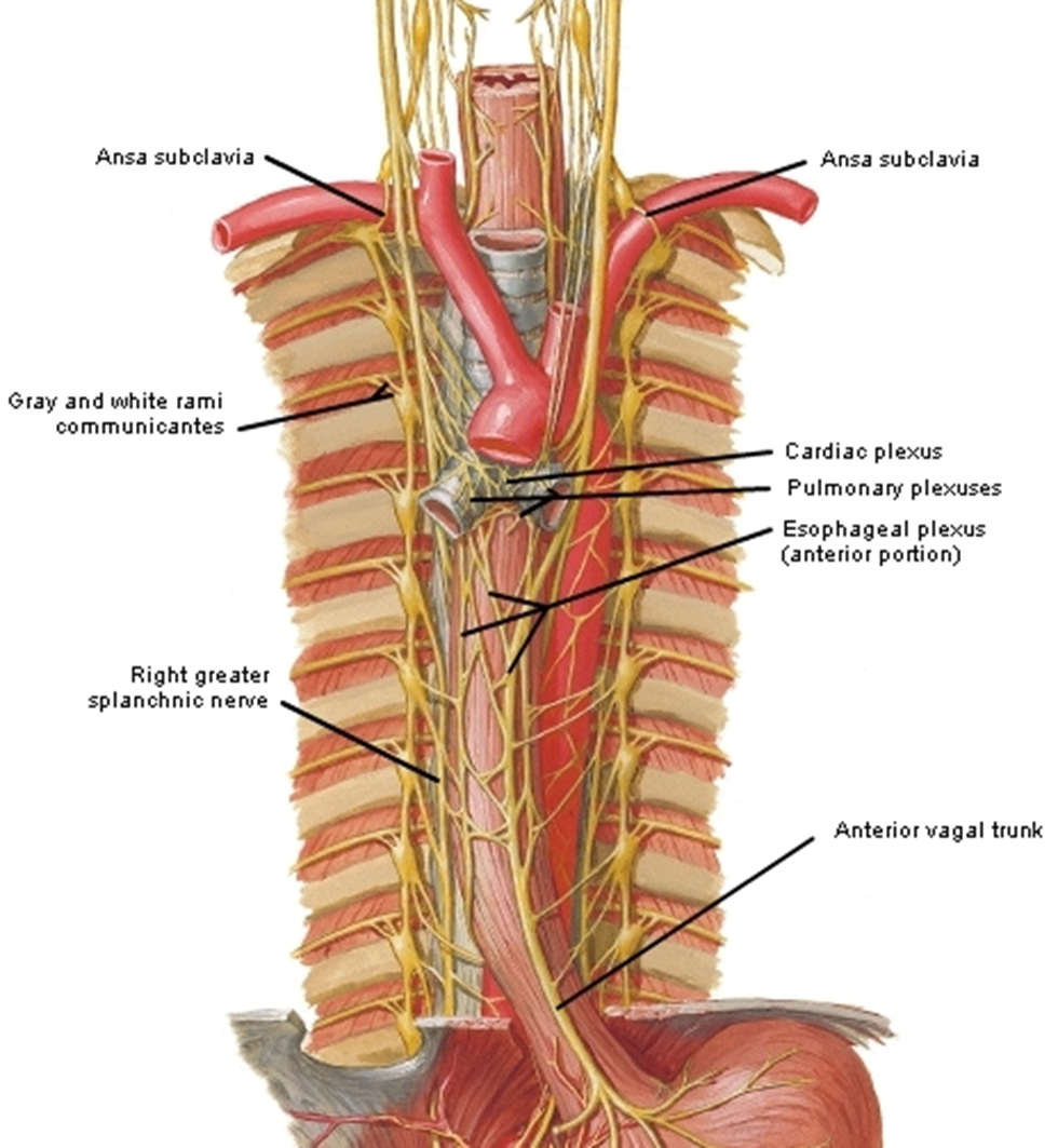
esophagus & nerve plexus
Esophagus is fibromuscular tube from pharynx to the stomach. Lies anterior to vertebral bodies. Passes through superior to posterior mediastinum.
Esophageal plexus: Branching fibers from both left and right vagus nerves mix in the esophageal plexus and then form trunks.
The anterior vagal trunk arises from the left vagus nerve.
The right vagus nerve gives rise to the posterior vagal trunk.
esophageal nerves
Parasympathetic innervation is from vagus nerves.
Sympathetic innervation is from upper thoracic ganglia.
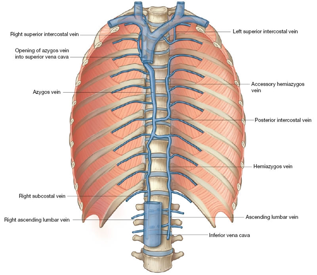
axygous venous system
Azygos vein forms a collateral pathway between SVC and IVC, and drains posterior wall of thorax and abdomen.
Forms from junction of subcostal and ascending lumbar vein at the L1 or L2 vertebral level.
Receives intercostal, accessory, and hemiazygos veins.
Arches over root of right lung and drains to the SVC.
Accessory hemiazygos: Crosses at T8 to empty into the azygos.
Hemiazygos: crosses at T9 to empty into the azygos.
Clinical note: azygos vein can provide a route for venous return to the heart if either SVC or IVC is obstructed.
thoracic duct
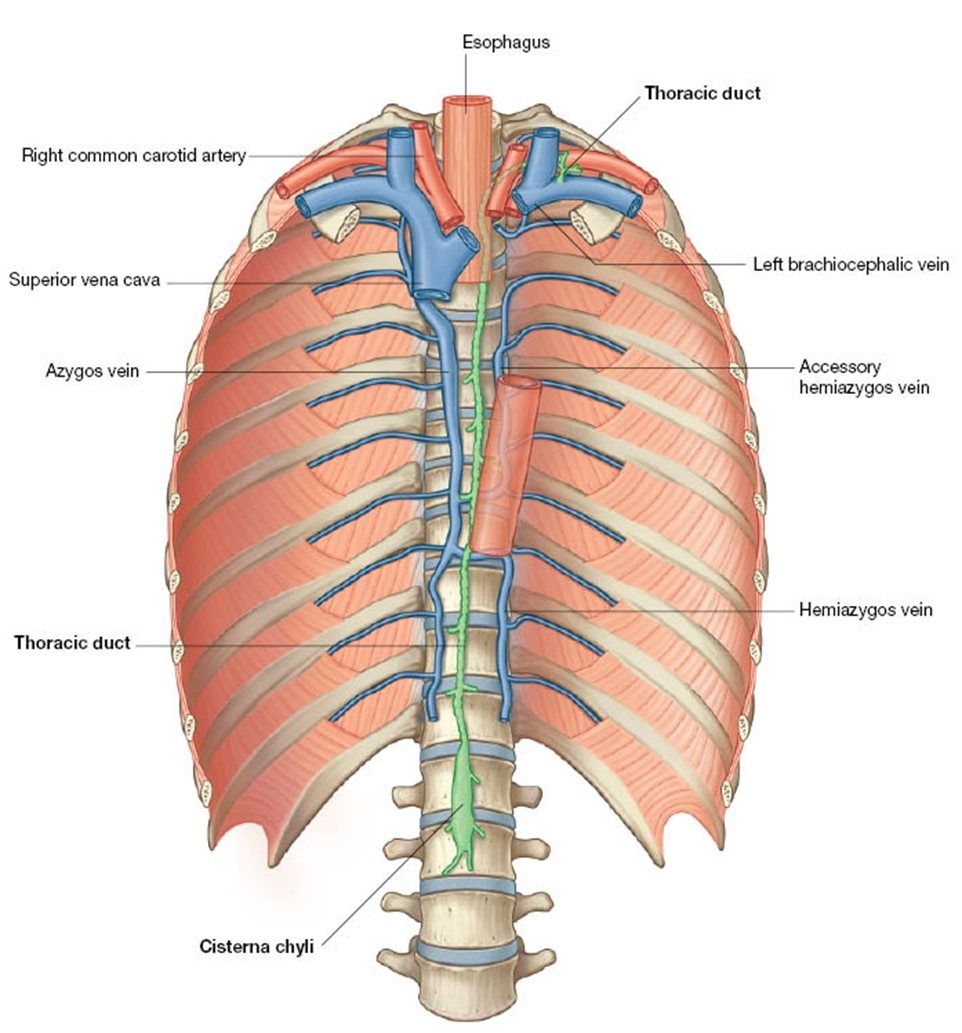
The thoracic duct (largest lymphatic vessel) begins at the cisterna chyli (L2 vertebral level).
Appears beaded due to valves.
Ascends to the right of midline between azygos vein and the aorta.
Traverses the diaphragm via the aortic hiatus.
At vertebral level T4, T5, or T6 it crosses to the left of midline and enters the superior mediastinum.
Empties on the left side at the junction of the internal jugular and left subclavian veins.
Clinical note: thoracic duct injury can lead to chylothorax (lymph in pleural cavity)
thoracic & right lymphatic duct
right lymphatic duct empties into the junction of the internal jugular and subclavian.
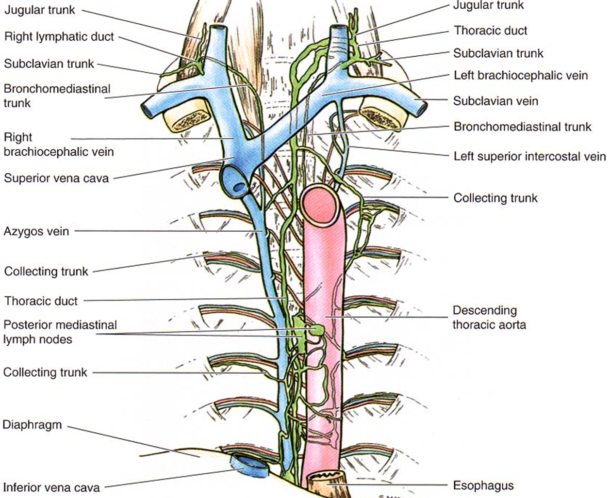
The right lymphatic duct drains right upper limb, head, neck, thorax.
Thoracic duct carries lymph drained from the rest of the body.
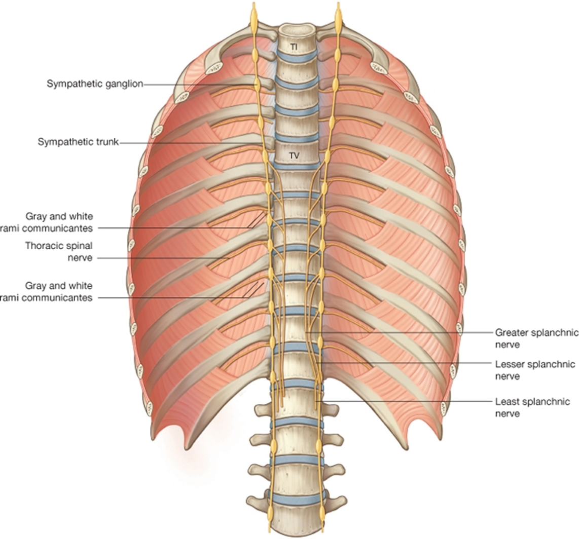
sympathetic trunks
Splanchnic nerves pass into the abdominal cavity carrying preganglionic sympathetic fibers to prevertebral ganglia.
Sympathetic ganglia and trunks. The ganglia are not in the posterior mediastinum.