Unit Three: Shoulder Complex
Unit Three: Shoulder Complex
Definitions
Shoulder complex: Refers to the entire shoulder region, encompassing multiple bones, joints, and muscles.
Shoulder girdle: Bony structure formed by the clavicle and scapula that connects the upper limb to the trunk.
Scapula & clavicle: The main bones of the shoulder girdle.
Shoulder joint: The glenohumeral joint, where the humerus and scapula connect.
Humeral head in glenoid fossa: The ball-and-socket arrangement of the shoulder joint, with the head of the humerus fitting into the glenoid cavity of the scapula.
Bones of the Shoulder Girdle
Clavicle:
Shape: "S" shaped bone connecting the axial skeleton to the appendicular skeleton at the upper extremities (UE).
Superior Surface: Smooth surface that serves as a muscular attachment site.
Inferior Surface: Contains multiple sites for ligament attachments.
Sternal End: Attaches to the manubrium of the sternum.
Acromial End: Attaches to the acromion of the scapula and serves as an attachment site for the upper trapezius (lateral 1/3).
Sternum
Parts of the sternum:
Manubrium
Body
Xiphoid process
Ribcage Structure
Connection of ribs: Ribs connect anteriorly and posteriorly (more details in Unit 4).
Costal Angles: The angles at which the ribs change direction from posterior to anterior.
Costal Tubercles: Points where ribs connect posteriorly to the thoracic vertebrae.
Scapula Anatomy
Scapula Characteristics:
Triangular shaped bone located on the posterior thorax.
Resting Position: Lies between ribs 2-7, with its medial border approximately 2-3 inches from the spinous processes.
Posterior Surface of Scapula:
Slightly convex with a scapular spine extending from the medial border to the acromion.
Attachment sites:
Middle trapezius
Lower trapezius
Superior Angle: Most superior portion of medial border, attachment for the levator scapulae muscle.
Inferior Angle: Point where medial and lateral borders meet, important for scapular rotation and palpation.
Supraspinous fossa: Area above the spine, serves as the attachment site for supraspinatus muscle.
Infraspinous fossa: Area below the spine, serves as the attachment site for the infraspinatus muscle.
Anterior Surface of Scapula (Costal Surface):
Slightly concave to fit on ribcage.
Subscapular fossa: Attachment site for subscapularis muscle.
Coracoid Process: Palpable from the front of the body, inferior to the clavicle; attachments for:
Pectoralis minor
Short head of biceps
Coracobrachialis
Acromion Process: Broad, flat area of the spine, attachment site for upper trapezius.
Medial Side of Scapula (Vertebral Side):
Base of scapular spine (root) and vertebral border; attachment sites:
Serratus anterior
Rhomboids
Lateral Side of Scapula (Axillary Side):
Lateral Border: Between glenoid fossa and inferior angle; attachment site for teres minor.
Glenoid Fossa: Socket of the shoulder joint; features:
Infraglenoid tubercle: Attachment site for triceps.
Supraglenoid tubercle: At superior border of glenoid, attachment site for long head of biceps.
Articulations of Shoulder Girdle
Sternoclavicular Joint:
Type: Synovial gliding joint, only direct attachment of shoulder girdle to trunk.
Movements: Elevation/depression, protraction/retraction, rotation. These occur in conjunction with shoulder blade motions.
Acromioclavicular Joint:
Type: Synovial gliding joint linking lateral clavicle to lateral scapula.
Movements: Elevation/depression, protraction/retraction, rotation (same conjunction with shoulder motions).
Glenohumeral Joint:
Type: Synovial ball-and-socket joint.
Movements: Flexion/extension/hyperextension, abduction/adduction, internal/external rotation, horizontal abduction/adduction.
Scapulothoracic Joint:
Not a true joint. Scapula rests on ribs, crucial for glenohumeral motion. Many shoulder issues arise from scapulothoracic dysfunction. Importantly discussed movements occur later.
Ligaments of Shoulder Girdle
Sternoclavicular Ligaments:
Anterior/posterior sternoclavicular ligament limits anterior/posterior sliding.
Costoclavicular ligament connects the first rib to the clavicle, limits clavicular elevation.
Interclavicular ligament is found between the two clavicles, limits clavicular depression.
Note: While informative, not tested on these details.
Acromioclavicular Joint:
Weak joint capsule.
Inferior/superior acromioclavicular ligament: Holds clavicle and acromion together, prevents dislocation. This is essential knowledge for assessments.
Coracoclavicular Ligament:
Connects the clavicle to the scapula, stabilizes and supports the AC joint.
It consists of:
Trapezoid Ligament: Lateral portion.
Conoid Ligament: Medial/deeper portion. Also essential for assessments.
Coracoacromial Ligament:
Connects lateral coracoid process to inferior acromion, forming a "roof" over the humeral head to prevent upward translation of forces Important for assessments.
Coracohumeral Ligament:
Connects the scapula to the humerus, from coracoid process to bicipital groove, and enhances upper joint capsule strength. Critical knowledge for assessments.
Motions of the Shoulder Girdle
Scapular Motions:
Elevation: All parts of the scapula move upwards on thorax.
Depression: All parts of the scapula move downwards on thorax.
Protraction: All parts move anteriorly away from vertebrae.
Retraction: All parts move posteriorly towards vertebrae.
Upward Rotation: Angular motion where the inferior angle and glenoid rotate up/down posteriorly and away from vertebral column, primarily involves:
Upper trapezius
Lower trapezius
Serratus anterior (force couple).
Downward Rotation: Returns the scapula to anatomical position; force couple involves:
Levator scapulae
Rhomboids
Pectoralis minor, accounting for gravity.
Tilt: Occurs during hyperextension of GH joint; results in posterior movement of inferior angle off ribcage with anterior tilt of superior border.
Winging: A pathological condition where the medial border lifts off ribcage, often caused by weakened serratus anterior.
Muscles of the Shoulder Girdle
Scapular Movers (Posterior):
Trapezius: Has upper, middle, and lower sections.
Rhomboids: Assist with retraction and stabilization of the scapula.
Levator Scapulae: Elevates the scapula.
Deltoid: Provides abduction and other motions.
Scapular Movers (Anterior):
Pectoralis Minor: Assists in protraction and depression of the scapula.
Scapular Movers (Lateral):
Serratus Anterior: Protracts and stabilizes the scapula against the ribcage.
Force Couples for Scapular Motion:
Upward Rotation Force Couple:
Upper trapezius
Lower trapezius
Serratus anterior
Downward Rotation Force Couple:
Levator scapulae
Rhomboids
Pectoralis minor
Concept Checks:
If upward rotation is impaired, explore possible causes.
Discuss intervention strategies as Physical/Occupational Therapy Assistants (PTAs/OTAs).
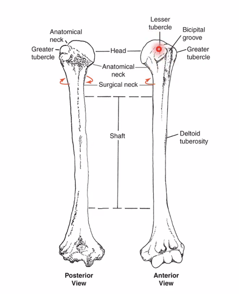
Ligaments and Supporting Structures of Shoulder Joint
Glenohumeral (GH) Joint Capsule:
Thin-walled, spacious ligament "sac" from the rim of the glenoid fossa to the anatomical neck of the humerus.
Outer fibrous layer and inner synovial lining for lubrication.
For full range of motion (ROM): Must maintain sufficient slack in ligaments; varies with arm positions.

Glenoid Labrum:
Fibrous cartilage ring around the glenoid, enhances stability, deepens the socket, providing further support to the shoulder as an inherently unstable joint.

Bursae:
Significant in the shoulder joint, providing protection to tendons against bone.
Commonly inflamed leading to "bursitis."

Motions of Shoulder Joint
Shoulder Movements:
Flexion
Extension
Abduction
Adduction
Internal Rotation (Medial)
External Rotation (Lateral)
Horizontal Abduction
Horizontal Adduction
Scaption (functional motion).
Concept Check: Understand the planes and axes of each shoulder movement.
Scapulohumeral Rhythm
Definition: The coordinated movements of the scapulothoracic and glenohumeral joints critical for full shoulder function, which occurs during flexion or abduction.
Phases of Scapulohumeral Rhythm:
Phase 1: 0-30 degrees - Purely GH joint movement with the involvement of the supraspinatus and deltoid.
Phase 2: 30-90 degrees - Combination of GH and scapular motion, approximately 40 degrees from humerus and 20 degrees from scapular upward rotation involving scapular upward rotators and deltoid.
Phase 3: 90-180 degrees - A combination of 60 degrees abduction and 30 degrees upward rotation by scapular rotators and deltoid.
Ratio: For every 2 degrees of GH motion, there must be 1 degree of scapular upward rotation, establishing a 2:1 ratio.
Glenohumeral Movements
Concave-Convex Rule:
Describes how the shape of the joint influences movement:
Convex surface moving on fixed concave surface moves in the opposite direction to body segment; e.g., as humerus elevates (roll), the humeral head depresses (glide) on the fixed glenoid.
Highlights why ligament capsule laxity is critical for function.
Concept Checks: During abduction and adduction, determine the directions of roll and glide.
During abduction, the humeral head rolls superiorly while gliding inferiorly on the glenoid, whereas during adduction, the humeral head rolls inferiorly and glides superiorly.
Muscles of the Shoulder Joint
Rotator Cuff Muscles:
Tendon structures blending together superficially to the capsule, providing crucial stabilizing support to the joint; collectively referred to as "SITS":
Supraspinatus
Infraspinatus
Teres Minor
Subscapularis
Shoulder Muscles (Scapula to Humerus):
Rotator Cuff Muscles:
Supraspinatus
Infraspinatus
Teres minor
Subscapularis
Deltoid Muscle Groups:
Anterior, middle, and posterior compartments.
Teres Major: Assists shoulder extension.
Coracobrachialis: Aids shoulder movements.
Muscle Grouping by Actions:
Evaluate shoulder muscles categorized by their functions:
Flexors: Scapular elevation.
Extensors: Scapular depression.
Abductors: Scapular protraction.
Adductors: Scapular retraction.
Horizontal Abductors/Adductors: Affect shoulder positioning.
Medial Rotators: Rotate shoulders inward.
Lateral Rotators: Rotate shoulders outward.
Innervation of Upper Extremity
Brachial Plexus Overview:
Comprised of spinal nerves from C5, C6, C7, C8, and T1 that combine and branch into major nerves innervating the upper extremities.
Brachial Plexus Structure:
Shows division into:
3 trunks
3 anterior divisions
3 posterior divisions
Cords and terminal branches linking to various upper extremity muscles.
Innervation Details:
Innervation of Muscles of the Shoulder (Lateral):
Axillary Nerve (C5, C6): Innervates:
Teres Minor
Deltoid
Rotator Cuff (RCF) Muscles:
Suprascapular Nerve (C5, C6): Innervates:
Supraspinatus
Infraspinatus
Subscapular Nerves:
Upper & lower subscapular nerve (C5-C7): Innervates subscapularis.
Lower subscapular nerve (C5, C6): Innervates teres major.
Posterior Muscles:
Dorsal Scapular Nerve (C5): Supplies:
Levator Scapulae
Rhomboids
Long Thoracic Nerve (C5-C7): Supplies serratus anterior and is crucial to avoid scapular winging, a dysfunction from long thoracic nerve damage.
Key Concepts
Understanding the complex interactions between the bones, ligaments, motions, and muscles of the shoulder girdle is vital.
Comprehension of scapulohumeral rhythm is crucial for optimizing shoulder function.
Recognizing the anatomical landmarks and innervation pathways will enrich assessment skills in clinical settings.