Respiratory Anesthesia Considerations
Quick Respiratory Anatomy
Flow from nostrils through: Nasal turbinates —> Vestibule (nasal cavity) —> Pharynx (naso/oro) —> Larynx —> Trachea —> Carina —> Bronchi —> Bronchioles —> Alveoli
Sinuses and nasal cavities drain into cervical lymph nodes.
Epiglottis: Covers the trachea to prevent entry of foreign bodies into the trachea/lungs.
Thyroid Cartilage: Also known as the Adam’s apple; this is where vocal cords are attached anteriorly.
Cricoid Cartilage: Located below the thyroid gland, indicating where the trachea begins.
Larynx Location: Spans from C3 to C6 vertebrae.
Trachea Location: Extends from C6 to T4-5 vertebrae.
Carina Location: Located at T4-5, at the sternal angle (Angle of Louis).
Alveoli: Form a tightly packed mesh near pulmonary capillaries where gas exchange occurs with blood.
Breathing Mechanism:
Spontaneous rhythm originates in the medulla.
Innervation: Diaphragm and intercostal muscles via Cranial Nerves IX & X for inspiration.
Expiration results from elastic recoil of muscles
Voice Production:
Muscles of the larynx control movement of thyroid and cricoid cartilage, facilitating movement of vocal cords to produce sound.
Innervation by Cranial Nerve X.
Static Compliance:
Definition: Pressure inside the lung when air isn’t moving
This is what keeps the lungs inflated.
Can be affected by disease processes and external factors.
Surfactant:
Produced by alveolar cells by 35 weeks gestation.
Function: Decreases surface tension on small airways to prevent alveoli from collapsing and promote gas exchange.
Anesthesia Considerations for Normal Respiratory Anatomy
Nasotracheal Intubation:
Tube should be directed into the nostril and advanced along the floor of the nasal cavity (not directed upwards towards turbinates).
Risks: Prolonged intubation can lead to sinus obstruction, infection, and fever; should be avoided if possible.
Note: Nasal infections increase the risk of meningitis due to communication between nasopharyngeal and intracranial circulation.
Measurements:
Incisor to larynx = 13 cm in adults.
Trachea: flexible; remember “Hose follows the Nose” to avoid accidental extubation risk factors.
Incisor to carina = 26 cm in adults.
Anesthesia Considerations for Normal Respiratory Anatomy (continued)
Breathing Function: “C3,4,5 keeps the diaphragm alive”
Spinal cord injuries above C5 require mechanical ventilation.
Static Compliance Impact:
Decreased pressure affects lung inflation; possible causes include fibrosis, Acute Respiratory Distress Syndrome (ARDS), or external compression.
Increased pressure means lungs are easy to deflate, leading to collapse of bronchioles and alveoli; causes can include emphysema.
Surfactant Production:
Can be hastened for pre-mature delivery by administering glucocorticoids antenatally or directly via neonatal airways to prevent Respiratory Distress Syndrome (RDS).
Respiratory Complications & Effect on Anesthesia
Obstructive Sleep Apnea (OSA)
Characterized by chronic hypoxia due to mechanical obstruction during sleep caused by relaxation of pharyngeal muscles, resulting in cessation of breathing for at least 10 seconds.
Risk Factors:
Most common, significant predisposing factor: Obesity.
Associated with increased risk of atherosclerosis, hypertension (HTN), stroke, diabetes mellitus (DM), postoperative complications, and morbidity.
Symptoms: Habitual snoring, fragmented sleep, daytime drowsiness.
Screening Tool: STOP BANG questionnaire.
Treatment Options: Weight loss and Continuous Positive Airway Pressure (CPAP).
Surgical Recommendations:
Use of positive-pressure oxygenation pre- and post-operation.
Conduct careful airway assessments: excessive neck tissue may complicate intubation and accelerate desaturation during induction.
Use pre-operative sedation cautiously.
Anticipate longer Post-Anesthesia Care Unit (PACU) stays and prolonged SpO2 monitoring.
Consider ICU admissions post-operation.
Evaluate the necessity of avoiding same-day discharge to home post-surgery.
Chronic Obstructive Pulmonary Disease (COPD)
Chronic hypoxia results from inflammatory responses of lung tissue that drive progressive, irreversible limitations of airflow.
Comprises both Chronic Bronchitis (defined as chronic mucous production for most days for at least three months/year over two years) and Emphysema (characterized by the destruction of lung parenchyma and alveoli).
Most common predisposing factor: Smoking.
Symptoms: Chronic cough, chronic sputum production, dyspnea upon exertion.
Screening Tool: BODE index.
Treatment Options: Smoking cessation, inhaled β2 Agonists, corticosteroids, anti-cholinergics, supplemental oxygen to maintain SpO2 > 88%.
Surgical Recommendations:
Assess resting SpO2 readings prior to surgery.
Conduct Pulmonary Function Tests (PFTs).
Be prepared for prolonged intubation and ventilation due to increased risk of Acute Respiratory Failure post-operation.
Consider regional anesthesia as an alternative to avoid airway manipulation/need for ventilation.
Avoid neuraxial anesthesia above T6 to prevent compromised coughing ability and anxiety from sensations of dyspnea.
Scans/Imaging
ETT in Chest X-ray
CXR for ETT placement is used to verify placement and rule out iatrogenic complications
15% of ETT may be malpositioned by inexperienced providers
Tip of ETT should be 5-7 cm above carina
Sometimes the carina is not easily visible, but you should know it is in the vicinity of T5-T6 and T6-T7 disk spaces
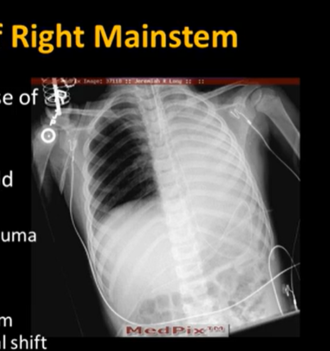
Right mainstem endobronchial intubation
Opacification of contralateral lung field
Due to atelactasis/collapse and hemothorax from trauma
Atelectasis: not always seen, but can include diaphragm elevation, mediastinal shift (towards the collapsed lung)
**more likely to occur than L mainstem
Left mainstem endobronchial intubation
ETT follows bronchus
Common to see opacification of contralateral lung field
Look for atelectasis and mediastinal shift
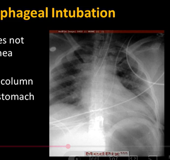
Esophageal intubation
The ETT does not follow the trachea
Secondary hypodense column
Distended stomach
Other complications of ETT:
ETT cuff over-distention leads to tracheomalacia
This risk if most likely if the cuff is 1.5x the diameter of the airway
barotrauma
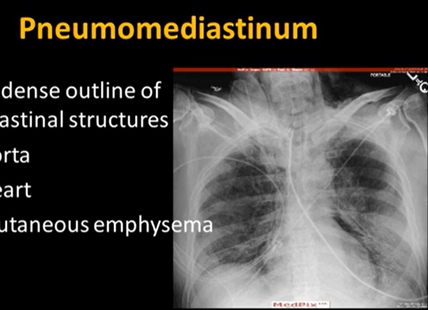
Pneumomediastinum
Hypodense outline of mediastinal structures
Aorta and heart
SQ emphysema is also present
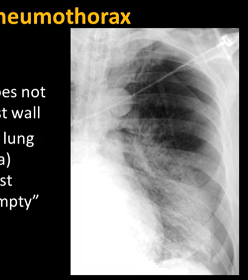
Pneumothorax
Pulmonary vasculature does not extend to chest wall
dense edge of lung (visceral pleura) outlined against hypodense “empty” thorax
Result of barotrauma
Main goals:
rule out tip misplaced
overdistention of cuff
barotrauma
Interpreting Chest X-ray
X-rays are less likely to penetrate denser materials
Like photographic negatives
darkest parts of the film are areas of the lungs (where photons can penetrate more areas)
sharp white areas are where dense bone material blocks photons from getting through
Checklist: ABCDEFG
1) Assessment: Begin assessing patient and exam data. Inspect image quality (no excess rotation of the patient, should see 10th or 11th posterior ribs for full inspiration, check that you can see fine markings)
Also Air where it shouldn’t be (e.g., pneumothorax, pneumomediastinum, pneumoperitoneum, and SQ emphysema). Even includes deviated trachea!
2) Bones. Check there are no deformities and you can see 12 ribs.
Also Body wall (there should be soft tissue outside of the chest). Can have swelling or masses.
3) Cardiac silhoutte and size (there should be a visible atrial appendage and Right atrium and Left ventricle)
The heart diameter should be 50% of the entire ribcage (from inner lung borders). Otherwise, could indicate cardiac pathology.
4) Diaphragm (it should be symmetric, not flat)
You can estimate diaphragm flatness in lateral view when it is >1.5 cm above the line connecting front to back lung walls
5) Equipment: lines, tubes, and wires (e.g., ETT should be in middle of trachea, NGT should have tip in stomach)
Pleural Effusion (causes a blunt angle of the normally sharp costafrenic angle)
6) Lung fields (should look symmetric with no haziness, white dots, or blotches)
7) Great vessels (should all be in right location and right size. Highest should be aortic arch/pulmonary artery. Deviation can be a result of congenital disease.