Bio test 6.1-6.3
What is going to be on the 6.1-6.3 test
The functions of the stomach: the large and small intestines
The control of the heart beat
Why Antibiotics are effective against bacteria
What type of sugar lactose is
How the structure of the villus is adapted for absorption
Know the need for enzymes in digestion
Be able to explain the structure of arteries and veins and how they are adapted for their function
Be able to distinguish between coronary thrombosis and atherosclerosis and list two factors that increase their risk
Be able to draw and label the diagram of the digestive system of the heart
Be able to discuss the causes, transmission, and social implications of AIDS
6.1 Digestion and absorption
The process by which food is broken down into smaller components that can be absorbed by the body, including mechanical and chemical digestion.
Functions of the stomach, small intestine, and large intestine
Stomach:
it is a muscular organ that stores and breaks down food.
it secretes (releases) gastric juice, containing:
Hydrochloric acid (HCI): this kills pathogens and provides the acidic pH for enzymes.
Pepsin (a protease): this enzyme breaks down proteins into smaller polypeptides.
Mucus: this protects the stomach lining from being digested by its own enzymes.
the stomach churns food into a semi-liquid mixture called chyme, which is gradually released into the small intestine.
The small intestine
the main site of digestion and absorption.
enzymes from the pancreas and intestinal wall continue digestion:
Amylase: this breaks down starch into maltose.
Lipase: this breaks down lipids into glycerol and fatty acids
Proteases (e.g., trypsin, peptidase): this breaks down proteins into amino acids.
bile (from the liver and stored in the gallbladder) breaks fat into smaller droplets for easier digestion.
villi and microvilli increase the surface area for absorption of nutrients into the blood and lymph.
The large intestine
absorbs water and minerals from undigested food, ultimately forming solid waste that is excreted from the body.
houses gut bacteria which aid digestion and synthesize vitamins like vitamin K and B12.
forms and stores feces before excretion.
Additionally, the large intestine plays a crucial role in maintaining electrolyte balance and supporting overall gut health by providing a habitat for beneficial microorganisms.
Need for enzymes in digestion
Why are enzymes necessary?
digestion is a slow chemical process, and enzymes speed it up by lowering activation energy.
they also ensure food is broken down into molecules small enough for absorption.
Examples of key digestive enzymes and their functions:
Enzyme | Produced by | Function |
amylase | salivary glands and pancreas | starch → maltose
|
maltase | small intestine | maltose → glucose
|
proteases (pepsin, trypsin) | stomach and pancreas | proteins → amino acids
|
lipase | pancreas | lipids → fatty acids + glycerol
|
The structure of the villus and adaptions for absorption
villi are tiny, finger-like projections lining the small intestine that increase surface area for nutrient absorption.
each villus contains:
Thin epithelium: a single layer of cells to minimize diffusion distance.
Capillaries: they transport absorbed glucose, amino acids, and other nutrients into the bloodstream.
Lacteal (lymph vessel): they absorb fatty acids and glycerol into the lymphatic system.
Microvilli: tiny projections on the surface of the epithelial cells that increase the surface area for absorption.
What type of sugar is lactose?
lactose is a disaccharide, meaning it consists of two simple sugars:
Glucose + Galactose
it is broken down by lactase in the small intestine.
people who are lactose intolerant lack lactase and cannot properly digest lactose, leading to digestive discomfort.
6.2 The blood system
The blood system is responsible for transporting nutrients, gases, hormones, and waste products throughout the body, ensuring that cells receive the necessary substances for metabolism and function.
Control of the heartbeat
the heart is myogenic, meaning it can beat without input from the brain.
the contraction is controlled by specialized pacemaker cells:
Sinoatrial Node (SA Node):
it is located in the right atrium.
it acts as the pacemaker, setting the rhythm of the heartbeat.
Atrioventricular Node (AV Node):
it delays the signal to ensure the atria contract before the ventricles.
Bundle of His & Purkinje Fibers:
it spreads the signal throughout the ventricles, causing them to contract.
Heart rate regulation:
the medulla (brainstem) adjusts the heart rate:
Sympathetic nerve → it speeds up the heart rate during stress or exercise.
Parasympathetic nerve → it slows it down during rest.
Adrenaline (epinephrine):
it is released by adrenal glands in response to stress which increases the heart rate.
Structure of arteries vs veins and their functions
Feature | Arteries | Veins |
Function | It carries oxygenated blood away from the heart.
| It carries deoxygenated blood back to the heart.
|
Pressure | High | Low |
Wall thickness | Thick to withstand the high pressure. | Thin, since the pressure is lower. |
Lumen size | Narrow to maintain the high pressure. | Wide to allow smooth blood flow and maintain low blood pressure. |
Valves | No valves, as high pressure prevents backflow. | Valves to prevent backflow. |
Coronary thrombosis vs atherosclerosis + risk factors
Atherosclerosis:
it is the buildup of fatty plaques (cholesterol deposits) in arteries that narrows them.
this can lead to high blood pressure and block blood flow.
Coronary Thrombosis:
it is a blood clot that forms in the coronary arteries, blocking blood flow to the heart.
this can cause heart attacks.
Risk factors:
smoking
high-fat diet
high blood pressure
diabetes
lack of exercise
Digestive system and heart drawing and labels
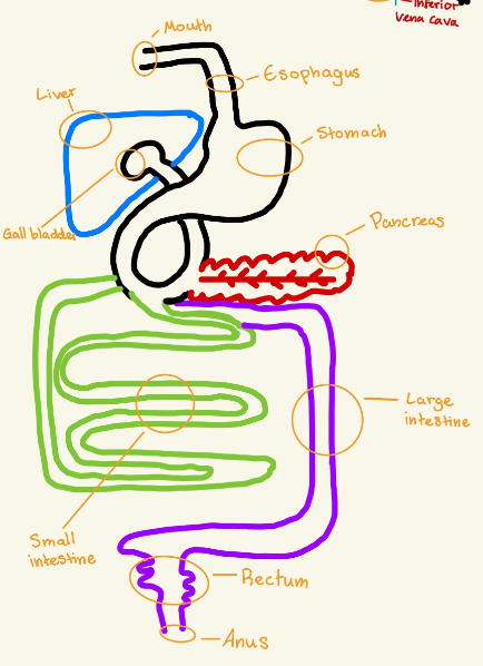
Mouth (Mechanical Digestion): Chews food and mixes it with saliva, which moistens food to make swallowing easier.
Esophagus: A muscular tube that moves food from the mouth to the stomach using wave-like contractions (peristalsis).
Liver: Makes bile, which helps break down fats in the small intestine. Also processes nutrients and removes toxins.
Stomach (Mechanical & Chemical Digestion): A muscular organ that churns food and uses acid and enzymes to break down proteins.
Pancreas: Produces enzymes to help digest carbs, proteins, and fats in the small intestine. Also releases hormones to control blood sugar.
Gall Bladder: Stores bile made by the liver and releases it into the small intestine to help digest fats.
Small Intestine: This breaks down food further with enzymes and absorbs nutrients into the bloodstream.
Large Intestine: Absorbs water and minerals from leftover food and turns waste into solid feces.
Rectum: Stores feces until it's ready to be released from the body.
Anus: The opening where waste leaves the body.
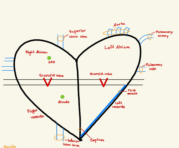
Superior Vena Cava: A large vein that carries oxygen-poor blood from the upper body to the right atrium.
Right Atrium: The top right chamber of the heart that receives oxygen-poor blood from the body.
SA Node (Sinoatrial Node): The heart’s natural pacemaker that sends electrical signals to make the heartbeat.
Tricuspid Valve: A one-way valve between the right atrium and right ventricle that prevents blood from flowing backward.
AV Node (Atrioventricular Node): A relay station that slows down the electrical signal before it moves to the ventricles, allowing the atria to contract first.
Right Ventricle: The bottom right chamber of the heart that pumps oxygen-poor blood to the lungs.
Inferior Vena Cava: A large vein that brings oxygen-poor blood from the lower body to the right atrium.
Aorta: The largest artery in the body that carries oxygen-rich blood from the heart to the rest of the body.
Pulmonary Artery: Carries oxygen-poor blood from the right ventricle to the lungs to pick up oxygen.
Left Atrium: The top left chamber of the heart that receives oxygen-rich blood from the lungs.
Pulmonary Vein: Carries oxygen-rich blood from the lungs to the left atrium.
Bicuspid Valve (Mitral Valve): A one-way valve between the left atrium and left ventricle that prevents blood from flowing backward.
Thick Muscle: The left ventricle has the thickest muscle because it needs to pump blood to the whole body.
Left Ventricle: The bottom left chamber of the heart that pumps oxygen-rich blood to the body through the aorta.
Septum: A wall of muscle that separates the left and right sides of the heart.
6.3 Defense against Infectious Diseases
The immune system plays a crucial role in protecting the body against pathogens by using various mechanisms such as physical barriers, immune responses, and the production of antibodies.
Why do antibiotics work against bacteria but not viruses?
Antibiotics kill bacteria by:
Blocking cell wall synthesis: Bacteria have a protective outer layer (the cell wall) that helps them keep their shape and survive. Antibiotics can stop the bacteria from building this wall, causing it to burst open.
Inhibiting protein synthesis: Bacteria need to make proteins to survive. Antibiotics can block their ability to make proteins, which means the bacteria can’t produce important enzymes or structures to stay alive.
antibiotics only kill bacteria and don’t work against viruses, and overuse can lead to bacteria becoming resistant to treatment.
Why don’t antibiotics work on viruses?
No cell wall: Bacteria have a protective layer (called a cell wall) that antibiotics can destroy. Viruses don’t have this, so antibiotics can’t attack them.
No ribosomes: Ribosomes are like factories in cells that make proteins. Bacteria have their own ribosomes, but viruses use the host cell's ribosomes to make new viruses. Antibiotics can’t stop this because viruses don’t have their own.
No metabolism: Bacteria can turn food into energy on their own, but viruses can’t. They need a host cell to do this. Since antibiotics target how bacteria use energy, they don’t work on viruses because viruses don’t have their own energy process.
Since viruses don’t have these things, antibiotics don’t have anything to attack or stop.
Antibiotic resistance
overuse: if we take antibiotics too often, then some bacteria can get used to them.
survival of the fittest: some bacteria already have tiny mutations that make them more resistant to medicine. These bacteria survive the treatment and then multiply, creating more bacteria resistant to that medication.
overtime, more and more bacteria can become resistant to the antibiotics, making the medicine less effective.
An example of this is MRSA (a bacteria that doesn’t respond to normal antibiotics). It happens because resistant bacteria keep surviving and spreading due to the overuse of antibiotics. Antibiotic resistance happens when bacteria learn to fight back, and the medicine doesn't work as well anymore.
Causes of AIDS
AID stands for Acquired Immunodeficiency Syndrome. It is caused by a virus called HIV (Human Immunodeficiency Virus).
HIV attacks a type of white blood cell called helper T cells. These cells are important because they help the body’s immune system recognize and fight off infections.
When HIV destroys enough helper T cells, the body’s immune system gets weak, making it harder for the person to fight infections. This stage is called AIDS.
Transmission of HIV
HIV spreads in a few different ways:
Unprotected sex: when a person has sex without a condom with someone who has HIV, the virus can be passed through bodily fluids like semen or vaginal fluids.
Sharing needles: if two people use the same needle (like for drug use), they can pass HIV through infected blood.
Blood transfusions: in the past people could get HIV from receiving infected blood but now blood is tested before being used so this is very rare now.
Mother-to-child: a mother with HIV can pass the virus to her baby during childbirth or through breastfeeding.
AIDS is caused by HIV, which weakens the immune system.
HIV spreads mainly through unprotected sex, sharing needles, blood transfusions, and from mother to child.
Socially, HIV/AIDS can cause stigma, lead to economic problems, and is addressed with education to prevent the virus from spreading.
Economic burden: HIV/AIDS requires long-term treatment and care, which can be expensive. The cost of healthcare for people with HIV puts a financial strain on both families and healthcare systems.
Keywords
Digestive system
Absorption: The process where tiny food molecules pass through the small intestine into the blood or lymph to be used by the body.
Amino Acid: The smallest building block of proteins. Proteins are broken down into amino acids so the body can use them to build new proteins.
Bile: A yellow-green liquid made by the liver that breaks large fat droplets into smaller ones, making it easier for lipase to digest them.
Capillary: The smallest type of blood vessel, which allows nutrients and oxygen to move into body cells and wastes to move out.
Chyme: A thick, soupy liquid made of partially digested food and stomach acid, which moves from the stomach to the small intestine.
Digestive Enzyme: A type of enzyme that cuts large food molecules into smaller pieces so the body can absorb them. Example: Lipase breaks fats into glycerol and fatty acids.
Disaccharide: A sugar made of two linked simple sugars. Example: Lactose is a disaccharide made of glucose and galactose.
Emulsification: The process where bile breaks fat droplets into smaller ones, making digestion easier.
Epithelium: A thin layer of cells that covers the inside of organs, such as the small intestine, helping with absorption.
Fatty Acid: A building block of fats that can be absorbed into the bloodstream and used for energy.
Glycerol: A part of fats that, along with fatty acids, is absorbed and used for energy.
Lacteal: A tiny lymph vessel inside a villus that absorbs fats (fatty acids and glycerol) and carries them into the lymphatic system.
Lipase: A digestive enzyme that breaks down fats into glycerol and fatty acids.
Microvilli: Tiny projections on villi that increase the surface area for absorbing nutrients.
Monosaccharide: The smallest type of sugar that can be absorbed. Example: Glucose is a monosaccharide that provides energy.
Peristalsis: A wave-like squeezing movement made by muscles in the digestive tract to push food forward.
Polypeptide: A chain of amino acids that is cut into smaller pieces by enzymes like proteases.
Protease: A type of enzyme that breaks down proteins into smaller polypeptides and amino acids. Example: Pepsin cuts proteins into polypeptides in the stomach.
Villus (plural: Villi): A finger-like structure in the small intestine that absorbs nutrients into the bloodstream.
Blood system
Adrenaline (Epinephrine): A hormone released by the adrenal glands that tells the heart to beat faster during stress or excitement.
Aorta: The largest artery in the body, which carries oxygen-rich blood away from the heart to the rest of the body.
Atherosclerosis: A disease where fat builds up inside arteries, blocking blood flow and increasing heart disease risk.
Atrioventricular Node (AV Node): A small group of heart cells that receives the signal from the SA node and delays it so the atria can finish contracting before the ventricles pump.
Atrium (plural: Atria): The top two chambers of the heart that collect blood before it moves into the ventricles.
Blood Clot (Thrombus): A solid mass of blood cells and proteins that can block blood flow in arteries and veins.
Capillary: The smallest type of blood vessel, which allows oxygen, nutrients, and waste to pass between blood and cells.
Coronary Arteries: The arteries that supply the heart muscle with oxygen and nutrients.
Coronary Thrombosis: A blood clot in the coronary arteries, which blocks blood flow to the heart and can cause a heart attack.
Lumen: The hollow space inside a blood vessel where blood flows.
Myocardial Infarction: The medical term for a heart attack, caused when blood flow to the heart is blocked.
Myogenic: A property of the heart that means it can beat on its own, without needing a signal from the brain.
Pacemaker: A group of heart cells (SA Node) that sends electrical signals to make the heartbeat.
Sinoatrial Node (SA Node): The natural pacemaker of the heart, which starts the heartbeat and sets the rhythm.
Stroke: A condition where the blood supply to the brain is blocked, often caused by a clot or a broken blood vessel.
Valve: A flap in the heart or veins that prevents blood from flowing backward.
Ventricle: The bottom two chambers of the heart that pump blood out to the lungs and body.
Defense against disease
Acquired Immunodeficiency Syndrome (AIDS): A disease where the immune system is too weak to fight infections, caused by HIV.
Antibiotic: A medicine that kills bacteria by stopping their growth or breaking their cell walls.
Antibiotic Resistance: When bacteria develop ways to survive antibiotics, making infections harder to treat.
Antibody: A Y-shaped protein made by white blood cells that attach to pathogens to help destroy them.
Bacteria: Single-celled microorganisms that can cause infections but can be killed by antibiotics.
Helper T Cell: A type of white blood cell that activates the immune system to fight infections. HIV destroys helper T cells, making it harder to fight disease.
HIV (Human Immunodeficiency Virus): A virus that attacks helper T cells, weakening the immune system and leading to AIDS.
Immune System: The body's defense system that fights infections and destroys harmful microbes.
Lymphocyte: A type of white blood cell that makes antibodies to attack pathogens.
Pathogen: Any microorganism that invades the body and causes disease (e.g., bacteria, viruses, fungi, parasites).
Phagocyte: A type of white blood cell that engulfs and digests pathogens.
Plasma Cell: A type of white blood cell that makes antibodies to fight infections.
Retrovirus: A type of virus (like HIV) that inserts its genetic material into the DNA of host cells, making it harder to cure.
Virus: A tiny infectious agent that invades host cells and forces them to make more viruses. Unlike bacteria, viruses cannot be killed by antibiotics.