Structure and Functions of Major Body systems
Integumentary System
The following make up the integumentary system.
Skin: Responsible for protection, temperature regulation, sensation, excretion, and vitamin D production
Epidermis layer: Outermost layer of epithelial tissue, covers the external surface of the body
Dermis layer: Thick layer beneath the epidermis that contains arteries, veins, nerves
Subcutaneous layer: Loose, connective tissue composed of adipose tissue and lipocytes
Hair follicles: Generate hair
Sebaceous (oil) glands: Produce sebum to keep skin and hair soft and prevent bacteria from growing on the skin
Fingernails and toenails: Protect the ends of fingers and toes
Sudoriferous (sweat) glands: Produce sweat to aid in cooling the body
The largest organ of the body is the skin. Hair, nails, and glands are accessory organs of this system. The skin has several functions, which the accessory organs also aid in.
Skeletal System
The skeletal system includes the following.
Axial skeleton: The adult axial skeleton has 80 bones, including the skull, vertebrae, and ribs.
Appendicular skeleton: The adult appendicular skeleton has 126 bones, including arms, legs, and pelvic girdle.
Ligament: The ligaments attach bone to bone for joint stability.
Tendons: The tendons join muscles to bones that help in moving extremities. Tendons in the muscular system help avoid muscle injury by absorbing some of the impact muscles take.
Connective Tissue/Cartilage: This tissue maintains, protects, and gives form to other tissues and organs. Cartilage is a part of connective tissue found in the larynx and respiratory tract. It also covers and protects the end of long bones.
The skeletal system gives the body structure and posture, as well as protecting the soft internal organs from injury. The skeletal system also plays a key role by serving as attachment points for muscles in the body. This symbiotic relationship between bones and muscles often results in the systems being referenced as one (the musculoskeletal system). Bones of the skeletal system are classified by shape.
Muscular System
The muscular system is made up of the following.
Skeletal muscle: Responsible for body movement; also called voluntary muscle or striated muscle
Smooth muscle: Found within the walls of hollow organs and blood vessels and in the iris of the eye; also called involuntary muscle
Cardiac muscle: Found only in the heart; cross-fibered to allow the heart to contract from the top and bottom to pump blood
Tendons: Ends of skeletal muscles that attach the muscle to a bone
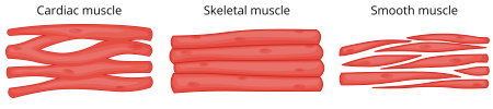
Skeletal muscle types include cardiac skeletal, and smooth.
Types of muscle
Muscles are responsible for movement, both voluntary (like walking) and involuntary (like digestion). The heart muscle is made of specialized fibers that allow it to function as a pump. The muscles and skeleton work together to provide posture, movement, and other essential body functions.
Immune and Lymphatic Systems
The lymphatic system includes the following.
Lymph nodes: Small, glandular structures concentrated in the neck, axilla, and groin that produce and store lymphocytes. Lymph nodes are home to macrophages that filter lymph.
Lymph nodules: Masses of lymphoid tissue comprised of macrophages and lymphocytes. Lymph nodules are not encapsulated like lymph nodes.
Thymus: Located posterior to the sternum. The thymus is large in children and atrophies (shrinks) after adolescence. It is responsible for the production and maturation of T-cells.
Spleen: Largest lymphoid organ, located in the upper-left quadrant of the abdomen. It is home to macrophages that filter the blood.
Interstitial fluid: Tissue fluid found between cells. Once collected and filtered, it is called lymph.
The immune system relies on the lymphatic system to prevent infections in the body. When a pathogen is detected, the lymphatic system begins activating the body’s defenses. A major component of these defenses are B-cells and T-cells.
Antigen: Foreign substance within the body
Antibody: Protein the body creates in response to specific antigens
Immunoglobulins: Antibodies
B-cells: Type of lymphocyte that can recognize antigens and responds by turning into plasma cells; these plasma cells then create antibodies against specific antigens
T-cells: Type of lymphocyte that can recognize antigens and attaches to them to attack the invading cells directly
Monocytes: Engulf and destroy pathogens that have been coagulated with antibodies
Take Note
Creating immunity, or the ability to resist pathogens, is an essential function of the immune system. People are born with some immunity and develop more over time.
Some immunity occurs from being exposed to pathogens, and other types of immunity are developed through immunizations. There are four primary types of immunity.
Types of Primary Immunity
Naturally acquired active immunity: This occurs when a person has an infectious disease and then develops antibodies against the pathogen that caused the disease. The antibodies have a memory that prevents future infections by the same pathogen.
Artificially acquired active immunity: This type of immunity is the result of administering a vaccination. The antibodies are activated by the vaccine and develop memory to recognize the pathogen in the future.
Naturally acquired passive immunity: This is a short-lasting immunity transferred through the placenta and breast milk.
Artificially acquired passive immunity: Also a short-lasting immunity, this is created by giving an exposed person antibodies containing blood products, as in an immune globulin.
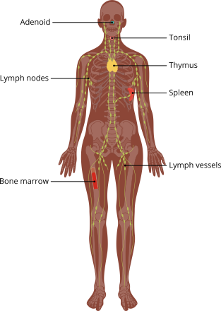
The organs of the immune system include the adenoids, lymph nodes, bone marrow, lymph vessels, spleen, thymus, and tonsils.
Cardiovascular System
The cardiovascular system is made up of the following.
Heart: Located within the central part of the chest (mediastinum) and functions as a pump to move blood throughout the body
Artery/Arteriole: Thick-walled vessels that carry blood away from the heart. They propel blood with each contraction of the heart and are associated with various pulse points on the body. Smaller branches are arterioles.
Vein/Venule: Vessels that carry blood toward the heart. They are thinner-walled than arteries and contain valves to prevent backflow. Smaller branches are venules.
Capillary: The smallest blood vessels, which connect arterioles to venules. They aid in the exchange of oxygen and nutrients between blood and body cells.
Endocardium: Innermost layer of cells that lines the atria, ventricles, and heart valves
Myocardium: Muscular layer of the heart
Pericardium: Outermost layer of the heart; a membrane that surrounds the heart and secretes pericardial fluid
The structures of the cardiovascular system work together to pump blood throughout the body. Blood carries essential oxygen and nutrients to cells and aids in eliminating cell waste. Blood travels to the heart to be pumped to the lungs for oxygen and then back to the heart to travel to the rest of the body.
Take Note
The average adult heart beats 60 to 80 times per minute while at rest.
The primary organ of the cardiovascular system is the heart. The heart is a muscle made up of three layers—the epicardium (outermost layer), myocardium (middle layer, thickest) and endocardium (inner layer, which is part of the electrical conduction system).
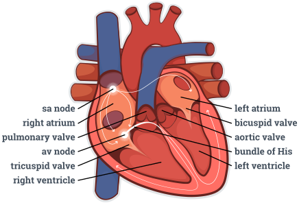
The heart
The heart contains four inner chambers. The right and left atria are the top chambers of the heart. The right atrium receives deoxygenated blood from the superior and inferior vena cava. The left atrium receives oxygenated blood from the pulmonary veins (the only veins in the body that carry oxygenated blood). The right and left ventricles are the bottom chambers of the heart. The right ventricle receives blood from the right atrium and sends deoxygenated blood through the pulmonary valve to the pulmonary artery and then to the lungs, where gas exchange occurs. The left ventricle receives blood from the left atrium and sends the oxygenated blood through the aortic valve to the aorta, which then branches off into smaller arteries that carry the blood to the body.
Between the right atrium and right ventricle is the tricuspid valve. Between the left atrium and left ventricle is the bicuspid (mitral) valve. The purpose of these valves is to prevent the backflow of blood into the atria when the ventricles contract.
Circulation of the blood occurs through two pathways—systemic and pulmonary circulation. Systemic circulation consists of arteries, arterioles, capillaries, venules, and veins in the body as a whole. Pulmonary circulation consists of arteries, arterioles, capillaries, venules, and veins going to, within, and coming from the lungs.
The heart contains its own electrical conduction system to keep the cardiac muscle contracting and blood flowing. This electricity can be mapped and analyzed to detect heart issues using an electrocardiogram (EKG).
The electrical impulse is generated by the sinoatrial (SA) node, also called the pacemaker of the heart.
From the SA node, the impulse travels to the atrioventricular node, also called the gatekeeper. From there, the impulse travels to the bundle of His and through the bundle branches located in the ventricular septum. Finally, the electrical impulse reaches the Purkinje fibers. These fibers cause the ventricles to contract and pump blood into the pulmonary artery and aorta. This entire process is the cardiac cycle.
Urinary System
The urinary system consists of the following.
Kidneys: Located on either side of the vertebral column at the level of the top lumbar vertebrae, the kidneys are responsible for removing waste from the blood and producing urine.
Ureters: These long tubes are responsible for carrying urine from the kidneys to the urinary bladder.
Urinary bladder: This small muscular sac located within the pelvic cavity is responsible for storing urine.
Urethra: This tube is responsible for carrying urine from the urinary bladder to the outside of the body and is longer in males due to pelvic shape and position of the prostate. The urinary meatus is located at the end of the urethra where urine exits the body.
The urinary system is primarily responsible for filtering blood to remove waste products. This waste is then prepared for elimination by combining with water to form urine. Urine is produced in the kidneys and then stored in the urinary bladder to await elimination.
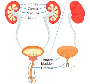
Gastrointestinal System
The gastrointestinal system is also known as the gastrointestinal tract. It begins with the mouth, or oral cavity. Digestion plays a vital role in the body’s ability to maintain homeostasis. Water and nutrients are essential for proper function of body systems, as well as organ, tissue, and cellular function. The primary organs of the digestive system collectively make up the alimentary canal. There are also accessory organs of the digestive system that aid in various digestive functions.
The following are part of the gastrointestinal system.
Mouth (oral cavity): This is responsible for initiating digestion, both mechanical (chewing) and chemical (saliva).
Pharynx: This includes the throat (the passageway for food between the oral cavity) and the esophagus (also part of the respiratory system).
Esophagus: This muscular tube connects the mouth to the stomach. It uses wave-like contractions called peristalsis to propel food into the stomach.
Stomach: Located below the diaphragm in the left upper quadrant (LUQ) of the abdominal cavity, the stomach receives food from the esophagus and continues breakdown using gastric juices. It then propels food to the small intestine. The stomach lining contains folds called rugae, allowing expansion.
Small intestine: This organ takes up most of the space within the abdominal cavity and is primarily responsible for absorption of nutrients. It is divided into the following sections.
Duodenum
Jejunum
Ileum
Large intestine: Also called the colon, the large intestine completes absorption and forms feces from solid waste products. It is divided into the following sections.
Cecum, which connects to the ileum, where the appendix is located
Ascending colon
Transverse colon
Descending colon
Sigmoid colon
Rectum: This is the end of the colon that stores feces until defecation.
Anus: This is the end of the rectum, which opens to the outside of the body to allow for elimination of feces.
Liver: This large organ is located in the right upper quadrant (RUQ) of the abdomen. It produces bile needed to break down fats.
Gall bladder: Located inferior to the liver, the gall bladder stores bile and connects to the duodenum.
Pancreas: Posterior to the stomach and connected to duodenum, the pancreas produces enzymes that aid with digestion
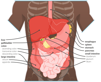
The digestive tract begins with the mouth and includes the esophagus, liver, stomach, gallbladder, pancreas, large and small intestines, cecum, appendix, rectum, and anus.
Gastrointestinal system
Respiratory System
The following are part of the respiratory system.
Nose: The nose is made of bones, cartilage, and skin. It contains small hairs called cilia to prevent large particles from entering.
Pharynx: During respiration, air enters through the nose and mouth into the pharynx. The pharynx is also part of the digestive system.
Larynx: Superior to the trachea, the larynx produces a person’s voice.
Trachea: Also called the windpipe, the trachea extends from the larynx and branches into bronchi. It is lined with cilia.
Lungs: These two cone-shaped organs are located in the chest. The lungs contain bronchi, alveoli, and many blood vessels. The right lung is larger and divided into three lobes. The left lung has two lobes. Both lungs are surrounded by a membrane called pleura.
The respiratory system functions by moving air into and out of the lungs, called respiration or breathing. The respiratory and cardiovascular systems work together to help deliver oxygen to the body via the blood and eliminate carbon dioxide. The exchange of oxygen and carbon dioxide within the lungs is external respiration, and the exchange within the hemoglobin of a red blood cell is internal respiration.
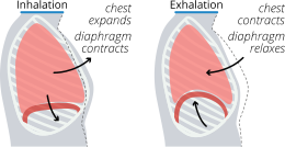
Breathing occurs when air is inhaled causing the lungs to expand and the diaphragm to contract, then exhalation when the chest contract and the diaphragm relaxes.
Breathing
Nervous System
The nervous system contains the following
Brain: The brain coordinates most body activities and is the control center for the body as well as thought, emotion, and judgment. It is divided into four lobes: frontal, parietal, occipital, and temporal.
Spinal cord: The spinal cord provides a pathway for nerve impulses travelling to and from the brain and extends from the base of the brain to the lumbar vertebrae through the vertebral column.
Peripheral nerves: The peripheral nerves include 12 pairs of cranial nerves and 31 pairs of spinal nerves branching off from the spinal cord. They carry nerve signals between the body and the brain.
Neuron: The neuron is the functional unit of the nervous system.
Dendrites: The dendrites have multiple branching structures.
Nucleus: The nucleus directs cellular activities.
Cytoplasm: The cytoplasm produces neurotransmitters and energy for the neuron.
Axon: The axon stores neurotransmitters.
The nervous system controls all other body systems and is divided into two main sections—the central nervous system (CNS) and peripheral nervous system (PNS). The CNS includes the brain and spinal cord. The PNS is made of peripheral nerves found throughout the body.
The PNS is broken down further into two separate branches—the somatic nervous system and autonomic nervous system. The somatic nervous system controls the body’s voluntary (skeletal) muscles. Afferent nerve cells, called neurons, carry information about the body’s environment to the CNS. Efferent neurons carry responses from the CNS to the body to initiate action.
For example, if a person were to touch a hot stove, afferent neurons would carry the heat and pain sensations to the brain. The brain would process and respond using efferent neurons to signal the arm muscles to move the person’s hand away from the source of the pain. The autonomic nervous system controls the body’s automatic functions like breathing and digestion. The sympathetic branch controls the “fight or flight” response to stress. The parasympathetic branch returns the body to resting state after stress has been resolved and is responsible for maintaining homeostasis.
A neuron generates an electrical impulse when stimulated. The nervous system contains multiple neurotransmitters.
Endocrine System
The endocrine system is made of organs and glands that produce, store, and release hormones. Hormones are chemicals used by the body to increase or decrease activity of the hormone’s specific target cells. This aids the body in maintaining homeostasis. There are two types of glands within the system: exocrine and endocrine. Exocrine glands release hormones into a duct for delivery to the target cells. Endocrine glands release hormones directly into the blood stream. There are many different organs that make up the endocrine system throughout the body.
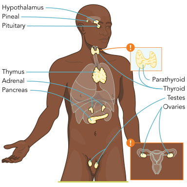
Endocrine system including locations of the hypothalmus, pineal gland, pituitary gland, thymus, adrenal gland, pancreas, parathyroid, thyroid, testes, and ovaries
Endocrine system
Reproductive Systems
The male and female reproductive systems work together to achieve fertilization and produce offspring. Some intersex variations can occur in sex development in which there is a discrepancy between the external genitals and the internal genitals. The female and male reproductive system each contain structures and functions to aid in the reproductive process.
Male Reproductive Organs
The following organs are part of the male reproductive system.
Testes: Produce sperm and testosterone; located below the pelvic cavity on the outside of the body, within the scrotum
Scrotum: A pouch of skin that houses the testes
Penis: External cylinder-shaped organ that moves urine and semen out of the body
Epididymis: Coiled tube located superior to each testis; responsible for maturation of sperm cells
Vas deferens: Connects the epididymis to the urethra
Seminal vesicles: Sac-like organs that secrete seminal fluid that stimulate muscle contractions in the female reproductive organs to aid in propelling sperm forward
Prostate gland: Surrounds the proximal urethra; contracts during ejaculation to aid in forward movement of sperm; secretes fluid that protects sperm within the vagina
Bulbourethral glands (Cowper’s glands): Inferior to the prostate gland; secrete fluid to lubricate the end of the penis to prepare for intercourse
Androgens: Group of male sex hormones
Testosterone: Most abundant and biologically active of male sex hormones
Female Reproductive Organs
The female reproductive system includes the following.
Ovaries: Pair of oval-shaped organs located within the pelvic cavity; produce ova, estrogen, and progesterone
Fallopian tubes: Muscular tubes with proximal opening near each ovary; connect distally to uterus; receive egg during ovulation
Uterus: Hollow muscular organ, lies low in pelvic cavity; receives fertilized egg, which implants into uterine wall for fetal development. If no egg has implanted into the uterine wall, the uterine lining sloughs off, causing menstruation. The lower portion of the uterus is the cervix, which creates a barrier between the uterus and vagina and dilates during childbirth.
Vagina: Muscular tube extending from the uterus to the outside of the body; expands during intercourse and childbirth
Labia majora: Folds of skin and adipose tissue that protect other external genitalia
Labia minora: Folds of skin within the labia majora, pinkish in color due to high blood circulation; form a hood over the clitoris
Clitoris: Highly sensitive erectile tissue located anterior to the urethra
Perineum: Area between the vagina and anus
Estrogen: Group of sex hormones
Progesterone: Hormone secreted by ovaries
Estradiol: Most abundant and biologically active female hormone
The female reproductive cycle refers to a monthly fluctuation of hormones that aids in reproduction and prepares the uterus for carrying a child. When fertilization does not occur, menstruation takes place, and the process begins again.