Cell Biology and Neuroscience: Exhaustive Study Guide on Glial Cells
Historical Foundation and Definition of Glia
Rudolf Virchow (1821-1902): Coined the term "glia" in 1856.
Key Publication: Die Cellularpathologie in ihrer Begründung auf physiologische und pathologische Gewebelehre (Twenty lectures given in February, March, and April 1858 at the Pathological Institute in Berlin).
Original Terminology: Virchow referred to "Neuroglia" as "Zwischensubstanz" (interstitial tissue) that serves as the connective substance within the brain and spinal cord.
Major Classes of Glial Cells and Their Locations
Central Nervous System (CNS):
Macroglia:
Astrocytes: Maintain CNS homeostasis. most numerous Glia in the brain
Oligodendrocytes: Myelinate and provide support to axons. found in the CNA, ‘many-branches’ cells
function: myelinate CNA neurons and inhibition of axon regeneration
Ependymal Cells: Line the ventricles and spinal canal, ‘outer garment’ cells found in the CNA
functions: lining brain ventricles and spinal cords central canal, cilia on apical membrane aid CSF movement, long processes on their basal surfaces extend and have many astrocyte like functions.
specialized versions of these cells form choroid plexus in the brain ventricles which secrete CSF
Microglia: Resident immune cells of the CNS, specialized microphages for CNS invaders
functions: inflammation response, phagocytosis of necrotic tissue microorganisms and foreign substances
Peripheral Nervous System (PNS):
Schwann Cells: Myelinate and support axons; facilitate repair.
function: form the myelin sheath, allow saltatory conduction, can have phagocytic properties and can promote axon regeneration
as cells grow around the axon during development, cytoplasm is squeezed our, multiple layers of cell membrane wrap around a part of the axon many times
Olfactory Ensheathing Glia: Located in the olfactory bulb and mucosa; provide lifelong regeneration of olfactory axons.
Enteric Glia: Support neurons in the enteric nervous system (submucosal plexus, circular muscle, mucosa, and lumen).
Developmental Origins of Glia
Ectoderm Origin:
Astrocytes, oligodendrocytes, and ependymal cells in the CNS.
Schwann cells, satellite cells, olfactory ensheathing cells, and enteric glia in the PNS.
Mesoderm Origin: Microglia (derived from bone marrow monocytes that migrate to the CNS).
Glial Functions in Nervous System Development
Early Stages:
Neuroepithelium proliferation and cell death.
Neuroepithelial transition timing
Neural stem cell proliferation.
Intermediate Stages:
Neuronal/glial specification.
Timing of neuronal differentiation.
Radial and tangential neuronal migration.
Clearance of ectopic neurons.
newborn neuronal survival
radial neuronal migration, tangential neuronal migration
Late Stages and Circuit Function:
Neurite outgrowth and axon pathfinding.
axo-dendritic specification
Dendritic spine growth and synaptogenesis.
Synaptic pruning and synapse stability.
Activity-dependent neurite remodeling.
Ion and neurotransmitter homeostasis.
Conduction velocity enhancement via myelination.
Metabolic support and neuronal plasticity.
Detailed Anatomy and Roles of Astrocytes
Morphological Classes:
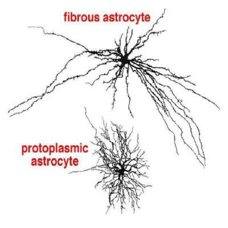
Fibrous Astrocytes: Found in white matter; primarily support axons.
Protoplasmic Astrocytes: Found in grey matter; perform homeostatic roles.
Structural and Regulatory Roles:
Blood-Brain Barrier (BBB) Integrity: Formed by endothelial cells, basal lamina (connective tissue), and astrocyte end feet. Astrocytes release factors to maintain selective permeability. selectively controls the entry and exit of substances from blood capillaries
Small Lipid-Soluble Molecules: Diffuse easily through the BBB.
Water and Charged Ions: Require specific transport proteins.
Proteins: Prevented from crossing to avoid abnormal neuronal stimulation or inhibition.
Metabolic Support (Brain Energy Metabolism):
Neurons take up glucose via to generate ATP.
Astrocytes take up glucose via ; it can be used for ATP or stored as glycogen.
Lactate Shuttle: If neuronal ATP declines, astrocyte pyruvate is converted to lactate, which enters neurons to be converted back to pyruvate for ATP production.
Maintaining Homeostasis:
Ion Buffering: During repolarization, voltage gated channels open causing ions exit neurons. at rest K+ leave neurons via leaky channels, Na+/K+ ATPase can pump some back into neurons, overtime there is build up of K+ ions → disturbs the concentration gradient increasing excitability of neurons
Astrocytes use specialized inwardly rectifying channels to siphon excess ions away from the neuron, preventing build up to toxicity and hyperexcitability.
Syncytium: Astrocytes are connected by gap junctions, allowing them to distribute ions across a network so no an individual astrocyte doesn’t increase its intracellular K+ too much.
Neurotransmitter Reuptake: Astrocytes take up excess glutamate to prevent neurotoxicity. Glutamate is converted to glutamine within the astrocyte. This glutamine is later used to synthesize Glutamate and GABA in neurons.
Glycogen reserve: glucose taken into neurons via GLUT-3, used to generate ATP. Enters astrocytes by GLUT-1, used to generate ATP and store as glycogen. If ATP levels decline, pyruvate in astrocytes can be converted to lactate and enter neuron, lactate converted to pyruvate to generate ATP
Additional Functions:
Regulation of the extracellular electrolyte balance (ions, water, pH).
Influence on endothelial cells and angiogenic factors.
Formation of the glia limitans.
Secretion of neurotrophic factors for neuronal survival and myelination.
Modification of the extracellular matrix (ECM) for neurogenesis and synaptogenesis.
Inhibition of axon regeneration in the CNS.
Immune modulation alongside microglia.
Areas with a Leaky Blood-Brain Barrier
Area Postrema: Triggers the chemo-trigger zone to induce nausea and vomiting in response to toxins.
Posterior Pituitary Gland: Releases hormones like oxytocin and Antidiuretic Hormone (ADH) directly into circulation.
Pineal Gland: Allows the release of melatonin into the bloodstream.
Median Eminence of the Hypothalamus: connects the hypothalamus to the pitutary gland, hormones produced collect here before entering the bloodstream
Oligodendrocytes and Myelination in the CNS
Function: One oligodendrocyte can myelinate multiple different axons (ranging from to axons).
Regeneration: Along with astrocytes, oligodendrocytes inhibit axon regeneration in the CNS.
Ependymal Cells
Location: Line the ventricular system (Lateral, Third, and Fourth ventricles) and the central canal of the spinal cord.
ventricles - fluid filled spaces in the brain with important roles
Functions:
Form the blood-cerebrospinal fluid (CSF) barrier.
secrete, monitor and aid in the circulation of CSF
Ependymocytes: Possess cilia and microvilli
Choroid Plexus: A network of capillaries and specialized cuboidal epithelium that secretes CSF.
Microglia and Immune Defense
Origin: Monocytes produced in the bone marrow migrate to the CNS and differentiate into microglia.
Function: immune defense and removal of cellular debris in the brain
Resting State: Characterized by long, branched processes (Iba1 positive).
Activated State:
Retract processes and become amoeboid/phagocytic.
Act as antigen-presenting cells to recruit T-cells.
Release proinflammatory substances: Nitric Oxide (), Reactive Oxygen Species (), free radicals, and cytokines.
Peripheral Nervous System (PNS) Glia
Schwann Cells:
Myelinate PNS axons (Spinal nerves and Cranial nerves III-XII).
Relationship: One Schwann cell myelinates exactly one axon segment (unlike oligodendrocytes).
Rapidly remove myelin debris via phagocytosis.
Promote Axon Regeneration: Produce permissive ECM components such as N-CAM, Nerve Growth Factor (), and laminin.
Satellite Cells: Provide structural and metabolic support by surrounding cell bodies in the dorsal root ganglia (DRG) and autonomic ganglia (sympathetic and parasympathetic).
Olfactory Ensheathing Cells (OECs): Derived from the ectoderm of olfactory placodes; resident glial cells of the primary olfactory nerves, they wrap primary olfactory axons to provide a pathway for growing axons
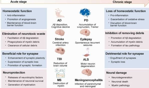
Glia in Disease and Pathology
Homeostatic Function (Acute/Normal Stage): Anti-inflammation, angiogenesis promotion, BBB maintenance, and debris removal.
Loss of Function (Chronic Stage): Pro-inflammation, oxidative stress, BBB disruption, and inhibition of debris removal.
Specific Pathologies:
Alzheimer's Disease (AD): Involved in (Amyloid-beta) deposition and degradation; synaptic engulfment.
Parkinson's Disease (PD): Accumulation of -synuclein.
Stroke: Cerebral artery infarction.
Epilepsy: Spontaneous recurrent seizures.
Amyotrophic Lateral Sclerosis (ALS): Motor neuron degeneration.
Multiple Sclerosis (MS): Destruction of myelin.
Meningoencephalitis: Infection of parenchymal and meningeal tissues.
Traumatic Brain Injury (TBI): Reduction of brain volume and synaptic loss.
Multiple Sclerosis (MS) Deep Dive
Nature: An autoimmune condition causing demyelination of white matter tracts.
Risk Factors: Females ( years old), Vitamin D deficiency, genetic markers (), and viral infections (, ).
Clinical Features:
CNS: Cognitive impairment, depression, dizziness, decreased memory.
Visual: Optic neuritis, nystagmus, diplopia, decreased visual acuity, color blindness.
Motor: Weakness, intention tremors, ataxia (coordination), spasticity, muscular atrophy.
Sensory: Paresthesia, numbness, increased pain sensitivity.
Autonomic: Bowel/urinary incontinence, constipation, impotence, dysphagia.
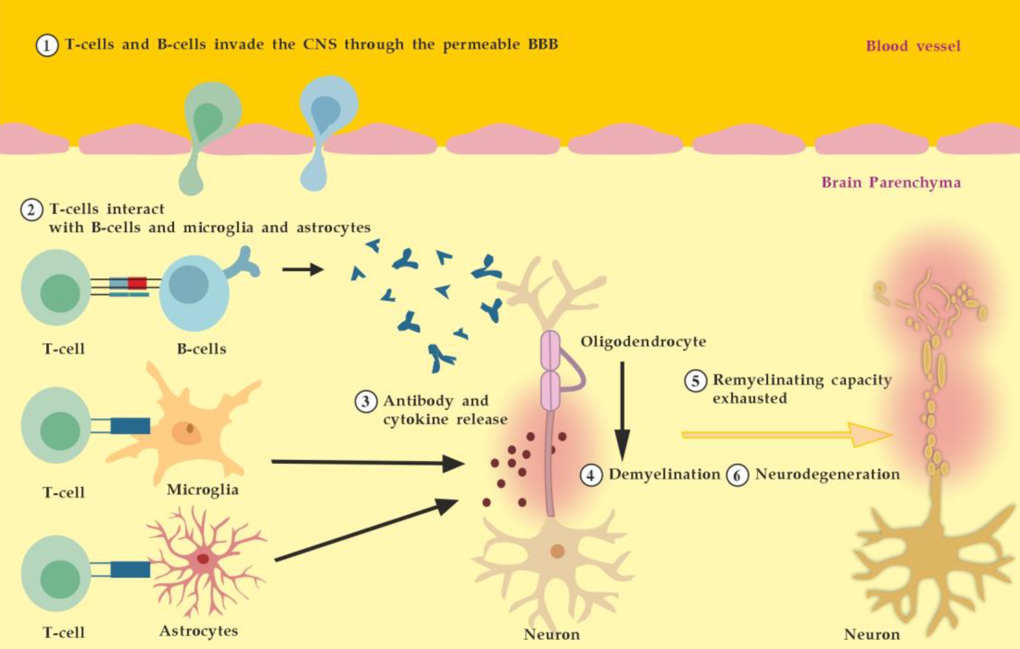
Pathophysiology:
1. T-cells and B-cells cross a permeable BBB.
2. T-cells interact with B-cells, microglia, and astrocytes.
3. Release of antibodies and cytokines lead to demyelination.
4. Remyelinating capacity is eventually exhausted, leading to neurodegeneration.
Treatments:
High-dose corticosteroids to suppress the immune system.
Plasmapheresis (plasma exchange).
Inhibition of T-helper cell cytokine release.
Future Directions: Glia and Stem Cells
Therapeutic Potential: Using stem cells to replace or support malfunctioning glia.
Targets:
Oligodendrocytes: For remyelination.
Astrocytes and Microglia: To halt neurodegeneration and provide neuronal support.
Neurons: Direct replacement of dead cells.