Spinal Cord & Spinal Nerves
Spinal Cord
Spinal Cord
Acts as a link btwn the brain and the rest of the body
Does some things independently from the brain
Spinal cord and spinal nerves
Provide a pathway for sensory and motor nerve signals
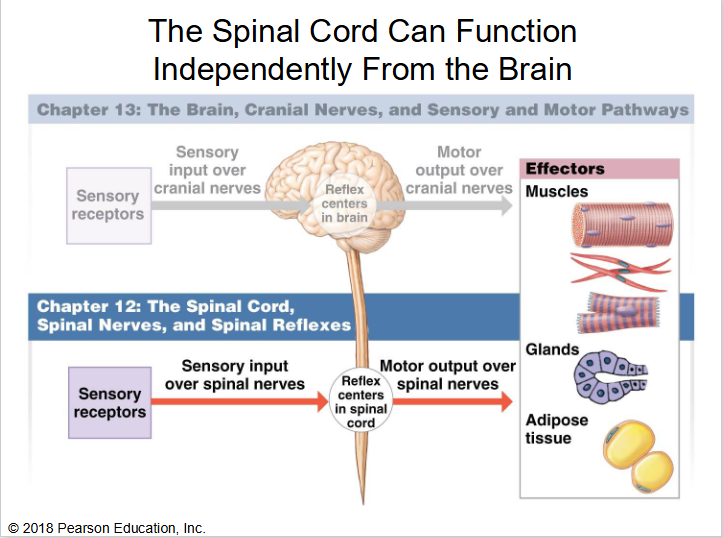
Important for spinal reflexes, which provide the quickest reactions to stimuli that are generally below the neck
Cranial nerve reflexes mostly involve the head and neck
CNS tissue from the foramen magnum to L1; protected by bone, meninges, and CSF
31 pairs of mixed spinal nerves (leading to PNS) escape btwn vertebrae and attach to the cord by paired roots; innervate the entire body except the head
Mixed—both sensory and motor
Spinal Cord Regions
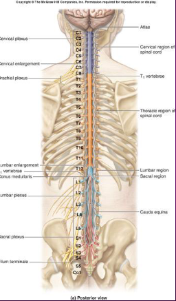
Named according to their point of issue
8 cervical (C1 - C8)
12 thoracic (T1 - T12)
5 lumbar (L1 - L5)
5 sacral (S1 - S5)
1 coccygeal (C0)
Conus medullaris—end of spinal cord
Cauda equina—nerves project inferior to the spinal cord
Filum terminale—strand of pia mater within the cauda equina that anchors the spinal cord to coccyx

Protection of Spinal Cord
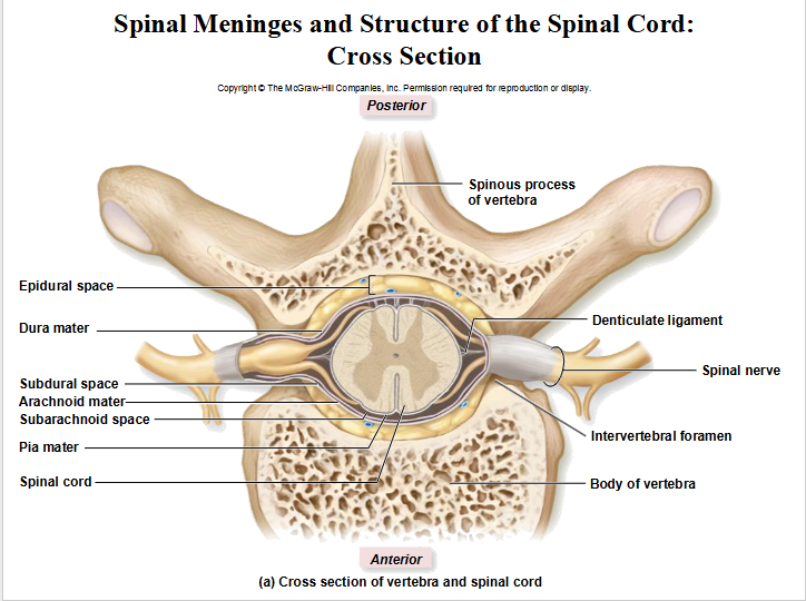
Structures surrounding the spinal cord, from innermost to outermost
Pia mater
Subarachnoid space
Arachnoid mater
Subdural space
Dura mater
Epidural space
Vertebra
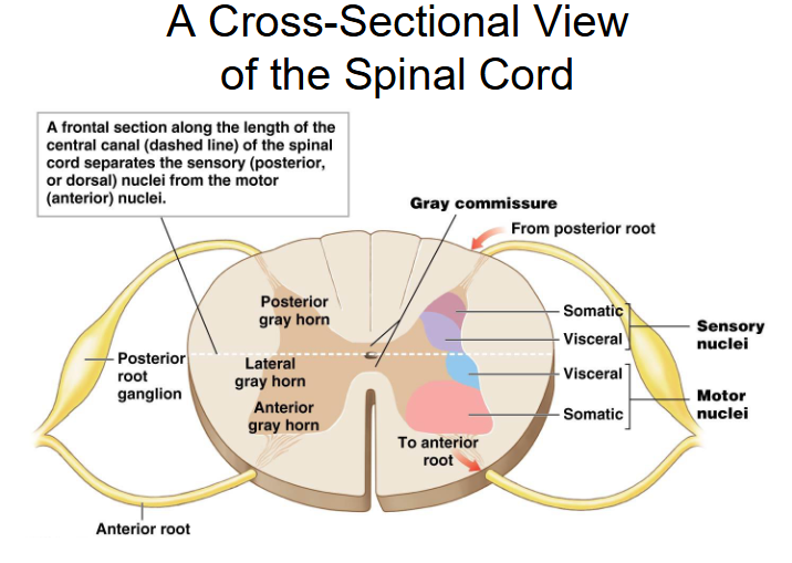
Spinal Cord Cross-Section
Gray Matter located toward center (gray commissure connects 2 sides)
White matter surrounds gray matter
Divisions
Posterior funiculus
Lateral funiculus
Anterior funiculus (White commissure connects 2 sides)
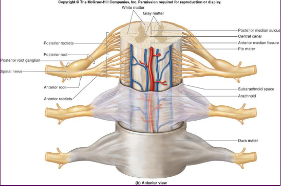
Central canal—carries CSF flowing inferiorly
Posterior median sulcus—divides posterior cord medially
Anterior median fissure—divides anterior cord medially

Spinal Cord Tracts
Ascending tracts—mainly relay sensory info up toward brain
Descending tracts—mainly relay motor info down brain

Spinal Roots Join to Form Spinal Nerves
Dorsal roots—carry sensory (afferent) neurons from spinal nerves to spinal cord
Ventral roots—carry motor (efferent) neurons from spinal cord to spinal nerves
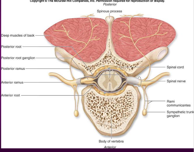
Spinal Nerves and Dermatomes
Dermatome—a bilateral section of skin innervated by a single spinal nerve (all nerves except C1)
The skin can be sectioned into a dermatome map

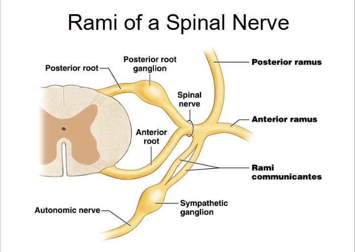
Spinal Nerves: Rami
The short spinal nerves branch into three or four mixed, distal rami
Smaller posterior ramus
Larger anterior ramus
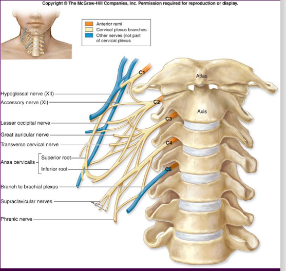
Nerve Plexuses
All anterior rami except T2 - T12 form interlacing nerve networks called plexuses
Plexuses are found in the cervical, brachial, lumbar, and sacral region
Each resulting branch of a plexus contains fibers from several spinal nerves
Fibers travel to the periphery via several different routes
Each muscle receives a nerve supply from more than one spinal nerve
Damage to one spinal segment cannot completely paralyze a muscle
Neural Reflexes
Reflex—an involuntary, rapid, predictable motor (muscle/gland) response to a stimulus
Reflexes may
Be inborn (intrinsic) or learned (acquired)
Involve only peripheral nerves and the spinal cord
Involve higher brain centers as well
The Spinal Cord Can Function Independently From The Brain
Reflex Tests
Performed clinically to check health of muscles, spinal nerves, and spinal cord regions
A reflex response may be normal, hypoactive, or hyperactive
Reflex Arc
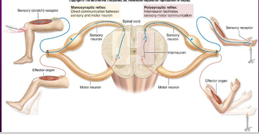
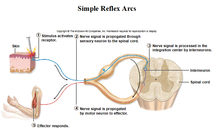
5 components of a reflex arc
Receptor: site of stimulus
Sensory neuron: transmits the afferent impulse to the CNS
Integration center: either monosynaptic or polysynaptic region within the CNS
Interneurons never leave the brain or spinal cord
Motor neuron: conducts efferent impulses from the integration center to an effector
Effector: muscle fiber or gland that responds to the efferent impulse
Ipsilateral v. Contralateral Reflex Arcs
Ipsilateral: the receptor organ is on the same side of the body as the effector organ
A hand poked by a pin will pull away from the pin
Contralateral: the receptor organ is on the opposite side of the body as the effector organ
If the left wrist is grabbed by a stranger, the left hand will pull back from the stranger while the right hand will extend to throw a punch
Monosynaptic v. Polysynaptic Reflexes
Monosynaptic—simple arcs w/o interneurons
Polysynaptic—more complex reflex arcs w/ more synapses and interneurons; incd delay time