Renal Structures and Functions

The Kidney
A bean-shaped, reddish brown organ.
They lie on either side of the vertebral column between the 12th thoracic and third lumbar vertebrae.
The right kidney is lower to make space for the liver.
It is surrounded by a capsule and a fat pad.
Each kidney has a superior and inferior pole.
Functions:
To filter blood
Homeostasis: a. maintain body water balance b. regulate quantity and extracellular fluid ions c. maintain plasma fluid and osmolarity d. Keep the pH of the circulation
Excretion: a. of foreign compounds b. also excretes end products of metabolism like urea from proteins, creatinine from the muscle, uric acid from DNA and RNA, and bilirubin from haemoglobin.
secretion: a. of erythropoietin, which it also produces b. renin, kallikrein, prostaglandins c. converts vitamin D into its active form
Erythropoietin stimulates the production of red blood cells.
vitamin D deficiency leads to a lack of calcium, resulting in problems with the bone.
kidney problems can lead to anaemia

The renal columns are extensions of the renal cortex
The renal papilla is the apex of the renal pyramids
The kidneys are rich in blood
25% of the blood from the circulation flows to the kidneys
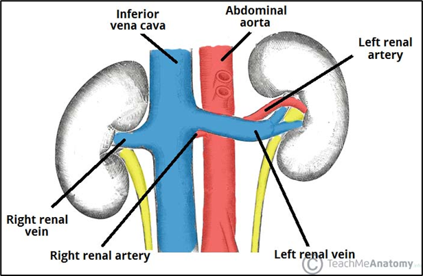
The renal arteries are both behind the renal veins.
If the renal vein is behind the renal artery, it will become compressed and won’t drain properly.
The left renal vein is longer as it crosses over the abdominal aorta.
The right venal artery is longer as it passes behind the inferior vena cava.
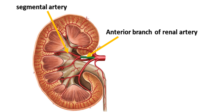
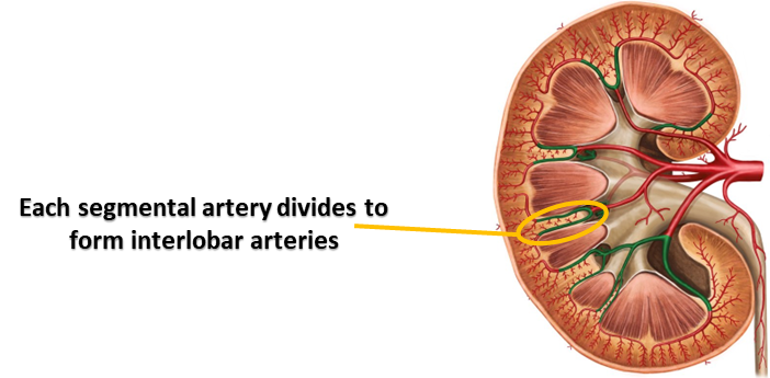
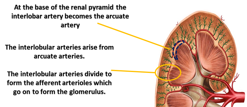
Aorta - 2. renal artery - 3. segmental artery - 4. interlobar artery - 5. arcuate artery - 6. interlobular artery - 7. afferent arteriole - 8. glomerulus (capillaries) - 9. efferent arteriole - 10. peritubular capillaries and vasa recta - 11. interlobular vein - 12. interlobar vein - 13. renal vein - 14. inferior vena cava
Below is a nephron which is a functional unit
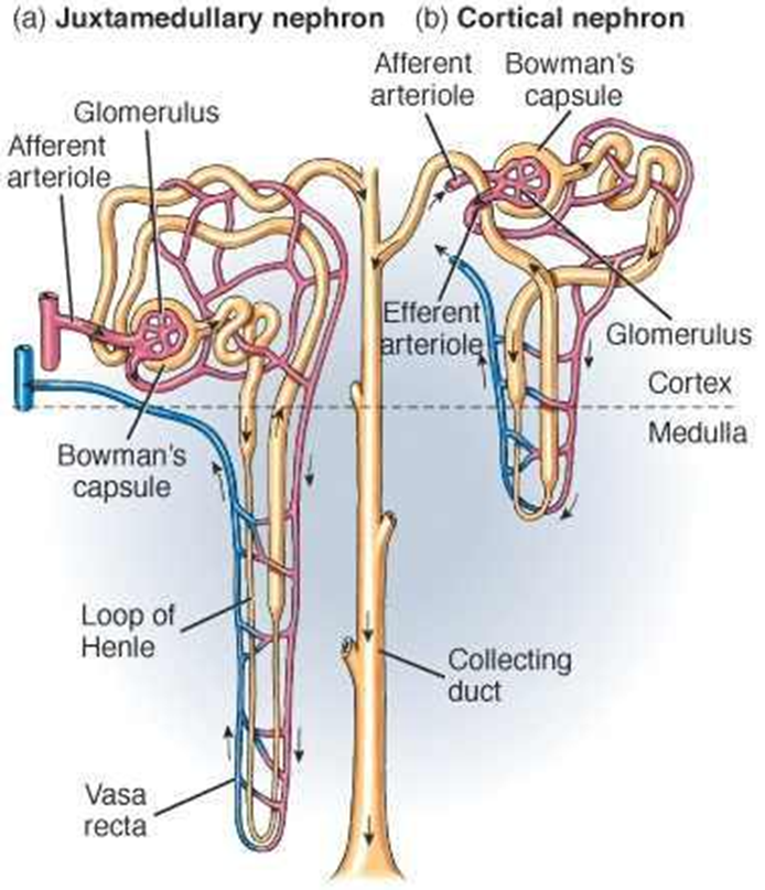
There are 1 million nephrons in each kidney.
20% of nephrons are juxtamedullary, which lie in the renal medulla
80% are cortical, which lie in the renal cortex.
The juxtamedullary nephrons concentrate urine.
They have thin and thick ascending limbs of the loop of Henle.
Has the vasa recta are hairpin-looking loops of Henle.
The cortical nephron is for filtration.
They also have ascending and descending limbs of the loop of Henle.
They have the peritubular capillaries.

Different nephrons connect to the main collecting duct.
The efferent arteriole is narrower due to back pressure.
The arterioles are the primary site for intrinsic and extrinsic regulation of the glomerular filtration rate (GFR).
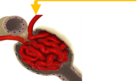
The Bowman’s capsule is a filtration barrier where ultrafiltration occurs.
The proximal convoluted tubule is the primary area for solute and water reabsorption (75%).
The loop of Henle sets up the osmotic gradient for concentrating and diluting urine.
The distal convoluted tubule is surrounded by peritubular capillaries. This is where solute exchange between tubules and the extracellular fluid occurs.
Basic Nephron histology:
Proximal convoluted tubule: Most reabsorption occurs here, has more microvilli to increase the surface area, is long and thin, and has lots of mitochondria.
Loop of Henle: has a thin and a thick portion, the ascending and descending limbs have different permeabilities, and they concentrate and dilute the filtrate.
Distal convoluted tubule: has hormonally regulated actions, has fewer microvilli and has a low water permeability.
The collecting duct: has intercalated and principal cells, has hormonally regulated actions such as ADH.