Weeks 1-2 Notes
Week 1- Taste Perception
Animals can possess 5 primary tastes, but there are more than 5 that they can taste.
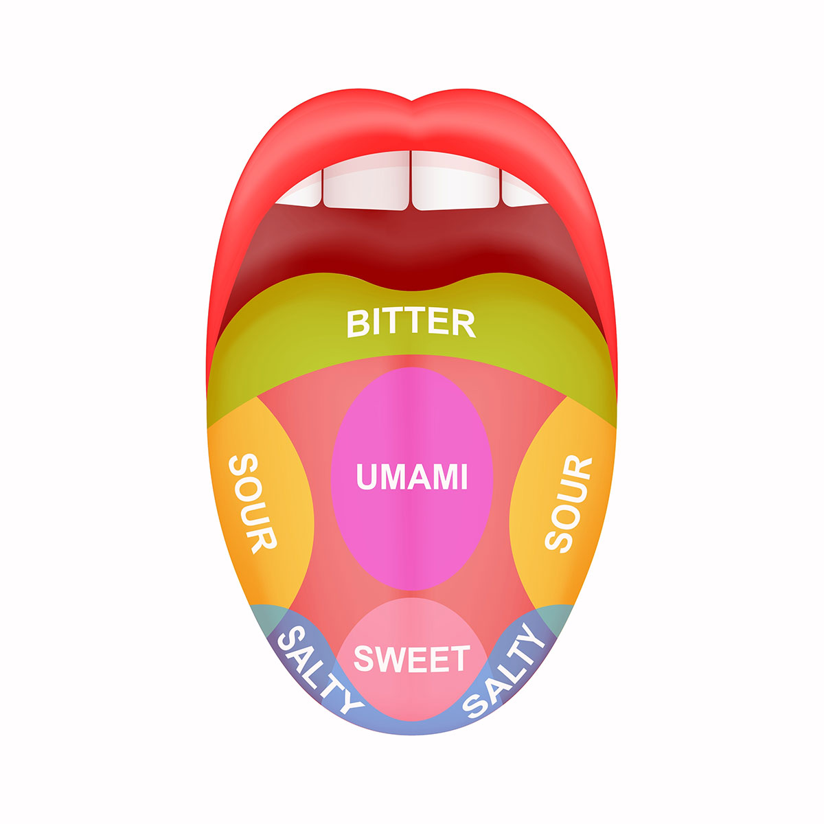
tongue map depicts which area of the tongue is used to sense a taste
can be tasted everywhere but some are better tasted in different areas
hypothesis: umami tastes better on the back of the tongue because it is the last chance to decide if you want to swallow it or spit it out
comparing the taste of sweet, sour, and salty, there is no difference between tasting it in the front vs the back of the tongue
there is a difference in the back vs the front for bitter and umami tastes, which is better detected in the back of the tongue
umami is the savory taste when eating meat
Why can we taste sweet?
relative sweeteners are centered around sucrose
high fructose is on the higher end of sweetness when compared to sucrose
fructose is way sweeter in terms of natural sugars
artificial sweeteners are even higher
naturally occurring sweeteners are on the lower end of the scale
sugars are metabolized to create energy
we evolved to taste sugars to know how much energy can be made from the food
when testing sweet intensity and sweet liking, the liking starts increasing with sweetness and drops when it becomes “sickeningly sweet”
similar results were found for umami and salty tastes
when there is too much umami we can convert them into fats and store them -which could lead to obesity- or there is so much nitrogen you need to excrete
when there is too much salt, we need to take in a lot of water, which could lead to hypertension or urination and cause dehydration
we like a little bit of sweetness but we don’t like too much
we want some energy in the food we are consuming but we don’t want too much energy because of the risk of obesity
obese animals are at higher risk of predation, not humans but other animals
Why can we taste bitter?
bitter compounds are toxic
protects ourselves against toxic things
buckley’s: “it tastes awful, and it works”
but medicine is often bitter and its supposed to be good for you
suppresses coughing mechanism
inhibits mucus production
because you don’t want to be coughing and sneezing but the body sees these as important mechanisms
cough reflex is important to clear unwanting things out of the respiratory system
mucus traps pathogens and particles in the air we breathe
cassava plant
tuber
has cyanogenic glycosides (toxic compounds) in the tuber because the tuber is an energy-storage structure
uses energy stored in tuber to generate sprouts
to prevent herbivory, it produces a toxic compound so herbivores eat it, get a bitter taste, and then leave it alone
when we consume it, cyanide (toxic compound) gets released and it inhibits ETC
the greater the cyanogenic glycoside the more bitter the taste
Why can we taste sour?
things that taste sour contain acid
some animals like sour taste while others don’t
wild pigs like sour → eat rotten fruit → only eat it if it tastes sour
if sour, contains lactic acid bacteria which is not pathogenic and kills pathogenic species
you can treat foods that contain pathogens with lactic acid and the pathogen will not be detected as lactic acid kills it
Thomas leaf monkey hate sour tasting things
foregut fermenter (like herbivores)
four-chamber
consume difficult-to-digest compounds
harbors gut bacteria to break down plants in stomachs
dislike sour because it affects their ability to digest the food
as pH is lower, the capacity to digest is more difficult
sour foods will reduce the acidity of the stomach to lower than normal → making it difficult to digest foods
Hindgut fermenter
ex. rabbits
cecum or large intestine for bacteria to digest plants
already have an acidic stomach and by the time the sour foods enter the cecum or large intestine they are already neutralized
How can animals taste?
tongue papillae are where tasting occurs
3 types of papillae
fungiform papilla
vallate papilla
foliate papilla
have taste buds somewhere on the papillae
3 types of cells make up a taste bud
taste cell → only cell that reaches the tongues surface and have microvilli (increase surface area) on the surface to maximize taste
supporting cell → structural cell
stem cells in green → replace any taste cells (epithelial cells that are exposed which get worn out and need to be replaced often)
tongue and soft palate contain taste buds that allow us to taste
in the brain the gust cortex is used for taste perception
tongue is involved in taste sensation
2 nerves involved in relaying information from tongue to brain
glossopharyngeal nerve (back 1/3 of tongue) → relays information from that location to gust cortex
facial nerve (2 branches → greater superficial petrosal and chorda tympani) → relay information to gust cortext
facial nerve also controls facial muscles as for expression
filiform papillae
4th papillae plays no role in taste perception
most numerous
pointy at the end → allows roughness on tongue
cats have more filiform papillae to groom themselves
used to keep ice cream on tongue
taste receptors
umami: T1R1 and T1R3
we don’t have receptors to taste nucleotides but if nucleotides are present in umami
sweet: T1R2 and T1R3
artificial sweeteners bind to different sites than natural sugars which is why they taste different
bitter: 30 T2Rs that detect toxic compounds
sodium: ENaC
sour acids: OTOP1
sour carbonated drinks: CA IV
Week 2 - Comparative Physiology of the Heart
Evolution of Animal Hearts
can be divided into 3 groups: no heart (diffusion only), peristaltic heart, chambered heart
No heart (diffusion only)
rely on diffusion to move oxygen, nutrients, and wastes throughout their bodies
no circulatory system
either very small, have a flat body or have a low metabolic rate (slow)
tardigrades (0.3-0.5mm), nematodes, sipunculans, echiurans, nemerteans, platyhelminthes, roitifers, ctenophores, cnidarians
vast majority of animal phyla lack a heart
diffusion works well 1mm or less
tardigrades have lost circulatory pumps
ancestors did have a heart, but tardigrades lost it over time
because they evolved from larger ancestors (needed heart)
flat body → no cell is more than 1mm away from the body’s surface
even large animals with low metabolic requirements don’t need a heart as they use nutrients and oxygen slowly
Peristaltic pumps
referred to as pumping vessels → just blood vessels that have become contractile
inefficient because they arent very good at preventing backflow
pumps forward and backward
common heart
cephalochordates, tunicates, echinoderms, hemichordates, onychophorans, arthropods, annelids
Chambered hearts
contain distinct chambers → specialized for particular function
4 basic chambers: conus arteriosus, atrium, sinus venosus, ventricule
atrium and sinus venosus are specialized for the inflow of blood (receiving blood)
conus arteriosus ad ventricule are specialized for outflow (pressurizing the blood and pumping it out back to all the tissues)
one way valves → prevent backflow
can be leaky when older, but efficient when working well
vertebrates and molluscs (two most intelligent)
evolved independently (homoplasy/ convergent evolution)
two most intelligent phylum groups
ex. humans and octopuses
chambered pump allows intelligence by providing nutrients to develop the nervous system
Peristaltic pumps (arthropods)
ex. insects
large structure on the back is dorsal vessel and contains the heart
insects have their hearts on the back side while vertebrates have them on the front
open circulatory system (don’t have a series of blood vessels)
the heart extends forward to the aorta (aorta and heart make up the dorsal vessel)
excurrent opening → opening at which the aorta ends
hemolymph makes up the rest of the insect body (hemocoel) and organs are just bathed by the hemolymph inside the hemocoel
peristalsis → wave in one direction
dorsal diaphragm (below the heart)
muscular for some insects (alary muscle)
contracts → expands volume of heart → increase in volume means decrease in pressure → creates pressure gradient → flow of hemolymph from outside to inside to balance pressure
hemolymph entered through openings called ostia (valve only allows one way)
then relax alary muscle → muscles contract → increase pressure inside heart → hemolymph leaves heart → out aorta → out excurrent opening → out the head
elastic for other insects (elastic ligaments)
researchers injected bubbles into the heart of an insect and followed movement of bubbles
width of heart gets shorter as you contract and wider as you relax
yellow bars (backward flow) is present
net movement forward but there is still inefficiency due to backward flow
backwards flow usually at the end of each contraction
dorsal muscle is good at getting circulating hemolymph within the hemocoel, but not good at getting it in appendages
mole cricket: challenge to get hemolymph into the antennas
ohm’s law: calculates the rate at which fluid can flow through a pipe
pressure gradient
resistance
poiseuille’s law: calculates the resistance
length
viscosity of hemolymph (similar to water)
the longer the pipe the greater the resistance
the smaller the radius the greater the resistance (the smaller it is, the harder it is for liquid to flow through it)
antennas are long, small radius structures → large resistance
evolved accessory pulsatile organs (accessory hearts)
at the base of each antennae
pumps hemolymph specifically through the antenna
ampulla (heart) → fill with hemolymph → contracts → forces hemolymph into antenna
dilator muscle (similar to alary muscle) → contracts → expands size of ampulla → decreasing pressure → draws hemolymph inside the ampulla
ostium → opening that hemolymph enters into
no physical connection between dorsal muscle and accessory pulsatile organ
they beat at different rates
antennas don’t have high energy demand compared to other parts
have other hearts for other appendages
insects are the “richest” in hearts
tunicates: have an open circulatory system
heartbeat reversal
pump in one direction → pause → pump in other direction
two pacemakers that trade-off
hypothesis: to even out metabolic processes
certain parts won’t get much nutrients without heartbeat reversal
collisions occur in isolated (during studies) hearts
rarely ever when they are in the body
Chambered hearts (molluscs)
closed circulatory system → nutrients have to diffuse out of blood vessels
have 3 hearts
systemic heart: pumps blood into peripheral vascular system which then brings blood to tissues
2 branchial hearts: from tissues, pump blood into the branchial vascular system which then pumps to gills to pick up oxygen again
why 3 hearts?
ohm’s law: rate at which fluid can flow through pipe
when the system heart contracts → pressurize the contents inside → creates gradient to drive blood flow through peripheral vascular system
every time blood flows through a capillary bed, it loses a lot of the pressure it contains
not enough blood flow to go through gill capillaries
branchial hearts re-pressurize it
systemic heart pressurizes the blood to a greater degree because the peripheral vascular system is longer so resistance is greater
double pressure single circuit
Fishes are inefficient because they only have one heart to pump through two capillary beds.
Week 2 - Lab
Neurogenic hearts
arthropods (insects and crustaceans)
pacemaker is a part of the nervous system (ganglion) located outside but still in association with the heart
Myogenic hearts
pacemaker in a specific region of the cardiac muscles within the heart
located in the sinoatrial node
regulated to quickly depolarize (fast heart rate) or slowly (slow heart rate)
vertebrates and molluscs (snails, octopuses, etc.)
Excitation within the pacemaker is what spreads to cardiac muscle fibres and causes contraction (systole).
Daphnia
exception that has myogenic heart
used to reduce ethical implications of working with vertebrates
Heart rate in vertebrates:
adrenaline (epinephrine) increases heart rate in vertebrates
binds to B1 adrenergic receptors
activates cAMP secondary messenger system
cAMP opens and closes channels (including T-type Ca2+ channels) → increases depolarization
caffeine increases heart rate in vertebrates
directly applied to the heart
acts as an inhibitor of phosphodiesterase (enzyme that degades cAMP)
allows cAMP to accumulate and continue to open channels that causes depolarization
ethanol increases heart rate in vertebrates
no clear mechanism
suggest that ethanol can bind to B1 adgenergic receptors and enhance activation of cAMP pathway
acetylcholine decreases heart rate in vertebrates
binds to muscarinic-type cholinergic receptors
causes opening of potassium channels/closing of Ca2+ channels
hyperpolarization → difficult for spontaneous depolarization to occur
atropine increases heart rate
blocks muscarinic-type colinergic receptors
nicotine
increases heart rate when administered outside of the heart (smoking) as it causes the release of adrenaline
decreases heart rate when applied directly to heart, suggesting that nicotinic-type cholinergic receptors may be in the heart allowing hyperpolarization
Lab procedure:
immobilize using vaseline
add drop of water
count heart rate for 2 minutes
count 10 seconds, wait 10 seconds, count 10 seconds, wait 10 seconds, count 10 seconds, and wait 10 seconds
repeat twice
measure heart rate
remove water using kimwipe
add drug
repeat