Midterm:
Tooth Structure, Wear, Development, and Bone Findings:
Bruxism
Grinding or clenching teeth together for non-functional purposes
Associated with attrition

Attrition
Slow process of tooth wear due to mastication

Abfraction
Wedge-shaped lesions at cervical areas of the teeth
Related to fatigue, flexure, and fracture from biomechanical forces

Erosion
Loss of tooth structure from chemical action
Influenced by diet, vomiting, and GERD

Abrasion
Pathologic wearing away of tooth structure from repeated mechanical force
Influenced by toothbrush technique

Anodontia
Definition: Congenital lack of ALL teeth (primary and permanent)
Treatment: prosthetic replacement of missing teeth
Diagnostic: clinical or radiographic

Hypodontia
Definition: lack of one or more teeth (developmental)
Permanent teeth MORE common
Common teeth: third molars (both arches), maxillary lateral incisors and mandibular 2nd premolars
Treatment: prosthetic replacement of missing teeth
Diagnosis: clinical or radiographic

Concrescence
Definition: two adjacent teeth are united by cementum ONLY
maxillary molars
Causes: developmental after tooth formation, crowding, or trauma
Treatment: none
Diagnostic: radiographic

Amelogenesis imperfecta
Definition: broad group of conditions that affect the structural formation of enamel
Divided into 4 types:
type I (hypoplastic)
type II (hypocalcified)
type III (hypomaturation)
type IV (hypoplastic-hypomaturation)

dens en dente (dens invaginatus)
Definition: tooth within a tooth → results when enamel invaginates into the crown before mineralization
Common in maxillary lateral incisors
Diagnosis: Clinical (tooth appears normal or peg-shaped) or radiographic
Treatment: replacement of prophylactic restoration (vital) or RCT (non-vital)

Compound odontoma
Definition: collection of numerous small teeth

resorption
Definition: observed radiographically when the apex of the tooth appears shortened or blunted and irregularly shaped
Two types: internal and external

traumatic bone cyst (simple bone cyst)
Definition: pathologic cavity in bone that is not lined with epithelium

stafne bone cyst (static bone cyst or lingual mandibular bone concavity)
Definition: radiolucency in the posterior region of the mandible inferior to the mandibular canal (NOT A TRUE CYST)
Cause: developmental concavity containing normal salivary tissue on lingual aspect of mandible
Diagnosis: radiographs
Treatment: none

Periapical cemento-osseous dysplasia
Definition: apical lesion that appears as a mass of fibrous connective tissue with spicules of cementum or a calcified mass resembling cementum
Found commonly in black women

Paget disease (osteitis deformans and leontiasis ossea)
Definition: chronic metabolic bone disease characterized by resorption, osteoblastic repair, and remineralization of involved bone

Gingiva, Mucosa, Healing and Clinical Signs
Gingival Enlargement:
Phenytoin (Dilantin) and nifedipine (Procardia)

Gingival hyperplasia
Definition: enlargement of the gingiva

Hyperplasia
Increase in number of normal cells due to stress

Hypertrophy
Increase in cell size due to stress

Atrophy
Decrease in size and number of normal cells due to stress

Necrosis
Definition: cell nucleus dies and releases enzymes → whole cell dies → membranes disintegrate → cell contents released into tissue causing inflammatory response
in short, premature death of cells or body tissue

Acute vs chronic inflammation
acute → early or immediate response
short duration (days)
several PMNs and monocytes
outcome: complete resolution, scarring (fibrosis), abscess formation, and progresses to chronic inflammation

chronic → response to persistent infection (can progress from acute)
long duration
several macrophages, lymphocytes, and plasma cells
outcomes: repair not complete until source of injury is REMOVED

frictional keratosis
Definition: chronic rubbing or friction against oral mucosal surface that may result in hyperkeratosis → appears as a white patch

Xerostomia
Definition: dryness of the mucous membrane
Causes: BY HYPOSALIVATION OR DECREASED SALIVARY FLOW

Operculum
Definition: flap of soft tissue covering a portion of the crown or partially erupted tooth

Exudate
Definition: inflammatory fluid formed as a reaction to injury of tissues and blood vessels

Fistula
Definition: Underlying tunnel that connects deep infection to that surface opening

Epulis fissuratum (denture-induced fibrous hyperplasia)
Definition: lesion caused by ill-fitting denture and located in the vestibule along the denture flange

pyogenic granuloma
Definition: intraoral lesion characterized by a benign growth of granulation tissue that occurs in response to injury
NO PUS and NOT TRULY A GRANULOMA
Treatment: excision and removal of local irritants
Causes: pregnancy-induced (pregnancy tumor)

erythema migrans/geographic tongue
Definition: erythematous patches surrounded by a white or yellow border
Area has no filiform papillae but has fungiform papillae
genetic factor or stress-induced
NO treatment

median rhomboid glossitis
Definition: appears as flat or slightly raised erythematous area in CENTER of tongue (extends to circumvallate papillae)
associated with Candida albicans
NO treatment, but antifungal may be used

lingual varicosities
Definition: appears as red-to-purple enlarged vessels that are on the ventral/lateral surface of the tongue
Common in older patients (60+)
Associated with smoking, CV disease, and varicosities in legs

Linea alba (treatment)
Definition: white line extends on the buccal mucosa along the occlusal plane
PROMINENT in clenching or bruxing habits

Fordyce granules
Definition: Sebaceous glands that appear as yellow-white papules on buccal mucosa and vermilion upper lips
Present in 80% of the population (normal, no treatment)

keloid
Definition: excessive scarring in skin
Appears raised and extends past injury
High occurrence in dark skin or family history

Angular Cheilitis
Definition: associated with deficiencies of B-complex vitamins; commonly a fungal condition OR Candida infection
located on the labial commissures
responds to antifungal cream and ointment

wickham striae
Definition: associated with lichen planus that appears as interconnecting lines

traumatic neuroma
lesion caused by injury to peripheral nerve

macule
area that is distinguished by a color different from that of the surrounding tissue
flat; does NOT protrude above tissue (freckle)

papule
small, circumscribed lesion that is elevated above the surface of surrounding tissues

lobule
segment or lobe that is a part of a whole
appeared fused

pedunculated
Attached by a stem-like base similar to that of a mushroom

coalescence
parts of a whole join together, or fuse, to make one

lymphadenopathy
Definition: abnormal enlargement of lymph nodes

urticaria (hives)
Definition: Localized redness and swelling due to increased vascular permeability

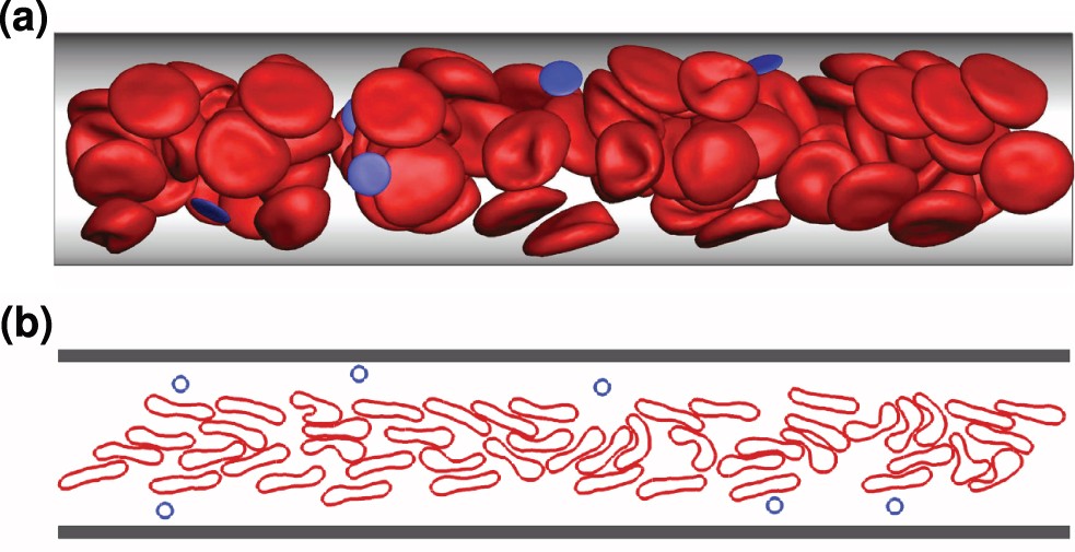
margination
movement of WBCs to vessel wall at the sight of injury
Pavementing
the adhesion of white blood cells to blood vessels during inflammation

Immunology, Infection, and Diagnostic Concepts
Plasma cells = B lymphocytes
B-lymphocytes differentiate into plasma cells
Make specific antibodies that bind to antigens

antigens
any substance able to induce a specific immune response

antibodies: natural, passive, acquired active and passive
natural active:
Antibodies produced due to exposure
natural passive:
Antibodies through the placenta to the developing fetus or via breast milk
acquired active:
antibodies produced due to vaccination
acquired passive:
antibodies injected

humoral immunity (antibody-mediated immunity)
type of immunity in which B-cell lymphocytes and antibodies they produce as plasma cells play predominant role

immunoglobin
proteins that, when secreted by plasma cells, serve as antibodies that respond to antigens

types of hypersensitivity
Type 1: immediate response to previously encountered antigen
Examples: allergies, urticaria, angioedema, and anaphylaxis

Type II: antibody-mediated
Examples: Rh incompatibility, pemphigus, pemphigoid, Graves disease, or myasthenia gravis

Type III: immune complex disease
Examples: SLE, serum sickness, fever, vasculitis, and arthralgias

Type IV: delayed reaction caused by T-cells
Example: contact mucositis or dermatitis

PPD skin test for TB:
Definition: antigen called purified protein derivative injected into the skin → if immune system has previously encountered antigen, positive skin reaction occurs → Type IV delayed-hypersensitivity reaction

incubation period
period between infection of an individual by a pathogen and manifestation of disease

latent period
period when an infectious agent is present but not producing obvious symptoms

parenteral administration of drugs
injection in which administration is more widespread and severe because the allergen is carried quickly

primary/secondary/tertiary healing
primary intention: wound margins are closely adapted to each other; promotes rapid, clean healing with minimal scar formation
most desired
example: cut with sharp knife

secondary intention: wound margins are NOT closely adapted; healing occurs from the base of the wound upward and inward
slower and less predictable
examples: tooth extraction

tertiary intention: wound cleaned and purposely left open to drain before closing
Used when infection is present

diagnostic processes
Clinical diagnosis → color, shape, location, and history
fordyce granules
torus palatinus
mandibular tori
melanin pigmentation
retrocuspid papillae
fissured tongue
geographic tongue/benign migratory glossitis
median rhomboid glossitis
white/black hairy tongue
circumvallate papilla

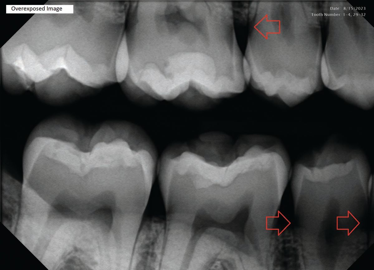
radiographic diagnosis → provides sufficient info to establish diagnosis
periapical pathosis
heavy interproximal calculus
external resorption
caries
compound/complex odontoma
supernumerary or impacted teeth
calcified pulp
nutrient canal
unusual findings (instruments, frames, piercings)
historical diagnosis → personal or family history, medical history, drug ingestion
presenting disease or lesion
amelogenesis imperfecta
dentinogenesis imperfecta

laboratory diagnosis
clinical lab tests (blood chemistries, organismal cultures, urinalysis)
helpful in determining the diagnosis of oral infections

microscopic diagnosis
main component of definitive diagnosis
certain pathology cannot be diagnosed on clinical appearance alone

surgical diagnosis
lingual mandibular bone cavity (static or Stafne bone cyst)

therapeutic diagnosis
nutritional deficiencies are common conditions
angular cheilitis (deficiency of B-complex vitamins)
necrotizing ulcerative gingivitis (responds to hydrogen peroxide)

differential diagnosis
decides which test is required to rule out conditions originally suspected and to establish definitive diagnosis
anaphylaxis
Definition: severe allergic reaction that can occur from exposure to an allergen

Disease and Conditions
Mucous membrane pemphigoid-symblepharon
Autoimmune disease that affects oral mucosa, conjunctiva, genital mucosa, and skin
Also called mucous membrane pemphigoid or cicatricial pemphigoid

Reiter Syndrome (Reactive arthritis)
Chronic disease compromising triads: arthritis, urethritis, and conjunctivitis

Aphthous ulcers (canker sore)
Definition: painful oral ulcers recurring in episodes
minor → canker sores with movable mucosa and red border (heals in 8-10 days) MOST common form
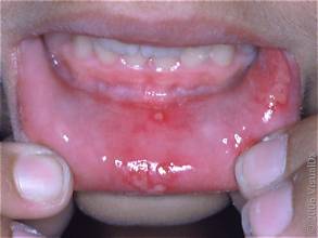
major → larger, appearing as multiple painful lesions, can be associated with HIV, (heals in a few weeks)

herpetiform → least common, resembles primary herpes and NOT related to HSV

fever blister = recurrent oral herpes = herpes labialis
Definition: most COMMON type of recurrent oral herpes simplex infection that occurs on the vermilion border of the lips

herpes zoster
Definition: reactivation of primary infection (varicella zoster/chickenpox) due to predisposing factors → immunosuppression, radiation, etc.
3 phases: prodrome, acute, and chronic
Treatments: antiviral drugs, pain medication, or vaccine
Diagnosis: clinical

varicella-zoster/chicken pox
Definition: highly contagious disease caused by varicella zoster virus that is characterized by vesicular and pustular eruptions of the skin and mucous membrane
Systemic symptoms: headache, fever, and malaise

Koplik spots/measles (rubeola)
Definition: small red macules with white necrotic centers seen intraorally with macules (Koplik spots)
caused by paramyxovirus

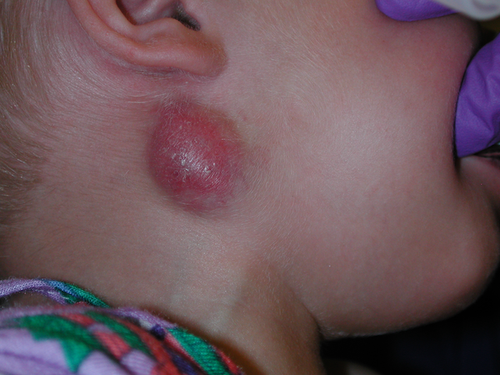
scrofula (tuberculous lymphadenitis)/TB
scrofula: TB that presents as enlargement of the submandibular and cervical lymph nodes
TB: infectious granulomatous disease caused by Mycobacterium tuberculosis that is the primary infection of the lung
treatment: antibiotics
diagnosis: PPD skin test
pseudomembranous candidiasis
white curd-like material is present on mucosal surface
underlying mucosa is red

minor erythema multiforme
self-limited disease that affects the skin and mucous membrane
cause is unknown but can be a hypersensitivity reaction

Behçet syndrome (pathergy test)
recurring autoimmune disease consisting of oral ulcers, genital ulcers, and ocular inflammation

infectious mononucleosis
Definition: Characterized by sore throat, fever, lymphadenopathy, enlarged spleen, malaise, and fatigue
Causes: Epstein-Barr virus
Treatments: resolves on its own

Scarlet fever and strawberry tongue
Definition: childhood disease caused by Group A beta-hemolytic streptococcus
characterized by red rash, strawberry tongue (fungiform papillae are red and prominent), sore throat, fever, enlarged lymph nodes

impetigo (treatment)
most commonly involves lesions on the skin of the face or extremities
APPEARS AS STICKY, HONEY-COLORED CRUST
treatment: topical antibiotics
diagnosis: clinical presentation

leukoplakia
Definition: white, plaque-like lesion on the oral mucosa that cannot be rubbed off or diagnosed as a specific disease

verruca vulgaris (common wart)
Definition: white papillary exophytic lesion caused by human papillomavirus

syphilis (treponema pallidum)
Definition: disease caused by the spirochete treponema pallidum that is transmitted from one person to another by 1. sexual contact 2. direct contact with blood 3. transplacental transmission
associated with Candida albicans

Candida albicans in newborns
associated with candidiasis, median rhomboid glossitis, and monillasis

chancre
lesion of the primary stage of syphilis

mucous patch
lesions of the secondary stage of syphilis that appear as multiple, painless, grayish-white plaques
