1.1 - Embryo Development
Outline
Fertilisation occurs 2 weeks after final menstrual period
Week 1 - Cleavage and Implantation
Week 2 - Bilaminar
Week 3 - Neurulation and Gastrulation
Week 4 - Body Plan
The week numbers in this outline are all as per the date from fertilisation which is known as the embryonic age or the fertilisation age.
In clinical setting it is normally the gestational age which is quoted which is the date from the last menstrual period
Easier to use because expectant mother will know date of the final period but if intercourse is frequent will not be able to pinpoint date of fertilisation
Day 1 - Fertilisation
Penetrating Corona radiata
Hyperactive mobility of the sperm - sperm moves quickly to the egg
Capacitated sperm - sperm then becomes capable of penetrating the egg
Corona radiata - the outermost layer of the egg, which the sperm must penetrate to reach the egg
Penetrating the Zona pellucida
Zona pellucida is the outer layer of the egg which is penetrated by the sperm
Acrosomoal reaction - the sperm releases enzymes that break down the zona pellucida allowing it to penetrate
ZP3, Acrosomal enzymes are specific enzymes that are released by the enzyme to aid with this purpose
Cortical reaction - the egg releases granules that help strengthen the zona pellucida and prevent polyspermy (when multiple sperms fuse with the egg)
Stimulated by an influx of calcium ions
Cortical granules and cortical enzymes released by the egg help strengthen the zona pellucida
Hyaline layer and zona reaction - the zona pellucida becomes more rigid and impermeable to prevent other sperm from entering
Pronuclei fusion
Male pronuclei - nucleus of the sperm
Femal pronuclei - nucleus of the egg
Zygote formation - the sperma and egg nuclei combine to form a single cell
Week 1 - Cleavage and Implantation
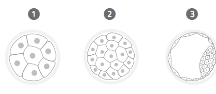
Cleavage
MTZ transition (8-cell stage)
The zygote undergoes several cell divisions to form an 8-cell embryo
Zygotic genome activation - the genertic material from the sperm and egg begins to get expressed
Maternal mRNA (the genetic material from the egg) is still being used to support the development of the embryo
Morula formation (16+ cell)
The 8-cell stage embryo undergoes several more cell divisions to form a morula, a compact cluster of cells
Blastomeres - the cells of the morula are called blastomeres. These will eventually form the embryo
Compaction
100+ cells - the morula undergoes further cell divisions to form a compact cluser of cells called a blastocyst
Embryoblast (Inner cell mass) - this is the actual cellular part of the blastocyst and is what will develop into the proper embryo
Blastocoel - this is a cavity within the blastocyst that will eventually form the amniotic cavity
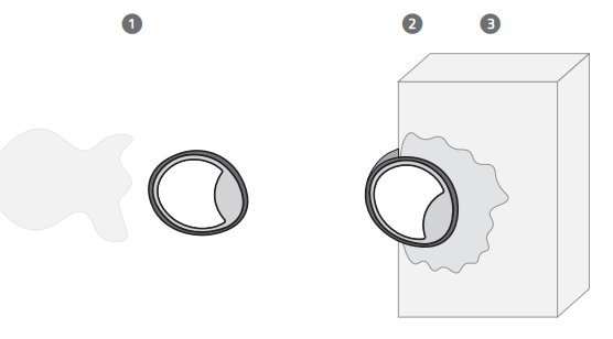
Implantation
Fertilisation has occurred in the ampulla region of the female reproductive system and next it will need somewhere for it to implant itself into
Hatching
Blastocyst hatches out of the zona pellucida layer.
Trophoblast - the outer layer of the blastocyst, which will eventually form the placenta and other supporting tissues
Blastocyst - compact cluster of cells that form the embryo
Zona Pellucida - outer layer of the egg, which the blastocyst must break through to implant in the uterus
Invasion
The blastocyst, more specifically the trophoblast invades the endometrium and upon invasion some of the trophoblasts move out and become syncytiotrophoblasts
Cytotrophoblast - the inner layer of the trophoblast, which will eventually form the placenta.
Syncytiotrophoblast: The syncytial layer of the trophoblast, which will eventually form the placenta. Body’s are all fused together with multiple nuclei
Really good at invasion.
The cells surrounding these are cytotrophoblasts
Endometrium - the lining of the uterus which the blastocyst will eventually implant itself into
HCG Secretion
Syncytyotrophoblast releases a hormone called HCG
HCG is the hormone that is detected on pregnancy test strips
HCG is what tells the mother that there has been an implantation event
Switches the mother’s physiology, ensuring that the endometrial lining stays and the pregnancy is carried forward
HCG tells the corpus luteum to release progesterone.
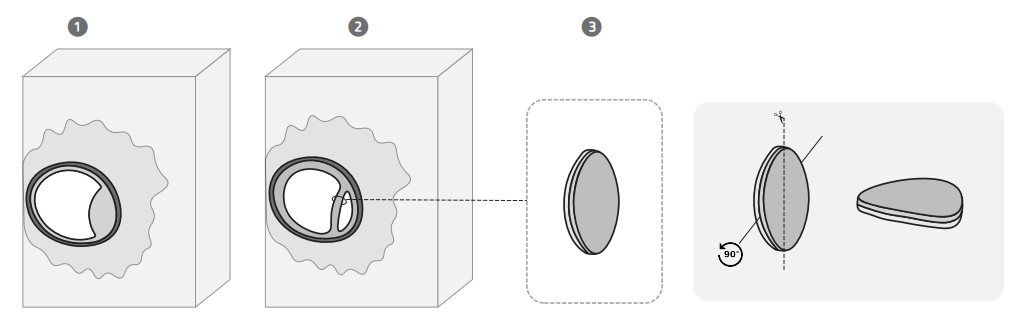
Week 2 - Bilaminar Disc Formation
Yolk sac formation
Yolk sac is the membrane that surrounds the embryo and provides it with nutrients. Formed from the proliferation of endodermal cells
Extracoelomic cavity - the blasteoceol becomes the extraceolomic cavity. This is the space that forms between the yolk sax and the embryo and is what will eventually become the amniotic cavity.
Cells lining the blasteoceol differentiate to become the Heuser’s membrane
Heuser’s membrane is what forms between the yolk sax and the embryo, helping to keep the two components separate.
Amniotic cavity formation
Amnioblasts are the cells that line the amniotic cavity. These help regulate the amount of fluid in the cavity and provide nutrients to the embryo,
Amniotic cavity: The amniotic cavity is a fluid-filled space that surrounds the embryo, and is formed by the proliferation of endodermal cells and helps to protect the embryo from the outside environment.
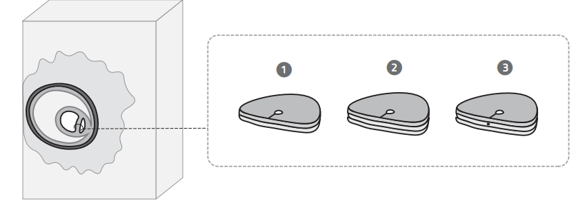
Bilaminar disc formation
As there are two cavities present, there is a small layer of cells dividing them. This layer of cells gets flattened out into a disc
This is called the bilaminar disc
Epiblast - layer of cells that forms the outer layer of the bilaminar disc. Made up of columnar cells which will eventually give rise to ectoderm
This is the disc side to the amniotic cavity
Hypoblast - layer of cells that forms the inner layer of the bilaminar disc, and is made up of cuboidal cells that will eventually give rise to the endoderm
This is the disc side to the yolk sac
Week 3 - Gastrulation and Neurulation
Gastrulation
Primitive Streak formation
Primtive streak - linear structure that forms along the anterior-posterior axis
Primitive streak = primitive node + pit + groove
Primitive node - group of cells that form at the anterior end of the embryo (the circular bit in the diagram below)
Primitive pit - depression that forms at the end of the embryo (the pit inside the primitive node)
Primitive groove - groove that forms along along the anterior posterior axis
Trilaminar disc formation
Ectoderm: The ectoderm is a layer of cells that forms the outermost layer of the embryo. It is thought to give rise to the skin, nervous system, and other structures.
Invagination: Invagination is the process by which the ectoderm layer folds inward to form a cavity. This cavity will eventually give rise to the amniotic cavity.
Mesoderm: The mesoderm is a layer of cells that forms between the ectoderm and endoderm. It is thought to give rise to the muscles, bones, and other structures.
Endoderm: The endoderm is a layer of cells that forms the innermost layer of the embryo. It is thought to give rise to the lining of the digestive tract and other structures.
Notochord formation
Thickening of cells right along the primative streak in the mesoderm layer - this is known as the notochord.
Lateral mesoderm: The lateral mesoderm is a layer of cells that forms on either side of the embryo. It is thought to give rise to the muscles and other structures.
Intermediate mesoderm: The intermediate mesoderm is a layer of cells that forms between the lateral mesoderm and endoderm. It is thought to give rise to the kidneys and other structures.
Paraxial mesoderm: The paraxial mesoderm is a layer of cells that forms along the anterior-posterior axis of the embryo. It is thought to give rise to the muscles and other structures.
Neurulation
Neural plate formation
Thickening happens on the ectoderm forms the neural plate
Folds happening with cells coming towards each other.
Neural fold formation
Neural folds converage and form the neural groove
Neural folds are the edges of the neural plate that fold inwards to form the neural tube
Cephalic flexure is a bend that forms the neural tube and is what separates the forebrain from the midbrain
Neural groove is a groove that forms the neural plate
Neural tube formation
Caudal neuropore - opening at the posterior end of the neural tube
Cranial neuropore - opening at the anterior end of the neural tube
Neural tube - tube-like structuret hat forms the precursor to the central nervous sytem and is formed by the folding of the neural plate.
Week 4 - Body Plan
Somitogenesis
Somites (thickening of cells) occurs at paraxial mesoderm
Sometimes diferentiate into 3 things (dermatome, myotome and scelrotome)
Embryo folding
Longitudinal plane folding
Transverse plane folding
Yolk sac squeezes at the top and folds down into the gut tube
Folding of embryo to form a basic body plan
Gut tube formation
The formation of the gut tube which will eventually lead to the formation of the digestive system
Week 5 - Organogenesis
Organs start forming
Gut tube
Neural tube
Limb bud formation - the buds of the limbs
Week 6 to 8
Week 6 - face structures form, heart formed, brain vesicles
Week 7 - ossification, fingers and toes, eyelids form
Week 8 - tail regression, skeleton develops, embryonic period end