Ch 5 - Pt 1: Integumentary System (Skin)
Components of the Integumentary System
Skin: The body's primary protective outer boundary.
Sudoriferous (sweat) glands: Exocrine glands responsible for sweat secretion.
Sebaceous (oil) glands: Exocrine glands responsible for sebum secretion.
Hairs: Filamentous structures growing from the epidermis.
Nails: Hard keratinized epidermal structures at the tips of digits.
Subcutaneous tissue: Deep connective tissue layer beneath the skin.
Structural Layers of Skin and Subcutaneous Tissue
Epidermis:
Outermost protective shield of the body.
Composed entirely of epithelial tissue (specifically keratinized stratified squamous epithelium).
Avascular (devoid of blood vessels), receiving nutrients via diffusion from underlying dermal blood vessels.
Dermis:
Underlies the epidermis and constitutes the bulk of the skin's mass.
Composed primarily of tough, flexible dense connective tissue.
Vascularized (supplied with blood vessels).
Contains nerve fibers, blood vessels, lymphatic vessels, epidermal hair follicles, sebaceous glands, and sudoriferous glands.
Fibers in the the matrix bind the body together - forms the animal "hide" that is processed into leather.
Subcutaneous Tissue (Hypodermis or Superficial Fascia):
Not technically a layer of the skin, but shares key functions and is integrated into the integumentary system.
Composed mostly of adipose tissue along with some areolar connective tissue.
Functions:
Loosely anchors the skin to underlying structures, primarily deep fascia and muscles, while allowing the skin to slide relatively freely.
Acts as a shock absorber to protect internal organs from trauma.
Functions as a thermal insulator to prevent body heat loss.

Cellular Composition of the Epidermis
Keratinocytes:
The dominant cell type forming the majority of the epidermis.
Produce fibrous keratin, a tough structural protein that gives the skin its protective, durable, and waterproof properties.
Millions slough off daily from the surface; continuous mitotic division creates a completely renewed epidermis every .
Melanocytes:
Spider-shaped cells positioned in the deepest layer of the epidermis (stratum basale).
Synthesize the pigment melanin, which is packaged into membrane-bound granules called melanosomes.
Melanosomes are transferred along cellular processes to nearby keratinocytes, accumulating as a pigment shield over the keratinocyte nucleus to protect cellular DNA from ultraviolet (UV) radiation damage.
Dendritic Cells (Langerhans Cells):
Star-shaped immune cells (macrophages) that patrol the deep layers of the epidermis.
Ingest foreign material and debris, serving as essential activators of the immune system.
Tactile Epithelial Cells (Merkel Cells):
Located at the epidermal-dermal junction.
Associated with sensory nerve endings to serve as light touch sensory receptors.
Strata of the Epidermis
The epidermis comprises four or five distinct layers (strata):
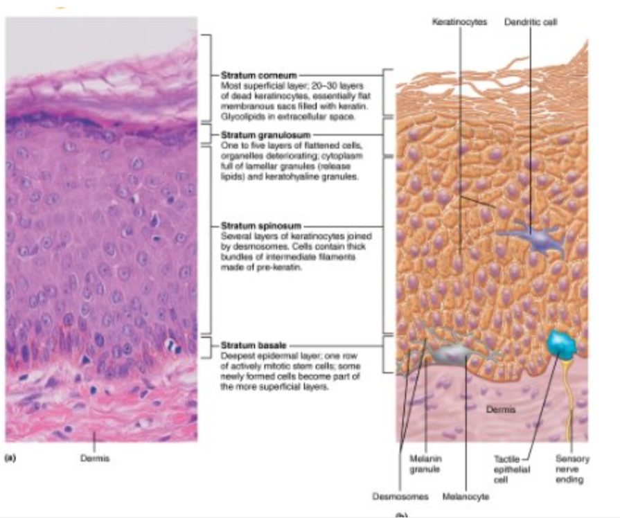
Stratum Basale (Basal Layer or Stratum Germinativum):
The deepest epidermal layer, firmly attached to the underlying dermis along a wavy borderline.
Consists of a single row of actively dividing (mitotic) stem cells.
Mitosis yields two daughter cells:
One daughter cell travels upward through the epidermal layers to the surface, taking , dying as it moves further from dermal nutrient sources.
The other daughter cell remains in the basal layer to continue functioning as a stem cell.
Termed stratum germinativum ("germinating layer") due to continuous cell division.
Composed of melanocytes, interspersed among the stem cells, along with occasional tactile epithelial cells.
Stratum Spinosum (Prickly Layer):
Several cell layers thick.
Contains a web-like network of intermediate pre-keratin filaments attached to desmosomes.
Desmosomal junctions allow the layer to resist mechanical tension and lateral pulling forces.
Keratinocytes shrink during tissue preparation while retaining desmosome attachments, causing them to appear spiky ("prickle cells").
Contains abundant melanosomes and dendritic cells scattered among the keratinocytes.
Stratum Granulosum (Granular Layer):
Four to six cell layers thick, but thin overall due to cellular flattening.
Drastic changes in cell appearance occur:
Cells flatten, and their nuclei and organelles disintegrate.
Keratinization begins.
Cells accumulate keratohyaline granules, which assist in assembling tough keratin fibers in upper strata.
Cells accumulate lamellar granules, containing a water-resistant glycolipid that is secreted into extracellular spaces to slow water loss across the epidermis.
Cells above this layer die because they are positioned too far from dermal capillaries to receive nutrients via diffusion.
Stratum Lucidum (Clear Layer):
Present only in thick skin.
Located superficial to the stratum granulosum and deep to the stratum corneum.
Consists of a thin, translucent band of 2 to 3 rows of clear, flat, dead keratinocytes.
Stratum Corneum (Horny Layer):
The most superficial epidermal layer.
Consists of 20 to 30 rows of flat, anucleated, keratinized dead cells (membrane-bound sacs filled with keratin).
Accounts for approximately three-quarters () of total epidermal thickness.
Functions:
Protects deeper layers from environmental damage.
Prevents transepidermal water loss.
Protects against physical abrasion and mechanical penetration.
Serves as a robust barrier against biological, chemical, and physical agents.
Regional Thickness Variations of Epidermis:
Thick Skin: Features all five epidermal strata (including stratum lucidum); located on the palms of hands, fingertips, and soles of feet.
Thin Skin: Contains only four strata (lacks stratum lucidum); covers the rest of the body.
Note: Thick and thin skin categories refer specifically to epidermal thickness, not overall skin thickness (e.g., the skin on the upper back possesses the thickest dermis and highest total thickness, but is classified as thin skin due to its four-layered epidermis).
The Dermis and Its Structural Layers
Layers of the Dermis:
Papillary Dermis:
Thin, superficial layer composed of areolar connective tissue featuring loose, interlacing collagen and elastic fibers heavily invested with blood vessels.
The loose, open structure allows patrolling phagocytes to move freely and ingest invading microorganisms.
Dermal Papillae: Peg-like projections extending into the overlying epidermis. Contain capillary loops, free nerve endings (pain receptors), and touch receptor’s called tactile corpuscles (aka Meissner's corpuscles).
Reticular Dermis:
Deep layer making up approximately of total dermal thickness.
Composed of coarse, dense irregular connective tissue.
Elastic fibers provide stretch-and-recoil capabilities; collagen fibers provide structural strength, resiliency, and bind water to maintain tissue hydration.
Named for its network (reticulum) of collagen fiber bundles, not for an abundance of reticular fibers.
Dermal Vascular Plexus: Network of blood vessels supplying the skin, situated between the reticular layer and the subcutaneous tissue.
Dermal Modifications and Skin Markings
Friction Ridges:
Occur in thick skin on fingers, palms, and soles.
Formed by dermal papillae resting atop larger dermal ridges, which force the overlying epidermis to form epidermal ridges.
Functions:
Enhance gripping ability on wet or smooth surfaces.
Amplify tactile sensations by transmitting vibrations to deep receptors (lamellar corpuscles).
Ridge patterns are unique to each individual, determined jointly by genetics and random fetal movements in the womb.
Sweat gland ducts open along the crests of friction ridges, leaving film traces known as fingerprints.

Cleavage (Tension) Lines:
Represent separations or less dense regions between underlying collagen fiber bundles in the reticular dermis.
Run parallel to the skin surface; externally invisible.
Arrange circularly around the trunk and longitudinally along the limbs.
Surgical importance: Surgical incisions made parallel to cleavage lines gap less and heal much faster than incisions made across collagen bundles.
Flexure Lines:
Dermal folds occurring at or near joints where the dermis is tightly anchored to deeper underlying fascia.
Because the skin cannot slide easily during joint flexion, deep creases are formed.
Clearly visible on the hands, wrists, fingers, soles, and toes.
Clinical Homeostatic Imbalances
Striae ("Stretch Marks"):
Extreme stretching of the skin (e.g., during pregnancy or rapid growth) tears collagen fibers in the dermis.
Leaves dermal scars visible externally as silvery white lines.
Blisters:
Fluid-filled pockets separating the epidermal and dermal layers.
Result from acute, short-term mechanical trauma (e.g., intense friction or burns).
Skin Color Pigmentation and Diagnostic Signs
Three Pigments Determining Skin Color:
Melanin:
The only pigment synthesized within the skin (by melanocytes).
Packaged into melanosomes and transferred to keratinocytes to shield nuclear DNA from harmful UV rays.
Increased exposure to sunlight stimulates melanocytes to produce more melanin.
Occurs in two primary forms ranging in shade from reddish-yellow to brownish-black.
Variations in human skin color reflect the specific types and quantities of melanin produced, rather than differences in the total number of melanocytes.
Freckles and pigmented moles represent localized, concentrated accumulations of melanin.
Carotene:
Yellow-to-orange pigment obtained through dietary intake.
Most noticeable in areas where the stratum corneum is thickest, such as the palms and soles.
Accumulates in the stratum corneum and subcutaneous adipose tissue.
Can be converted by the body into vitamin A, vital for normal vision and epidermal maintenance.
Hemoglobin:
Oxygen-binding protein in red blood cells within dermal capillaries.
Gives fair skin its pinkish tint due to lower melanin levels allowing the red hue of oxygenated blood to shine through translucent epidermis.
Diagnostic Color Changes in Clinical Practice:
Cyanosis (Blueness): Indicates poorly oxygenated hemoglobin; a diagnostic sign of severe respiratory or cardiovascular distress.
Pallor or Blanching (Paleness): Caused by emotional stress, low blood pressure, or anemia (decreased oxygen-carrying capacity of blood).
Erythema (Redness): Caused by embarrassment, fever, localized inflammation, or allergic reactions.
Jaundice (Yellowness): Indicates accumulation of bilirubin due to liver disorders.
Bruises/Hematomas (Red/Purple/Green/Yellow Marks): Result from blood escaping surrounding capillaries and clotting in subcutaneous or dermal spaces after vessel trauma.
Hyperpigmented / Dark Patches: Velvet-like, hyperpigmented, thickened skin in body folds (e.g., axilla or neck) can indicate underlying endocrine disorders.