Microscope
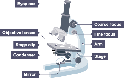
Light microscope:
use light and lenses to form a magnified image of a specimen
images of cells and large subcellular structures are visible
used to study living cells and for regular use when relatively low magnification and resolution is enough
Practical - preparing light microscope slides
Put a thin sample of tissue (e.g. onion skin) onto a microscope slide.
Add a few drops of a suitable stain/dye (e.g. iodine).
Place a coverslip on top of the tissue and place the slide onto the microscope stage.
Use the objective lens with the lowest magnification, and focus on the sample.
Increase the magnification and refocus to see different features of the cell.
Electron microscope:
Electron microscopes use beams of electrons, rather than light, to visualise specimens
The wavelength of an electron beam is much smaller than that of visible light, which gives electron microscopes a much higher resolution and magnification
provide higher magnifications and higher resolution images but cannot be used to view living cells
There are 2 types of electron microscope:
the transmission electron microscope (TEM) is used to examine thin slices or sections of cells/tissues
the scanning electron microscope (SEM) has a large depth of field so can be used to examine the surface structure of specimens
