Comprehensive Study Guide: Human Anatomy, Chemical Biology, and Cellular Physiology
Anatomical Terminology, Body Cavities, and Serous Membranes
Anatomical Planes of the Body:
Sagittal Plane: A vertical plane dividing the body into right and left portions.
Coronal Plane: A vertical plane dividing the body into anterior (front) and posterior (back) portions.
Axial / Transverse Plane: A horizontal plane dividing the body into superior (upper) and inferior (lower) portions.
Directional Terminology:
Superior: Toward the head or upper structure.
Inferior: Toward the feet or lower structure.
Anterior: Toward the front of the body.
Posterior: Toward the back of the body.
Medial: Toward the midline of the body.
Lateral: Away from the midline of the body.

Subdivisions of the Posterior (Dorsal) Body Cavity:
Cranial Cavity: Encased by the skull; houses and protects the brain.
Vertebral Cavity: Runs along the vertebral column; houses and protects the spinal cord.
Subdivisions of the Anterior (Ventral) Body Cavity:
Thoracic Cavity: Superior cavity enclosed by the ribs, thoracic muscles, and sternum. Subdivided into:
Superior Mediastinum: Central region containing major vascular trunks, esophagus, and trachea.
Pleural Cavity: Bilateral cavities containing the lungs.
Pericardial Cavity: Located within the mediastinum; surrounds the heart.
Diaphragm: Muscular partition dividing the superior thoracic cavity from the inferior abdominopelvic cavity.
Abdominopelvic Cavity: Subdivided into:
Abdominal Cavity: Contains digestive organs including the stomach, small intestine, large intestine, liver, and spleen.
Pelvic Cavity: Inferior cavity containing reproductive organs, urinary bladder, and rectum.
Serous Membranes (Serosa):
Definition: Thin, double-layered membranes lining the walls and organs of closed body cavities (thoracic and abdominopelvic cavities).
Pleura: The serous membrane associated with the lungs and pleural cavities.
Pericardium: The serous membrane associated with the heart within the pericardial cavity.
Peritoneum: The serous membrane associated with the abdominal organs (e.g., stomach, small intestines).

Anatomical Layers of Serous Membranes (Pericardium Example):
Visceral Layer (Visceral Pericardium): Inner layer covering the external surface of the organ directly.
Parietal Layer (Parietal Pericardium): Outer layer lining the cavity wall.
Serous Cavity (Pericardial Cavity): Fluid-filled potential space/air space between the visceral and parietal layers, acting like a double-walled balloon filled with fluid to reduce friction.
Chemical Organization of the Body and Atomic Structure
Matter and Mass:
Matter: Anything that occupies space and has mass.
Mass: A measure of the quantity of matter in an object, standardly measured in grams () or kilograms ().
Elements: Pure chemical substances with unique chemical and physical properties that cannot be broken down into simpler substances through ordinary chemical reactions.
Chemical Symbols of Elements:
Designated by single capital letters or combinations of two letters.
Derived from English terms: Carbon (), Calcium ().
Derived from Latin roots:
Sodium (): Derived from natrium.
Potassium (): Derived from kalium.
Clinical Terminology for Concentration Imbalances:
Prefix Hyper-: Indicates excessive or above-normal levels.
Prefix Hypo-: Indicates deficient or below-normal levels.
Hypernatremia: Pathological condition characterized by excessive sodium levels in the blood.
Hypokalemia: Pathological condition characterized by insufficient potassium levels in the blood.
Composition of Living Organisms:
Major Elements: Four primary elements comprise approximately of human body mass:
Oxygen ()
Carbon ()
Hydrogen ()
Nitrogen ()
Trace Elements: Essential elements present in minor quantities (e.g., Calcium [], Phosphorus [], Iron []).

Atomic Structure:
Atom: The fundamental, smallest unit of matter retaining all chemical properties of an element.
Atomic Regions:
Nucleus: Dense central region containing positively charged Protons ( charge) and neutral Neutrons ( charge).
Electron Cloud: Surrounding area containing negatively charged Electrons ( charge) orbiting in defined orbitals/shells.
Atomic Number: Defined exclusively by the number of protons in the nucleus; dictates element identity.
Neutral Atom Balance: In an uncharged neutral atom, the total number of electrons equals the number of protons.

Organization of the Periodic Table:
Elements are arranged sequentially by increasing atomic number.
Groups (Columns) and Periods (Rows) reflect valence shell electron configurations and elemental chemical reactivity.
Electron Shells and Energy Levels:
Electrons occupy distinct energy shells located at varying distances from the nucleus.
Shells closer to the nucleus possess lower potential energy levels than outer shells.
Energy dynamics: Transitioning an electron to an outer shell requires energy absorption; dropping an electron to an inner shell releases energy.
Valence Shell and Valence Electrons:
Valence Shell: The outermost electron shell of an atom.
Valence Electrons: Electrons residing in the valence shell that dictate chemical reactivity and bond formation.
Octet Rule: Most biologically critical elements require valence electrons in their outermost shell to achieve structural stability.
Ion Formation Mechanisms:
Cation Formation (Sodium Example):
Sodium () atom: protons (), electrons (), with valence electron.
To satisfy the octet rule, sodium readily loses its single valence electron, becoming a positively charged cation () with protons and electrons.
Anion Formation (Chlorine / Chloride Example):
Chlorine () atom: protons (), electrons (), with valence electrons.
Chlorine accepts electron into its valence shell, forming a negatively charged anion called chloride () with protons and electrons.

Physiological Concentrations of Key Electrolytes:
Extracellular Fluid (ECF / Outside Cell):
Sodium (): High concentration ().
Chloride (): High concentration ().
Calcium (): High concentration ().
Potassium (): Low concentration ().
Intracellular Fluid (ICF / Inside Cell):
Potassium (): High concentration ().
Organic Anions (): High concentration (large intracellular proteins/molecules).
Sodium (): Low concentration ().
Chloride (): Low concentration ().
Calcium (): Extremely low concentration (<1\,\mu\text{M}).
Organic Compounds and Macronutrients
Carbon Chemistry and Hydrocarbons:
Carbon contains valence electrons.
Rather than gaining or losing electrons, carbon forms stable covalent bonds by sharing four electron pairs.
Creates elongated, complex carbon skeletons.
Hydrocarbons: Organic molecules composed exclusively of carbon and hydrogen atoms.
Macronutrients:
Definition: Nutrients required by the body in large quantities to furnish metabolic energy and structural framework.
Three Primary Classes:
Carbohydrates
Lipids (Fats)
Proteins
Carbohydrates:
Definition: Organic biomolecules composed of carbon, hydrogen, and oxygen (termed "hydrated carbon").
Generic Formula:
Stoichiometry: Hydrogen and oxygen exist in a atomic ratio, identical to water.
Saccharides (Sugars) Classifications:
Monosaccharides: Single monomer units.
Disaccharides: Molecules composed of two covalently bonded monosaccharide monomers.
Polysaccharides: Complex polymers composed of hundreds to thousands of monosaccharide units.

Monosaccharides:
Hexose Sugars (Six-Carbon Monosaccharides, ):
Glucose: Primary cellular metabolic fuel source.
Fructose: Hexose sugar found in fruits.
Galactose: Hexose sugar found in dairy products.
Pentose Sugars (Five-Carbon Monosaccharides, / ):
Ribose: Sugar component of RNA.
Deoxyribose: Sugar component of DNA.

Disaccharides:
Sucrose (Table Sugar): Composed of Glucose + Fructose.
Lactose (Milk Sugar): Composed of Galactose + Glucose.
Maltose (Malt Sugar): Composed of Glucose + Glucose.
Polysaccharides:
Starches: Long-chain glucose polymers synthesized by plants; serving as easily digestible plant storage carbohydrates.
Glycogen: Highly branched glucose polymer synthesized by animals; stored predominantly in skeletal muscle and liver tissue.
Lipids:
Properties: Water-insoluble organic molecules composed mostly of nonpolar hydrocarbons, rendering them hydrophobic.
Major Biological Lipids:
Triglycerides
Phospholipids
Steroids / Sterols

Triglycerides:
Abundance: Most prevalent dietary lipid class and major energy storage form in body tissues.
Physical States: Fats (solid at room temperature) and Oils (liquid at room temperature).
Molecular Structure: One Glycerol backbone (a three-carbon molecule) linked to three long-chain Fatty Acids.
Dehydration Synthesis: Chemical reaction bonding three fatty acids to glycerol with the simultaneous removal and production of three water () molecules.
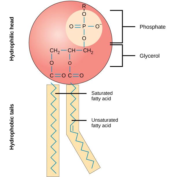
Phospholipids:
Molecular Structure: Diglyceride structure consisting of a glycerol backbone attached to two nonpolar fatty acid chains and one charged phosphate group (often coupled with a nitrogenous group).
Amphipathic Nature:
Polar / Hydrophilic Head: Contains the charged phosphate group; attracted to aqueous environments.
Nonpolar / Hydrophobic Tail: Contains neutral fatty acid chains; repelled by water.

Behavior of Phospholipids in Water:
Planar Bilayer: Spontaneous alignment into a two-layer sheet roughly thick, with polar heads facing water and nonpolar tails sequestered inside.
Liposomes: Spherical vesicles formed by a lipid bilayer enclosing a central aqueous interior, surrounded by an aqueous exterior.
Steroids / Sterols:
Molecular Structure: Composed of four interlocking hydrocarbon rings.
Cholesterol: Precursor molecule from which all body steroids (such as steroid hormones) are synthesized.

Proteins:
Elemental Composition: Always contain Carbon, Hydrogen, Oxygen, Nitrogen (), and frequently Sulfur ().
Monomer: Amino Acids.
Structural and Functional Roles: Structural framework (e.g., Collagen), catalytic enzymes (e.g., Digestive Enzymes), defense (e.g., Antibodies).
Fundamental Amino Acid Anatomy:
Central Carbon () atom.
Hydrogen () atom.
Amino Group ( / ).
Carboxyl Group ( / ).
Variable Side Chain ( Group): Confers specific chemical properties to each amino acid.
Structural Hierarchy of Proteins:
Peptide: Short chain containing to amino acids joined by peptide bonds (e.g., Oxytocin, a -amino-acid hormone).
Polypeptide: Chain of more than amino acids (e.g., Glucagon, a -amino-acid polypeptide).
Protein: Functional macromolecule consisting of one or more polypeptides ranging from to thousands of amino acids folded into complex three-dimensional structures (e.g., Collagen, featuring over amino acids arranged in a triple helix).
Cellular Structure and Organization
The Cell:
Fundamental Definition: The basic structural, functional, and biological unit of all living organisms; the smallest unit of life.
Physiological Principle: Cell activity dictates tissue and organ performance (e.g., Hepatocytes execute liver metabolic functions; Myocytes shorten to provide muscle contraction).
Specialized Cell Types:
Nerve Cells (Neurons)
Muscle Cells: Striated Muscle (voluntary skeletal), Smooth Muscle (involuntary), Cardiac Muscle.
Bone Cells (Osteocytes)
Gland Cells
Blood Cells: Red Blood Cells (Erythrocytes) and White Blood Cells (Leukocytes: Lymphocytes, Monocytes, Neutrophils, Eosinophils, Basophils).
Reproductive Cells (Gametes): Sperm and Ovum.

Animal Cell Subcellular Structures:
Plasma Membrane: Outer boundary enclosing cytosol and organelles.
Nucleus: Encloses genetic material. Contains:
Nucleolus: Site of ribosomal RNA synthesis.
Chromatin: DNA and histone protein complex.
Nuclear Envelope: Double membrane perforated by Nuclear Pores.
Cytoplasm & Cytosol: Intracellular fluid containing organellar machinery.
Ribosomes: Protein synthesis sites (free in cytosol or bound to Rough ER).
Rough Endoplasmic Reticulum (Rough ER): Membranous network studded with ribosomes; modifies and packages proteins.
Smooth Endoplasmic Reticulum (Smooth ER): Lipid and steroid synthesis, carbohydrate metabolism, and calcium storage.
Golgi Apparatus: Modifies, sorts, and packages macromolecules for secretion or delivery.
Mitochondria: Primary sites of ATP production via cellular respiration.
Lysosomes: Digestive organelles containing hydrolytic enzymes to break down debris.
Peroxisomes: Neutralize toxins and break down fatty acids.
Centrioles: Microtubule-organizing centers active during cell division.
Cytoskeleton: Microtubules and Microfilaments providing structural support and movement.
Cilia: Motile surface projections driving fluid across the cell membrane.
Structure and Dynamics of the Plasma Membrane
Plasma Membrane Architecture:
Semipermeable selective barrier separating intracellular fluid (ICF) from extracellular fluid (ECF).
Arranged as a Phospholipid Bilayer:
Hydrophilic Phosphate Heads: Face outward toward aqueous ECF and ICF.
Hydrophobic Fatty Acid Tails: Face inward toward the membrane core, establishing a hydrophobic barrier to water-soluble solutes.

Plasma Membrane Permeability Rules:
Small, Nonpolar Molecules (e.g., Oxygen [], Carbon Dioxide []): Easily cross the lipid bilayer by diffusing directly through the hydrophobic core.
Small, Polar Molecules (e.g., Water []): Can traverse the bilayer slowly without protein assistance.
Large, Polar Molecules (e.g., Glucose) and Charged Ions (e.g., , , , ): Unable to penetrate the nonpolar hydrophobic region without transport proteins.
Functional Classes of Membrane Proteins:
Integral Proteins: Proteins embedded directly within the lipid bilayer, spanning part or all of the membrane (transmembrane proteins).
Channel Proteins: Water-filled pores allowing specific solute flux.
Carrier Proteins: Transporters undergoing conformational shape changes.
Peripheral Proteins: Attached to inner or outer surface surfaces of the membrane.
Outer Surface Peripheral Proteins: Serve as cellular identification markers.
Inner Surface Peripheral Proteins: Anchor the plasma membrane to the internal cytoskeleton to maintain cell shape.
Glycocalyx (Glycoproteins and Glycolipids):
Glycoproteins: Carbohydrate chains conjugated to integral proteins; function as primary markers for self versus non-self cell recognition.
Glycolipids: Carbohydrate chains conjugated to membrane phospholipids; act as blood type surface markers.
Glycocalyx: Surface carbohydrate coat formed collectively by glycoproteins and glycolipids.
Membrane Fluidity Regulation by Cholesterol:
Cholesterol molecules insert between adjacent phospholipids within the bilayer.
High Temperatures: Restricts fatty acid chain movement, decreasing excessive fluidity and stabilizing the membrane.
Low Temperatures: Prevents fatty acid chains from packing tightly, preventing freezing and maintaining membrane fluidity.
Membrane Transport Mechanisms and Tonicity
Classification of Transport Mechanisms:
Passive Transport: Movement of substances across the membrane down their gradient without cellular energy () expenditure.
Active Transport: Energy-requiring () movement of substances across the membrane, often against their gradient.
Gradients:
Chemical Gradient: Concentration difference of a solute across a membrane.
Electrical Gradient: Difference in electrical charge across a membrane.
Electrochemical Gradient: Combined chemical and electrical forces driving ion movement.
Passive Transport Modalities:
Simple Diffusion: Unassisted passive movement of lipid-soluble particles, , and from high concentration to low concentration directly through the phospholipid bilayer.
Facilitated Diffusion: Passive movement of large polar molecules or ions down their concentration gradient via channel or carrier proteins (e.g., movement of glucose via Glucose Transporters [GLUT]).
Dynamic Equilibrium: Condition where solute molecules continue crossing the membrane at equal rates in both directions, yielding no net change in concentration over time.
Ion Channel Biophysics:
Pore Selectivity & Electrochemical Exclusion: Channel pores exclude ions based on charge and pore diameter.
Channels selective for cations (, , ) contain negatively charged amino acid side chains lining the pore.
Channels selective for anions () contain positively charged side chains lining the pore.

Gated Ion Channels:
Ligand-Gated Channels: Open or close when a specific chemical signal (ligand) binds to the receptor site (e.g., Acetylcholine [ACh] binding to open cation channels for , , and ).

Mechanically-Gated Channels: Open or close in response to physical membrane deformation (e.g., stretching, mechanical pressure, vibration, touch).

Voltage-Gated Channels: Open transiently in response to changes in membrane electrical potential.

Nongated (Leak) Channels:
Permanently open pores allowing continuous, passive ion leakage down electrochemical gradients.
Resting Membrane Potential Determination: At rest, open potassium () leak channels far outnumber sodium () leak channels. Consequently, passive efflux is the primary factor establishing resting membrane potential.
Osmotic Pressure and Osmosis:
Osmotic Pressure: The pulling force with which a solution draws water into itself across a semipermeable membrane; directly proportional to dissolved solute particle concentration.
Units: Osmoles or milliosmoles ().
Physiological Osmotic Baseline: Human ICF and ECF maintain an osmotic pressure of approximately .
Osmosis: Net diffusion of water across a semipermeable membrane from an area of low osmotic pressure (high water concentration) to high osmotic pressure (low water concentration).

Tonicity Dynamics:
Definition: The ability of an extracellular solution to alter cell shape and volume by inducing fluid movement.
Isotonic Solution: Equivalent osmotic pressure to ICF. Zero net water movement; cells retain normal shape and biological function.
Hypertonic Solution: Higher osmotic pressure than ICF. Water exits the cell rapidly via osmosis; cells shrivel and shrink (crenation).
Hypotonic Solution: Lower osmotic pressure than ICF. Water enters the cell via osmosis; cells swell and risk rupture/bursting (hemolysis).
Clinical Isotonic Intravenous (IV) Solutions (Target ):
Sodium Chloride ( / Normal Saline): .
Dextrose in Water (): .
Lactated Ringer's (): Contains , , , , and lactate (metabolized to bicarbonate): .
Clinical Isotonic Equivalence: .

Primary Active Transport:
Directly uses energy from hydrolysis to pump solutes against concentration gradients.
Sodium-Potassium Pump ( ATPase):
Stoichiometric Ratio: Expels ions out of the cell and imports ions into the cell per single consumed.
Maintains steep extracellular and intracellular concentration gradients.

Secondary Active Transport and Cotransport Mechanisms:
Secondary Active Transport: Uses the potential energy of an ion gradient created by primary active transport (e.g., gradient) to move another substance against its gradient.
Antiport (Counter-transport): Transports two distinct solutes in opposite directions across the membrane (e.g., moved out while is moved in).
Symport (Cotransport): Transports two distinct solutes in the same direction across the membrane (e.g., /Glucose symporter dragging glucose into the cell along with influx).
Vesicular Bulk Transport (Endocytosis):
Phagocytosis ("Cell Eating"): Endocytosis of large solid particles or invading pathogens by immune cells (forming a phagocytic food vacuole that fuses with lysosomes for enzymatic destruction).
Pinocytosis ("Cell Drinking"): Non-specific endocytosis bringing extracellular fluid and dissolved solutes into the cell within micro-vesicles.
Questions & Discussion
Question: Only sodium was capable of moving and it moved into the cell. The inside of the cell would become _________ __________.
Options:
A. more positive
B. more negative
C. less positive
D. less negative
Correct Answer: A. more positive
Physiological Explanation: Sodium () carries a net positive charge. Because extracellular sodium concentration () is much higher than intracellular sodium concentration (), opening a pathway for sodium allows positive ions to stream down their electrochemical gradient into the cytosol. This influx of positive charge causes the intracellular environment to become more positive (depolarized).