Stomach
Describe the physiology of gastric acid and other gastric secretions.
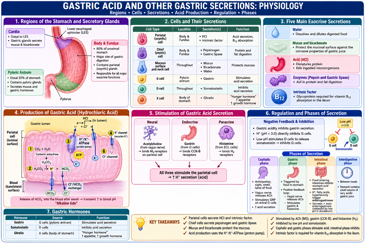
Regions of the Stomach and Secretory Glands
Cardia: Located distal to the lower esophageal sphincter, it contains gastric glands that secrete mucus and bicarbonate. (top part)
Body and Fundus: Comprises 80% of the proximal stomach and serves as the major site of gastric digestion. It contains parietal (oxyntic) glands, which are responsible for all major exocrine functions. (middle/whole)
Pyloric Antrum: Comprises the distal 20% of the stomach. It contains pyloric glands that secrete mucus and gastric hormones. (Buttom part)
Cells and Their Secretions
Parietal (oxyntic) cells: Secrete hydrochloric acid (HCl) and intrinsic factor.
Chief (peptic) cells: Secrete pepsinogen and gastric lipase.
Mucous surface and neck cells: Secrete mucus, bicarbonate, and water.
Five Main Exocrine Secretions of the Stomach
Water: Dissolves and dilutes digested food.
Mucus and bicarbonate: Protect the mucosal surface against the corrosive properties of gastric juice.
Acid (HCl): Denatures protein and kills ingested microorganisms.
Enzymes (Pepsin and Gastric lipase): Aid in protein and fat digestion.
Intrinsic factor: A glycoprotein required for vitamin B12 absorption in the ileum
Gastric Hormones
Gastrin: Secreted by G cells in the pyloric antrum.
Somatostatin: Secreted by D cells.
Ghrelin: Released by X cells in the body of the stomach. Known as the "hunger hormone," it signals the hypothalamus to increase appetite and stimulates the pituitary gland to release growth hormone
Production of Gastric Acid (Hydrochloric Acid)
Hydrogen ions (H+) are generated inside the parietal cell through the action of carbonic anhydrase, which catalyzes carbon dioxide and water into carbonic acid (H2CO3).
The carbonic acid then dissociates into H+ and bicarbonate (HCO3-).
H+ is pumped from the cytoplasm into the stomach lumen by the H+/K+-ATPase (proton pump), which uses ATP to pump H+ out in exchange for potassium (K+).
Potassium is recycled back into the lumen through K+ channels.
Bicarbonate exits the cell into the blood in exchange for chloride (Cl-) entering the cell via a Cl-/HCO3- exchanger at the basolateral membrane. This release of bicarbonate into the blood after meals causes a transient increase in blood pH known as the "alkaline tide".
Chloride is then secreted into the gastric lumen through chloride channels.
Finally, H+ and Cl- combine in the lumen to form hydrochloric acid (HCl).
Stimulation of Gastric Acid Secretion
Neural stimulation: Acetylcholine is released from the vagus nerve and binds to M3 receptors on the parietal cell.
Endocrine stimulation: Gastrin is released from G cells in the antrum and binds to CCK-B receptors.
Paracrine stimulation: Histamine is released from enterochromaffin-like (ECL) cells and binds to H2 receptors.
Regulation and Phases of Secretion
Negative feedback autoregulation: Gastric acidity inhibits gastrin secretion. Hydrogen ions (when pH is < 3.0) directly inhibit G cells.
Paracrine inhibition: Low pH stimulates D cells to release somatostatin, which inhibits G cells.
Cephalic phase: Occurs in response to the anticipation, sight, smell, and taste of food. The vagus nerve releases acetylcholine and stimulates gastrin-releasing peptide on antral G cells to increase acid secretion.
Gastric phase: Triggered by food in the stomach. A positive feedback loop occurs where the vagal nerve releases acetylcholine, stimulating gastrin and histamine.
Intestinal phase: Food entering the intestines inhibits acid secretion in the stomach. Luminal factors like H+, fatty acids, and hypertonicity trigger the release of enterogastrones. Secretin is the primary enterogastrone, released in response to low pH in the duodenum.
Interdigestive phase: The period between meals where the stomach contains a small volume of very acidic gastric juice.
Discuss the patterns of gastric motility and emptying.
Gastric Motility Patterns
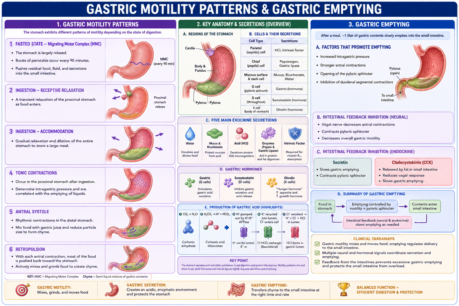
Fasted state: The stomach is largely relaxed, except for bursts of peristalsis that occur every 90 minutes. This is controlled by the Migrating Motor Complex (MMC), which serves to flush out the stomach and small intestine.
Ingestion (Receptive relaxation): A transient relaxation of the proximal stomach occurs with the arrival of each food bolus.
Ingestion (Accommodation): A gradual relaxation and dilation of the entire stomach during eating, which allows the stomach to store a large meal.
Tonic contractions: Occur in the proximal stomach after ingestion. These contractions determine intragastric pressure and are correlated with the gastric emptying of liquids.
Antral systole: Rhythmic contractions in the distal stomach. These contractions mix food with gastric juice and reduce particle size to create "chyme," which is a suspension of partially dissolved food particles.
Retropulsion: With each antral systole, there is a forceful reflection of most of the food back from the pyloric sphincter into the stomach. This mechanism actively mixes and grinds the food.
Gastric Emptying
After a meal, the stomach contains about 1 liter of material that slowly empties into the small intestine.
Emptying is brought on by: Increased intraluminal pressure, increased antral contractions, the opening of the pyloric sphincter, and the inhibition of duodenal segmental contractions.
Intestinal feedback inhibition (Neural): Feedback from the intestines can slow emptying. The vagal nerve decreases antral contractions, contracts the pyloric sphincter, and decreases overall gastric motility.
Intestinal feedback inhibition (Endocrine):
Secretin: Slows gastric emptying and acts to contract the pyloric sphincter.
Cholecystokinin (CCK): Responds to the presence of fat in the small intestine. It reduces the response of the vagal nerve and slows gastric emptying.
Describe the pathophysiology including the clinical presentation, epidemiology and risk factors, pathogenesis, pathology and clinical manifestations of the following disorders of the stomach:
Acid-peptic disease:
Acid-Peptic Disease (General Characteristics)
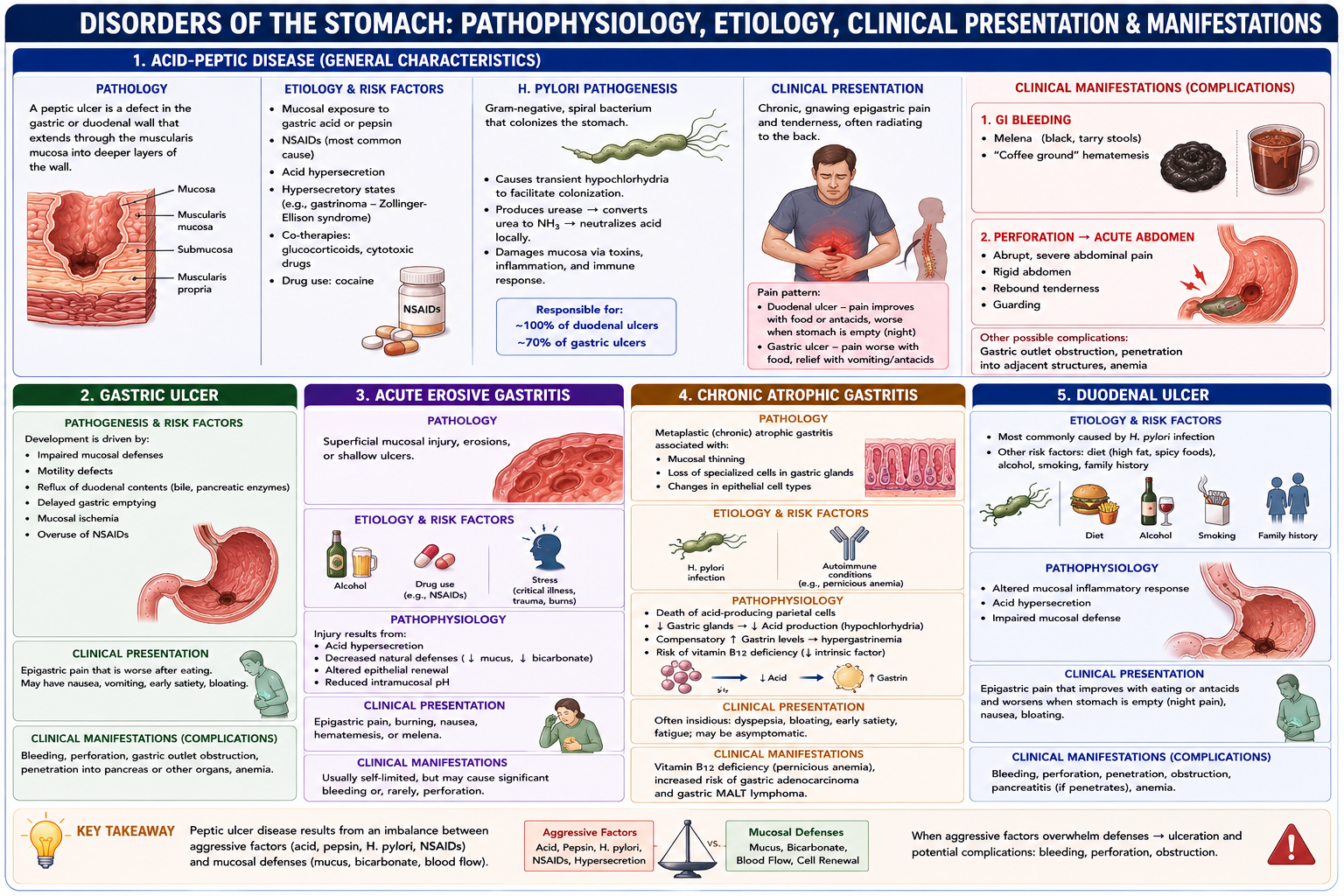
Pathology: A peptic ulcer is a defect in the gastric or duodenal wall that extends through the muscularis mucosa into deeper layers of the wall.
Etiology and Risk Factors: Primarily caused by mucosal exposure to gastric acid or pepsin, nonsteroidal anti-inflammatory drugs (NSAIDs), or acid hypersecretion. Other causes include hypersecretory states (like gastrinoma), co-therapies (glucocorticoids, cytotoxic drugs), and drug use like cocaine.
H. Pylori Pathogenesis: H. pylori is a gram-negative bacteria that causes transient hypochlorhydria to facilitate its colonization in the stomach. It is responsible for approximately 100% of duodenal ulcers and 70% of gastric ulcers.
Clinical Presentation: Patients typically experience chronic, gnawing epigastric pain and tenderness, which often radiates to the back.
Clinical Manifestations: Complications include GI bleeding (presenting as melena or "coffee ground" hematemesis) and acute abdomen if the ulcer perforates (presenting with abrupt abdominal pain, a rigid abdomen, rebound tenderness, and guarding)
Gastric ulcer:
Gastric Ulcer
Pathogenesis and Risk Factors: Development is driven by impaired mucosal defenses, motility defects, reflux of duodenal contents, delayed gastric emptying, mucosal ischemia, and the overuse of NSAIDs
Acute erosive gastritis:
Acute Erosive Gastritis
Pathology: Characterized by superficial mucosal injury, erosions, or shallow ulcers.
Etiology and Risk Factors: Commonly caused by alcohol, drug use, and stress.
Pathophysiology: Injury results from acid hypersecretion, decreased natural defenses, altered epithelial renewal, and reduced intramucosal pH
Chronic atrophic gastritis:
Pathology: A metaplastic (chronic) atrophic gastritis associated with mucosal thinning, the loss of specialized cells in gastric glands, and changes in epithelial cell types.
Etiology and Risk Factors: Can be caused by H. pylori infection or autoimmune conditions.
Pathophysiology: It is uniquely characterized by the death of acid-producing parietal cells, a subsequent reduction of gastric glands (leading to reduced acid production), and a compensatory increase in gastrin levels
Duodenal ulcer:
Etiology and Risk Factors: More commonly caused by H. pylori infection than gastric ulcers. Other risk factors include diet, alcohol, smoking, and family history.
Pathophysiology: Driven by an altered mucosal inflammatory response and acid hypersecretion
Gastroparesis:
Pathology and Pathophysiology: A syndrome defined by the objectively delayed gastric emptying of solids, specifically in the absence of any mechanical obstruction.
Etiology and Risk Factors: Causes can be idiopathic, diabetes-related, iatrogenic (medication-induced), or post-surgical. It is heavily linked to neurologic disorders that damage parasympathetic, sympathetic, or enteric nerves, such as diabetic neuropathy, Parkinson's disease, and multiple sclerosis.
Clinical Presentation: Symptoms include bloating, early satiety, abdominal pain, and epigastric distension or tenderness.
Clinical Manifestations: The symptoms can mimic or lead to an eating disorder, potentially resulting in avoidant/restrictive food intake
