Lecture 2
Lecture Outline
How common are glia in the CNS?
What are glia? Definitions beyond textbooks.
Revisiting the Blood Brain Barrier.
Understanding the Glymphatic System.
Learning Objectives
Compare and contrast glial cell types—anatomy and function.
Discuss modern findings challenging traditional textbook definitions regarding glial functions.
Describe the cellular response when an astrocyte network is activated.
Define hemodynamic response, outlining the signaling pathway from neural activity to blood flow increase.
Define the glymphatic system and its role in waste removal from the brain.
Glia: The Support Cells of the Nervous System
What Are Glia?
Glia, derived from the Greek word meaning 'glue', are various support cells in the nervous system.
Types of Glia
Astrocytes: Provide structural and regulatory support to neurons.
Myelinating Cells: Form myelin sheaths around axons.
Microglia: Act as immune cells in the CNS.
Additional Support Cells
Meninges: Membranes covering the brain and spinal cord.
Neurovascular Unit: Interplay among neurons, glia, and blood vessel cells to regulate physiological functions.
Myelinating Cells: Role and Function
Types of Myelinating Cells
Oligodendrocytes in the CNS and Schwann Cells in the PNS.
Different embryonic origins and interactions with neurons.
Myelin Characteristics
Myelin is a lipid-rich substance crucial for insulating axons.
Responsible for the appearance of white matter in brain regions with numerous axons.
Functions of Myelinating Cells
Myelin provides electrical insulation, enhancing conduction speed and energy efficiency.
Schwann cells proliferate post-PNS injury, assisting in axon regrowth.
Myelinating cells supply their axons with essential metabolites (e.g., glucose).
Myelination Patterns in the CNS
Findings on CNS Myelination
Serial electron microscopy shows non-uniform myelination along cortical neuron lengths.
Reference: Tomassy, G. S. et al. (2014).
Adaptive Myelination
Learning can enhance myelination of active axons, suggesting a link between myelination and cognitive function.
Reference: Shimizu et al. (2023).
Microglia: The Immune Cells of the CNS
Overview of Microglia
Reside within the CNS; develop during early life and replicate when needed.
Functions of Microglia
Perform innate immune functions: surveillance, scavenging, and mediating inflammation.
Not the sole immune defenders; outside white blood cells also play roles in CNS response to injury or infection. however they are bad at adopting a proper immune response despite popular belief.
The Role of Microglia in Brain Health
Engage in 'neural gardening'—promoting neuron growth and pruning synapses.
Consequences of Microglial Deletion
Research Spotlight-Scientists made mice that lacked microglia by deleting a specific gene promotoer (FIRE). The mice are born healthy and generally seem remarkably normal until about 8 months old.
Selective microglia deletion results in age-related pathologies.
Transplanting cultured microglia into newborn mice prevented development of similar pathologies.
Study shows potential for microglia toward maintaining neural health across lifespan.
Reference: Munro et al. (2024).
Understanding Astrocytes in Depth
Characteristics of Astrocytes
Most common and diverse glial cells in the CNS, resembling starbursts in structure.
Process interaction with neurons and blood vessels enhances communication within the CNS.
Functions of Astrocytes
Regulate extracellular environments, particularly K+ levels. distribute them to areas of lower concentration to prevent buildip and overexciation of neurons.
Regulating the environment of active neurons
Promoting and regulating neuron and synapse growth
Regulating the interaction between neuron and blood vessels.
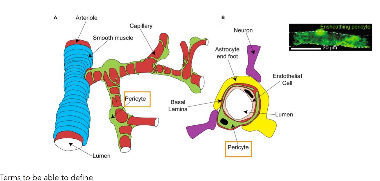
Often drawn as a part of the BBB, but they are not physically a part of it. The physical bbb is actuallt all in the specializations within the endothelial cells in the capillary itself. not in the astrocytes.
Tripartite Synapse Concept
Increasing debate on astrocyte involvement in synaptic transmission.
Some researchers propose astrocytes actively participate in neurotransmission (gliotransmission).
The Blood-Brain Barrier (BBB)
Astrocytes and the BBB
Astrocytes appear as integral to the BBB in illustrations, but they are not physically part of the barrier.
The BBB features tight junctions within endothelial cells that regulate permeability.
astrocytes contribute to bbb function by secreting signalling molecules such as ANG-1, SHh, retinoic acid, Wnt, growth factor, GDNF, FGH, IGF-1, and apoE.
Key Implications
Understanding astrocyte functions can provide insights into how they influence BBB integrity and function.
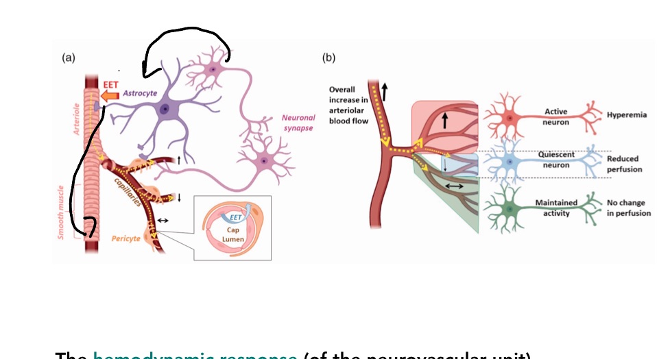
they are not electrically excitable,but they do respond to neuronal activity with Ca 2+ waves. (astrocytes respond to stimuli with elevations of cytoslic calcium levels and share them with their neighbours, causing waves of calculum increases to spread across active brain regions.
Brain capillaries are enmeshed by contractile pericytes which can locally restrict blood flow.
The Haemodynamic response leads to highly localized blood flow in response to neural activity.
The haemodynamic response refers to the change in blood flow and blood oxygen levels in the brain that happens when a specific region becomes active. in correct order: neuron-astrocyte-pericyte
Ependymal Cells:
Ependymal Cells: Ciliated glial cells that line the ventricles of the brain and the central canal of the spinal cord, involved in the production and circulation of cerebrospinal fluid (CSF).
They secrete and regulate CSF and ISF.
Tanycytes: special ependymal cells found lining parts of the third ventricle in the brain.They help transfer signals and substances between the brain and the blood in nearby capillaries.They also regulate hormones and control how molecules move between the cerebrospinal fluid (CSF) and the brain.
Blood CSF Barrier: a protective boundary that controls what can move between the blood and the cerebrospinal fluid (CSF).It’s made up mainly of cells in the choroid plexus that are tightly joined together so only certain molecules (like nutrients, not toxins) can pass through.
Choroid plexus: The choroid plexus is a network of blood vessels and ependymal cells found inside the ventricles of the brain.
Its main job is to make cerebrospinal fluid (CSF), which cushions the brain and removes waste.
Ventricular System: The ventricular system is a series of connected cavities (ventricles) deep inside the brain that are filled with CSF.
CSF is produced in these ventricles and flows through them, then around the brain and spinal cord.
Overview of Glymphatic Function
The Glymphatic System: The glymphatic system is the brain’s waste removal system. It uses cerebrospinal fluid (CSF) to wash away waste products like toxins and proteins from brain tissue.
The glymphatic system facilitates CSF pumping through brain tissue, aiding toxin removal.
It involves interactions between astrocytes, arterioles, and blood flow alterations.
Evidence of CSF Movement in the Brain
Imaging studies show the glymphatic system's activity, particularly during sensory stimulation.
stimulating the glympathic system using sensory stimulation helps the brain clear AD plaques.
Conclusive Insights
Summary Points
Glia play critical roles in supporting neuron function; they are not merely passive cells.
The neurovascular unit and meninges also interact dynamically with the nervous system.
New insights reveal that glial functions influence neural plasticity and repair mechanisms.
Final Thoughts
Increased understanding of glial cells suggests that glia may play pivotal roles in neurological health and disease.
Further Considerations on Glial Overview
Glia’s Population in the CNS
Estimates indicate glia constitute roughly 50% of CNS cell population, contrary to older claims of 90%.
Reference: Herculano-Houzel (2014).
Glial Research Advances
Continued research into glial cell functions is essential for a comprehensive understanding of brain physiology.
Notable Figures in Glial Research
Ben Barres (1954-2017) highlighted the significance of glial study, emphasizing glial cells' essential roles in cognition and brain health.
Learning Objectives with Answers
Compare and contrast glial cell types—anatomy and function.
Astrocytes: Most common and diverse glial cells in the CNS, resembling starbursts in structure. They provide structural and regulatory support to neurons, regulate extracellular environments (particularly K+ levels), provide and recycle nutrients/metabolites for neurons (notably facilitating lactate transport), and foster neuron and synapse growth and interaction with blood vessels. Astrocytes appear integral to the BBB in illustrations, but are not physically part of the barrier.
Myelinating Cells (Oligodendrocytes in CNS, Schwann Cells in PNS): Oligodendrocytes are in the CNS and Schwann Cells are in the PNS. They have different embryonic origins and interactions with neurons. Myelin is a lipid-rich substance crucial for insulating axons, responsible for the appearance of white matter. Myelin provides electrical insulation, enhancing conduction speed and energy efficiency. Schwann cells proliferate post-PNS injury, assisting in axon regrowth. Myelinating cells supply their axons with essential metabolites (e.g., glucose).
Microglia: Reside within the CNS; develop during early life and replicate when needed. They act as immune cells in the CNS, performing innate immune functions (surveillance, scavenging, mediating inflammation). They are not the sole immune defenders; outside white blood cells also play roles in CNS response to injury or infection. Microglia engage in 'neural gardening'—promoting neuron growth and pruning synapses.
Discuss modern findings challenging traditional textbook definitions regarding glial functions.
Adaptive Myelination: Learning can enhance myelination of active axons, suggesting a link between myelination and cognitive function (Reference: Shimizu et al. (2023)). This suggests myelin is more dynamic than previously thought.
Tripartite Synapse Concept: There is increasing debate on astrocyte involvement in synaptic transmission. Some researchers propose astrocytes actively participate in neurotransmission (gliotransmission), challenging the traditional view of synapses solely between neurons.
Glia’s Population in the CNS: Estimates indicate glia constitute roughly 50% of the CNS cell population, contrary to older claims of 90% (Reference: Herculano-Houzel (2014)). This challenges older numerical estimations.
Consequences of Microglial Deletion: Research suggests that selective microglia deletion results in age-related pathologies, and transplanting cultured microglia can prevent these (Reference: Munro et al. (2024)). This highlights a critical and active role for microglia in brain health, beyond just immune responses.
Describe the cellular response when an astrocyte network is activated.
When an astrocyte network is activated, cellular responses include:
Regulation of extracellular environments, particularly K+ levels.
Provision and recycling of nutrients/metabolites for neurons, notably facilitating lactate transport.
Fostering neuron and synapse growth and interaction with blood vessels.
Potential active participation in neurotransmission (gliotransmission) by some researchers.
Involvement in interactions with arterioles and blood flow alterations to facilitate CSF pumping through brain tissue (glymphatic system).
Define hemodynamic response, outlining the signaling pathway from neural activity to blood flow increase.
The provided notes mention "Define hemodynamic response, outlining the signaling pathway from neural activity to blood flow increase" as a learning objective and refer to the "Neurovascular Unit" as the "Interplay among neurons, glia, and blood vessel cells to regulate physiological functions." Astrocytes are noted to "interact with blood vessels" and their processes "enhance communication within the CNS." However, a detailed definition of the hemodynamic response itself or its explicit signaling pathway is not outlined in the provided text.
Define the glymphatic system and its role in waste removal from the brain.
Definition: The glymphatic system facilitates CSF pumping through brain tissue, aiding toxin removal. It involves interactions between astrocytes, arterioles, and blood flow alterations.
Role in waste removal: Its role is aiding toxin removal from the brain. Imaging studies show its activity, particularly during sensory stimulation.