bio-u2
UNIT 2: CELL STRUCTURE AND FUNCTION
LESSON 2.1: Prokaryotes and Eukaryotes
The Two Types of Cell
Like houses, cells come in different forms and sizes. A cell, the basic functional unit of life, is classified into two types: prokaryotic cell and eukaryotic cell.
Prokaryotic Cell
A prokaryotic cell is a type of cell that does not have a nucleus and membrane-bound organelles within its cytoplasm. This type of cell is like a studio-type condo unit that does not have several compartments. Prokaryotes are small, single-celled organisms that have prokaryotic cells. These organisms are metabolically diverse because they can utilize different nutrients and energy sources and they can inhabit all types of environment on Earth. All bacteria that include the organisms of domains Archaea and Bacteria are considered as prokaryotes.
Eukaryotic Cell
The other type of cell that is characterized by the presence of nucleus and membrane-bound organelles within its cytoplasm is called a eukaryotic cell. Membrane-bound organelles of eukaryotic cells provide compartmentalization in the cell. This is comparable to a mansion that has several rooms or compartments. Domain Eukarya which includes protists, fungi, plants, and animals are examples of eukaryotes. Eukaryotes are organisms that consist of eukaryotic cells.
Distinguishing Features of Prokaryotic and Eukaryotic Cells
In the Warm Up activity, you were tasked to spot the differences between the given pictures. To spot the differences, you had to look into the details of the pictures. Cells are classified into two types because scientists spot certain differences between them. The details that the scientists looked into are the distinguishing features of prokaryotic and eukaryotic cells.
Presence of Nucleus
The main feature that distinguishes eukaryotic cells from the prokaryotic cells is the presence of a nucleus with a nuclear membrane that encloses the genetic materials (DNA or deoxyribonucleic acid) as seen in Fig. 2.1.2.. In a prokaryotic cell, the genetic materials (DNA) are concentrated in a region of the cytoplasm called the nucleoid. The term prokaryotic comes from the Greek terms pro- which means before and karyon or kernel that refers to the nucleus. On the other hand, the term eukaryotic comes from the Greek terms eu- meaning true and karyon or kernel.
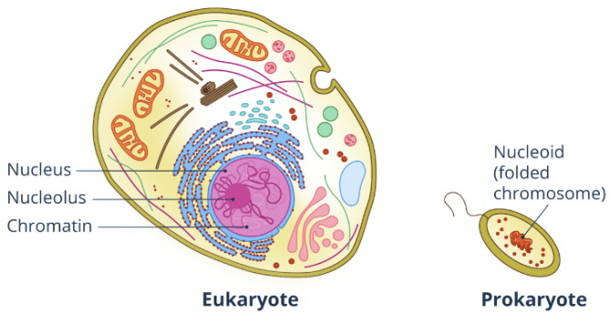
Fig. 2.1.2. The genetic material is enclosed in the nucleus of eukaryotes and in the nucleoid region of prokaryotes.
Cell Wall and Cell Membrane
The cell wall and the cell membrane share some functions in both prokaryotes and eukaryotes. The cell wall provides shape and rigidity to the cell and the cell membrane provides protection and plays an important role in the transport of materials. However, the cell wall is present in almost all prokaryotic cells, but not in most eukaryotic cells (these are not found in animals and most protists). Structurally, the cell wall of prokaryotes is made up of peptidoglycan – a complex sugar (polysaccharide) and a few amino acids. In eukaryotes, it is either made up of cellulose as in plants and chitin in fungi.
As for the cell membrane structure, prokaryotes do not have sterols in the cell membrane but have a sterol-like lipid component called hopanoid. In eukaryotic cells, the sterols that are present in the cell membrane are cholesterol (animals), phytosterol (plants), and ergosterol (fungi).
Endomembrane System and Other Organelles
Another distinguishing feature of the prokaryotic and eukaryotic cells is the presence of the endomembrane system. This refers to the interacting organelles between the nucleus and the cell membrane. The endomembrane system includes the rough and smooth endoplasmic reticulum, Golgi apparatus, lysosome, endosome, and vacuole as shown in Fig.2.1.3.. The endomembrane system is present in eukaryotic cells, but not in prokaryotic cells. Other organelles like mitochondria and chloroplast are also present in eukaryotic cells, but not in prokaryotic cells.
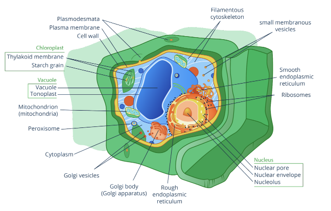
Fig. 2.1.3. Endomembrane system and other organelles of a plant cell, which is an example of a eukaryotic cell
The presence of organelles in eukaryotic cells provides compartmentalization. It increases the surface area-volume ratio of the cells, allows the occurrence of simultaneous cell activities without interference from each other, and allows separation of DNA in the nucleus, mitochondria, and chloroplast.
Ribosomes
Ribosomes are present in both prokaryotic and eukaryotic cells. These have the same function in both, which is protein synthesis. In terms of location and structure, however, they differ from one another. In a prokaryotic cell, all ribosomes are found in the cytoplasm. In eukaryotic cells, they can be found in the cytoplasm, outer nuclear membrane, rough endoplasmic reticulum, mitochondrion, and chloroplast. Generally, ribosomes are made up of large and small subunits. Each subunit is made up of rRNA (ribosomal ribonucleic acid) and proteins. Prokaryotic cells, mitochondria, and chloroplast contain 70S ribosomes while eukaryotic cells have 80S ribosomes. S or Svedberg unit is the unit of the sedimentation coefficient.
The Shape of DNA and Number of Chromosome
DNA or deoxyribonucleic acid as shown in Fig. 2.1.4. is the genetic material in prokaryotic and eukaryotic cells. It codes for proteins and RNA. It also transmits information from one generation to the next generation. In prokaryotic cells, DNA is found in the nucleoid region of the cytoplasm. Prokaryotes, as well as the mitochondrion and the chloroplast, have covalent, closed, circular DNA that is not coiled in protein “spools” called histones. Most prokaryotes have only one chromosome and an extrachromosomal DNA called a plasmid.
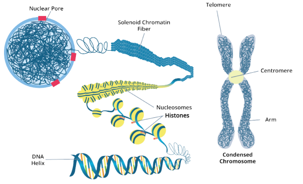
Fig. 2.1.4. In eukaryotes, the DNA is wrapped around histones to form nucleosomes.
On the other hand, in eukaryotic cells, the linear DNA is coiled in histones and is found inside the nucleus. Eukaryotes have more than one chromosome so histones are essential in packaging DNA into nucleosomes and helping it to condense into chromatin. Through this, several chromatins can fit into the nucleus without the high risk of mechanical damage.
Cell Size
Prokaryotic cells are smaller compared to eukaryotic cells. Typical prokaryotic cells can range from 0.1 to 5μm in diameter while eukaryotic cells are typically 10 to 100μm in diameter. Despite their larger size compared to prokaryotic cells, eukaryotic cells can still perform metabolic activities efficiently due to the compartmentalization of cellular parts as discussed previously.
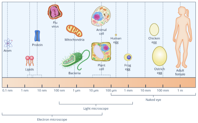
Relative sizes of bacteria (prokaryote), plant cell (eukaryote), and animal cell (eukaryote)
Prokaryotes are unicellular or are made up of only one cell. On the other hand, eukaryotes are either unicellular or multicellular. Most protists and yeast are unicellular eukaryotes. Plants, animals, most fungi and some protists are multicellular eukaryotes. Multicellular organisms are composed of more than one cell.
Mode of Reproduction
Most prokaryotic cells reproduce through binary fission and some reproduce through spores. Binary fission is a process of reproduction wherein one cell is divided into two new cells.
In eukaryotic cells, individual cells reproduce through mitosis and meiosis. Mitosis (somatic or body cell division) and meiosis (sex cell division) are types of cell division in eukaryotic cells. Mitosis is the type of cell division that involves somatic or body cells. This process produces two identical, diploid daughter cells. It is important for growth, development, and tissue repair. On the other hand, meiosis involves sex cells or gametes. This process produces four, non-identical, haploid daughter cells. It is also known as gametogenesis or gamete formation which is important in sexual reproduction. Take note that many eukaryotic organisms are multicellular, meaning that the mode of producing more cells is not necessarily the way that these organisms reproduce.
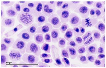
Eukaryotic cells undergoing mitosis
DNA Replication
DNA replication is the process of synthesizing a new DNA strand using an old DNA strand as a template. In both types of cell, it is an antiparallel and semiconservative process wherein both strands are replicated through complementary base pairing. However, the difference in the characteristics of DNA also result in different processes in prokaryotic and eukaryotic cells. DNA replication in eukaryotes takes a longer time than in prokaryotes because eukaryotes have more DNA than prokaryotes. In prokaryotic cells, DNA replication occurs in two opposing directions at the same time in the cytoplasm. On the other hand, eukaryotic cells have multiple points of origin and use unidirectional replication within the nucleus.
DNA replication in prokaryotic cells and eukaryotic cells also differs in terms of enzymes involved in the process. For example, in eukaryotes, an enzyme called telomerase is involved in the replication of telomeres of the eukaryotic chromosome. Prokaryotic cells do not have telomeres so telomerase is not present and involved in their DNA replication. Prokaryotes continuously replicate their short DNA while eukaryotes only replicate their DNA during the S-phase of interphase in cell division.
Transcription and Translation
Transcription refers to the synthesis of RNA using DNA as a template. Translation then refers to the process of protein synthesis. In prokaryotic cells, transcription and translation can be done at the same time in the cytoplasm. Aside from that, there is no post-transcriptional processing because the DNA of prokaryotes does not have a non-coding part called introns. In eukaryotic cells, the transcription occurs in the nucleus and the translation occurs in the cytoplasm. Eukaryotic DNA contains exons (coding part) and introns (non-coding part). Post-transcriptional processing is done in eukaryotes to remove introns and come up with the final RNA.
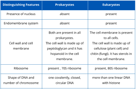
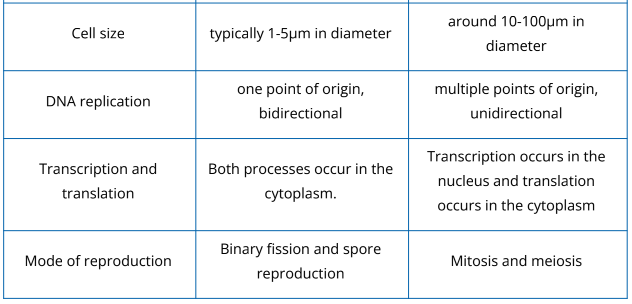
Prokaryotic and eukaryotic cells vary in different ways. Table 2.1.1. summarizes the distinguishing features of both prokaryotes and eukaryotes.
Table 2.1.1. Distinguishing features of prokaryotic and eukaryotic cells
Key Points
- Cells are classified into two types: prokaryotic cell and eukaryotic cell.
- Prokaryotic cells are found in prokaryotic organisms while eukaryotic cells are found in eukaryotic organisms.
- Examples of prokaryotes are Archaeans and Bacteria. Examples of eukaryotes are plants, animals, fungi, and protists.
- Prokaryotic and eukaryotic cells vary in terms of presence of nucleus, endomembrane system, cell wall and cell membrane, ribosome, shape of DNA andnumber of chromosome, cell size, DNA replication, transcription and translation, and mode of reproduction.
LESSON 2.2: Structures and Functions of Animal Cells
All organisms are made up of at least one cell. In complex multicellular organisms like animals, cells come in different structures and functions—they differ in terms of shapes and sizes, and they also have specialized functions.
Hierarchy of Biological Organization
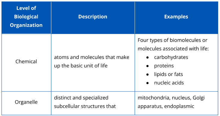
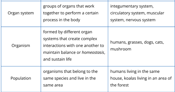
Biology is a vast field of study. To understand life from the molecular level up to the global scale of the entire living planet, biologists divide the enormous range into different levels of biological organization. The hierarchy of biological organization is shown in Fig. 2.2.1. The description and examples of each biological organization is shown in Table 2.2.1.
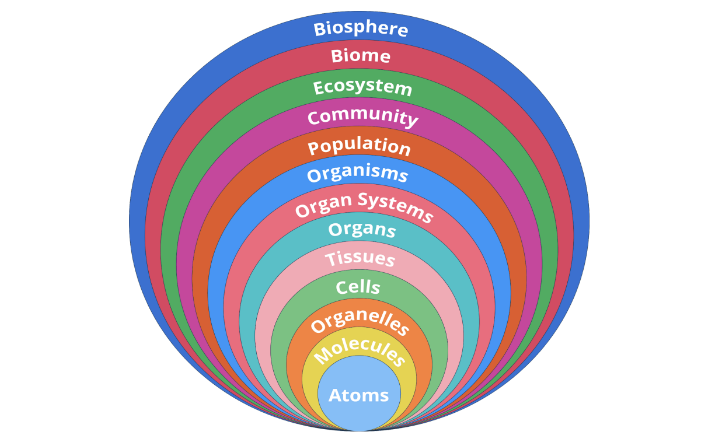
Fig. 2.2.1. The hierarchy of biological organization includes the assemblage of life from the smallest biomolecules to the interacting ecosystems of the biosphere.
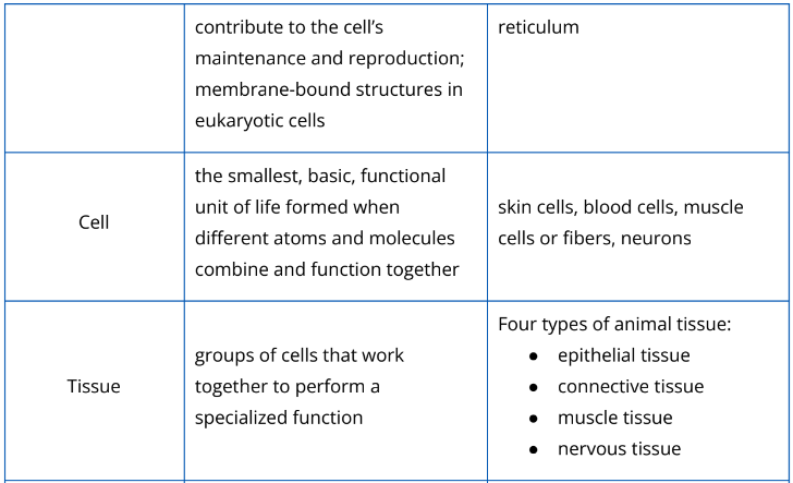
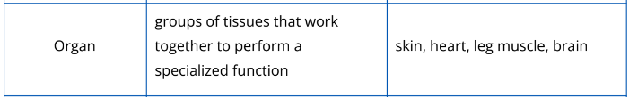
Table 2.2.1. Different levels of biological organization
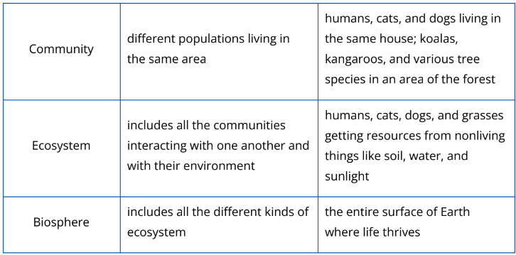
Types of Animal Tissues
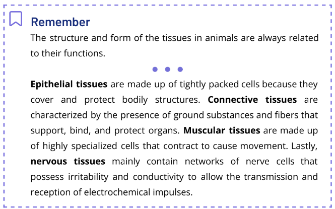
Tissues are groups of cells that are similar in structure and function. In animals, as shown in Fig. 2.2.2, there are four main types of tissue—epithelial, connective, muscle, and nervous tissues.
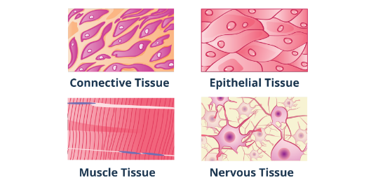
Fig. 2.2.2. The four types of tissues in animals vary significantly in structure and function.
Epithelial Tissues
Epithelial tissue or epithelium is a type of animal tissue that forms the inner and outer lining of organs, the covering in surfaces, and the primary glandular tissue of the body. In terms of structure, epithelial cells are closely packed to form continuous sheets. This kind of structure allows epithelium to form linings and impart protection to bodily structures. The presence of cell junctions like desmosomes and tight junctions, as shown in Fig. 2.2.3, permits the cells of epithelial tissue to absorb and filter different substances. One side of an epithelial cell is unattached and is exposed to the body’s exterior or to the cavity of an internal organ. This exposed part is called the apical surface. Some apical surfaces are smooth, but some have surface modifications, such as cilia or microvilli. Another important structure in epithelial tissues is the basement membrane. It is a structureless material secreted by cells in the lower surface of the epithelium. The basement membrane serves to adhere to the epithelium to the loose connective tissue underneath it.
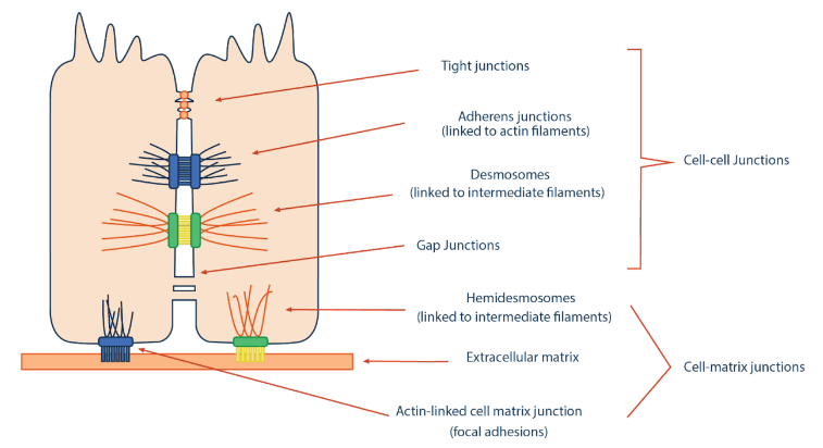
Fig. 2.2.3. The different types of cell-cell junctions characterize various epithelial tissues.
Epithelial tissues are avascular, which means that they do not have a blood supply of their own. They acquire nutrients and release waste materials through diffusion from the capillaries in the underlying connective tissue. Another distinct characteristic of epithelium is its ability to regenerate easily.
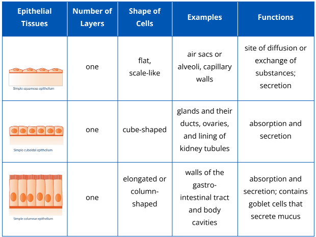
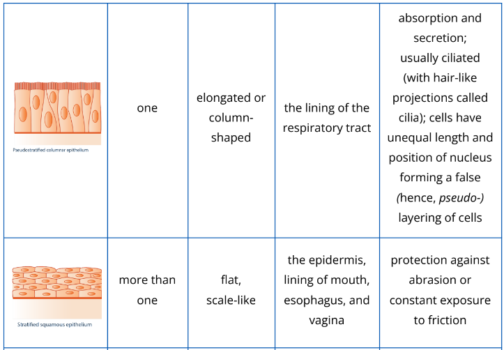
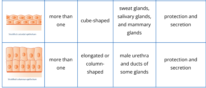
Epithelial tissues are classified based on cell arrangement or number of layers (i.e., simple and stratified epithelial tissues), and based on cell shapes (squamous, cuboidal, and columnar). They are given two names based on these two bases for classification. Table 2.2.2 below describes in detail the types of epithelial tissues in animals.
Table 2.2.2. Different types of epithelial tissues
Connective Tissues
Connective tissue is the most abundant tissue in the body that connects body parts. Unlike the avascularized epithelial tissues, most connective tissues are vascularized (with constant blood supply from blood vessels) except tendons and ligaments. If epithelial tissues have a basement membrane, connective tissues have an extracellular matrix as shown in Fig. 2.2.4. These are varying amounts of substances found outside the cells. It is made up of ground substance and fibers. The ground substance is mostly made up of water, adhesion proteins, and large polysaccharides or complex sugars. The amount of polysaccharide determines the consistency of the matrix. Also, the amount and type of fiber vary depending on the type of connective tissue.
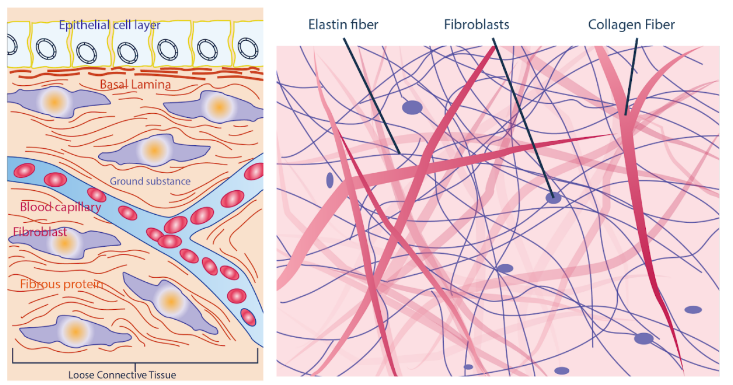
Fig. 2.2.4. The basic components of connective tissues vary according to their type (left, extracellular matrix; right, fibers and fibroblast).
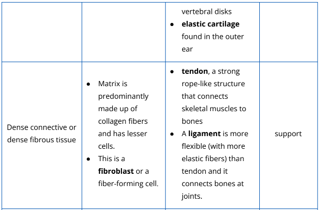
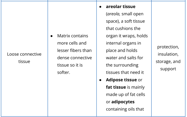
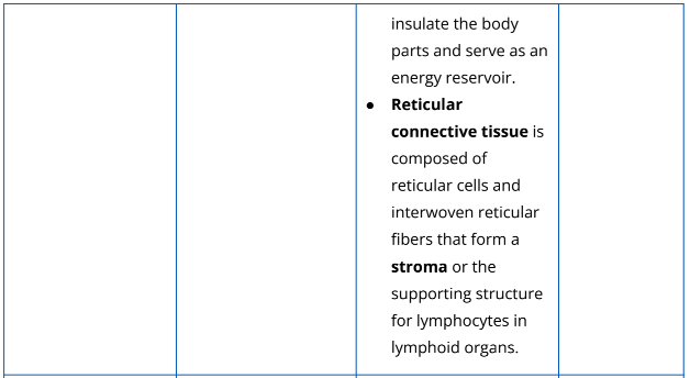
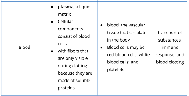
Fibers in connective can be elastin, collagen, or reticular fiber, as shown in Fig. 2.2.4. Elastin is a rubberlike protein which has the capacity to stretch and recoil. Reticular fibers are usually oriented randomly, forming mesh-like structures such as in the spleen. Collagen, the strongest and the thickest among the three fibers, has a strength comparable to steel. Connective tissues have different functions depending on their structural components. This tissue type supports, connects, and protects other body tissues. It also serves as a water reservoir because of its ability to absorb large amounts of water. Table 2.2.3 describes in detail the composition, functions, and examples of each type of connective tissue.
Table 2.2.3. The different types of connective tissues and their examples
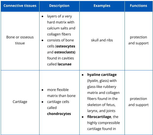
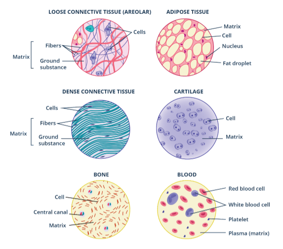
The different connective tissues vary with the composition of their extracellular matrices and the types of cells they consist of.
Muscular Tissues
Muscular tissue, or simply muscle (as shown in Fig. 2.2.5), is made up of specialized cells that can shorten or contract to produce movements. Muscle tissues consist of long and extensive muscle fibers. There are three types of muscle tissues that differ in structure and function.
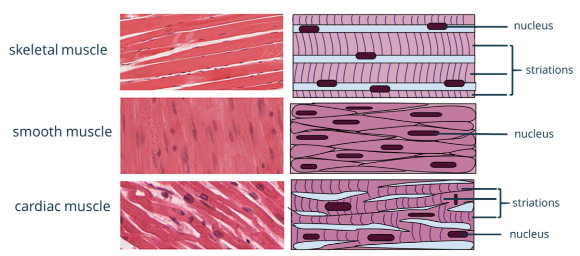
Fig. 2.2.5. The types of muscle tissues differ in their general structure but more or less perform the same function, i.e., to elicit movement.
- Skeletal muscle is a muscle tissue attached to the skeleton or bones. These muscles can be controlled consciously or voluntarily. Skeletal muscle cells are long, cylindrical, striated (with visible stripes), and multinucleated (with more than one nucleus). When they contract, they pull the bone and the skin to cause movement.
- Smooth muscle or visceral muscle is a type of muscle tissue commonly found in the walls of hollow organs such as intestines, stomach, bladder, blood vessels, and uterus. It involuntarily contracts slower than the other two types of muscle tissue. Smooth muscles are nonstriated, uninucleated, and spindle-shaped (have pointed ends) cells.
- Cardiac muscle is a muscle tissue found in the heart. Unlike a skeletal muscle, it is uninucleated (one nucleus) and it moves involuntarily (cannot be controlled consciously). However, it has striations like skeletal muscle. Cardiac muscle cells are branching together and fit tightly together at junctions called intercalated disks. These disks contain gap junctions that facilitate the rapid conduction of electrical impulses across the heart.
Nervous Tissues
Nervous tissue makes up the central nervous system and peripheral nervous system. It is composed of neurons and neuroglia or supporting cells. Each neuron serves as the basic structural unit of the nervous system. The two basic characteristics of neurons are irritability and conductivity. Irritability allows them to be sensitive and responsive to the various stimuli, while conductivity allows for the transmission of the reception and conduction of electrochemical signals from one part of the body to another. A neuron is made up of the cell body or soma, dendrite, and axon as shown in Fig. 2.2.6. The dendrite is the receiver of electrochemical signals from external stimuli or from adjacent neurons. It transmits incoming signals towards the cell body. The cell body or soma contains the nucleus and specialized organelles that produce molecules needed by the neuron. Electrochemical signals will be transmitted away from the cell body through the axon. The axon is surrounded by an insulating layer called the myelin sheath that allows impulses to transmit quickly and efficiently along the neuron. The periodic gaps between myelin sheaths on an axon are called Nodes of Ranvier. Between two neurons, or a neuron and a muscle or gland, there is a neural junction or synapse where the transmission of electrochemical signals occurs.
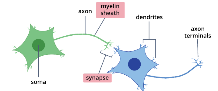
Fig. 2.2.6. Neuron, the basic unit of the nervous system, consists of structures that can conduct electrochemical signals as a form of information.
There are three types of neurons based on function. These are sensory neuron interneuron, and motor neuron, the structural variations of which are shown in Fig. 2.2.7. Sensory neurons are usually unipolar or pseudounipolar with an axon that branches into two extensions. The first one is connected to the dendrite that receives sensory input, and the other one transmits the information to the central nervous system. Interneurons are bipolar or multipolar neurons with one axon and multiple dendrites. It connects the sensory neuron to the motor neuron. Motor neurons are multipolar neurons that carry electrochemical signals from the CNS to the muscles or glands.
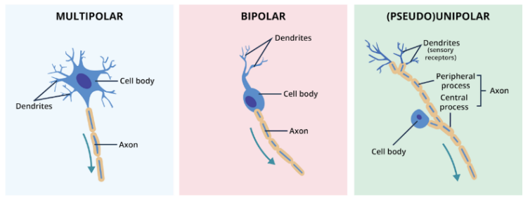
Fig. 2.2.7. Neurons may be classified based on their number of cellular processes.
Aside from neurons, nervous tissues also contain neuroglia or supporting cells. They do not conduct nerve impulses, but rather support, protect, or insulate neurons. There are six types of neuroglia or glial cells—four of them are in the central nervous system (CNS) and two in the peripheral nervous system (PNS). The CNS consists of the brain and the spinal cord, whereas the PNS consists of the nerves that are distributed throughout the body. The glial cells, as shown in Fig. 2.2.8., include the astrocytes, microglial cells, ependymal cells, and oligodendrocytes in CNS. Fig. 2.2.9. shows the satellite cells and Schwann cells in the PNS.
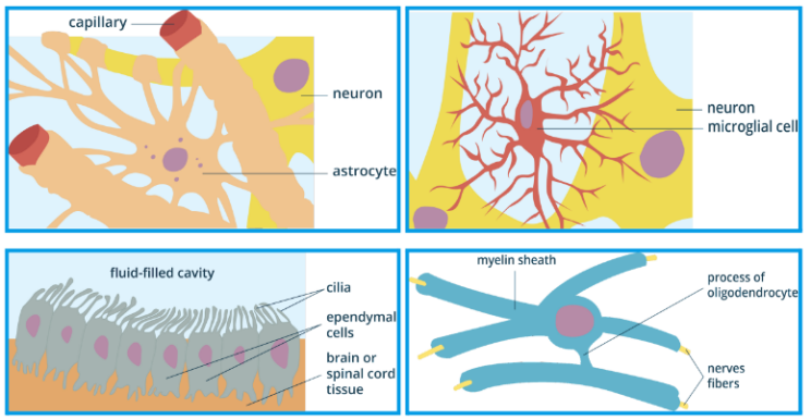
Fig. 2.2.8. Neuroglia or glial cells in the central nervous system
Astrocytes are star-shaped cells that support and control the chemical environment around the neurons. It is the most abundant glial cell in the CNS. Microglial cells are ovoid cells in the CNS that can transform into a phagocytic macrophage to clean neuronal debris and wastes. Ependymal cells are ciliated cells that line the central cavities of the brain and the spinal cord and form a fairly permeable membrane between the cavities with cerebrospinal fluid and the tissues of CNS. Oligodendrocytes are responsible for the production of the myelin sheath.
In the PNS, satellite cells surround the cell body of a neuron, and Schwann cells surround all the nerve fibers and produce myelin sheath similar to the oligodendrocytes.
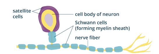
Fig. 2.2.9. Neuroglia or glial cells in the peripheral nervous system
Key Points
- There are different levels of biological organization, and these include the following (lowest to highest): chemical, organelle, cell, tissue, organ, organ system, organism, population, community, ecosystem, and biosphere.
- Animals have four types of tissue: epithelial tissue, connective tissue, muscle tissue, and nervous tissue. They all differ significantly in structures and functions.
- Epithelial tissue is composed of tightly packed cells that cover, line, and protect the body part. It can be classified based on cell arrangement (simple, stratified, pseudostratified) and cell shape (squamous, cuboidal, columnar).
- Connective tissue is made up of cells and an extracellular matrix that connects, protects, and supports body parts. Bone, cartilage, dense connective tissue, loose connective tissue, and blood are the types of connective tissue.
- Muscular tissue is composed of highly specialized muscle cells that contract to produce movement. It has three types: skeletal, cardiac, and smooth muscles.
- Nervous tissue is made up of neurons that receive and conduct electrochemical signals and supporting cells (glial cells) that support, protect, and insulate neurons.
LESSON 2.3 Structures and Functions of Plant Cells
Plant Tissues
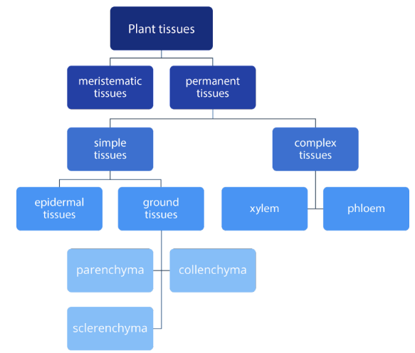
“All organisms are made of cells,” is one of the statements in cell theory which was formulated by Rudolph Virchow. Cells, in a particular part of a body, have specific functions. When a group of cells is performing the same function, they are considered as tissues. There are different types of cells found in an organism’s body. In plants, tissues can be classified as meristematic and permanent tissues. These tissues are generally responsible for growth, support, and transport.
Meristematic tissues are found in the growing areas of plants like roots and stems and are made of actively dividing cells. These tissues are classified according to location and type of growth they are responsible for. The major meristematic tissues in plants are apical meristem and lateral meristem. On the other hand, permanent tissues are composed of nondividing cells which can be found in stems, roots, flowers, and leaves. Permanent tissues are classified as simple and complex tissues. Ground tissues (parenchyma, collenchyma, and sclerenchyma) and epidermis are simple permanent tissues, while xylem and phloem are complex permanent tissues.
Meristematic Tissues
Meristematic tissues are composed of cells that give rise to another set of cells. The cells produced from meristematic tissues can either stay as meristematic cells to continually produce more cells or transform into specialized cells which will become parts of some tissues and organs of a growing plant. Meristematic tissues are mainly responsible for the growth of a plant. They also give rise to essential parts of a growing plant. Apical and lateral meristems are the major meristematic tissues found in plants. These meristems differ in location (as shown in Fig. 2.3.1) and function.
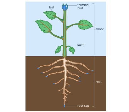
Fig. 2.3.1 The blue parts represent the location of the apical meristem, while the red ones represent the location of lateral meristems.
Apical Meristems
The height of plants is attributed to the work of apical meristems. These meristems are located on the shoots and roots of the plant. As apical meristems divide to produce new cells, they are also elongating the root and shoot systems by producing the primary plants’ body which includes the dermal, vascular, and ground tissues. Elongation of the root and shoot systems is referred to as primary growth in plants. Apical meristems, located at the root tips (as shown in Fig. 2.3.2) and terminal buds in a plant’s shoots, are continuously dividing to produce primary meristems. Primary meristems are derivatives of apical meristems consisting of protoderm, procambium, and ground meristem which will, later on, give rise to the plants’ three tissue systems-- epidermis, stele (xylem and phloem), and ground tissues, respectively.
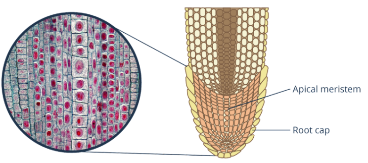
Fig. 2.3.2 Apical meristems located at the root tips (right) are responsible for the primary growth of plants. The microscopic view of cells (left) shows the apical meristems of Allium cepa.
Lateral Meristems
A plant does not only increase in height but also increases in diameter. In most plants, growth in diameter and girth is an essential factor for survival because it increases the plants’ rigidity and sturdiness. A growth in diameter and girth is called secondary growth which results from the continuous division of lateral meristems. Vascular cambium (shown in Fig. 2.3.3) and cork cambium are the lateral meristems found along the length of the plant which causes the increase in diameter and girth of plants.
Vascular Cambium
Vascular cambium gives rise to secondary xylem and phloem. Inward growth of vascular cambium produces xylem, while an outward growth produces phloem. As vascular cambium grows, layers of secondary xylem are added which becomes wood. When wood develops, plants become sturdy and rigid. Vascular cambium appears as a ring composed of two kinds of cells: ray initials and fusiform initials. Ray initials are composed of xylem and phloem rays that aid in radial transport of water and nutrients in woody stems. On the other hand, fusiform initials are responsible for producing secondary xylem and phloem.
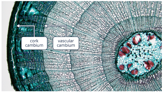
Fig. 2.3.3 Vascular cambium in woody dicot stem produces secondary xylem and phloem, while cork cambium produces cork cells.
Cork Cambium
Cork cambium (shown in Fig. 2.3.3), on the other hand, produces cork that replaces the epidermis of plants as they mature. Cork acts as a protection of plants from damage and disease-causing organisms. Moreover, cork releases suberin as a waxy protective coat to prevent water loss from the stems. The layers of cork cambium and cork are collectively known as periderm. Many of us know that the outer protective layer of woody plants is bark. It is still correct to refer to the outer protective layer of plants like bark; however, unlike periderm, bark consists of phloem, cork cambium, and cork.
Simple Permanent Tissues
When meristematic tissues differentiate into specialized cells, these cells become permanent tissues that do not have the capability to divide and give rise to a new set of cells. Permanent tissues, as its name suggests, are composed of fully matured cells that do not divide. Generally, these tissues provide support, aid transport of water and minerals, and act as storage of plant food. Permanent tissues in plants are either simple or complex. Simple permanent tissues are composed of one kind of cell, while complex permanent tissues are composed of two or more kinds of cells. Ground and epidermal tissues are considered as simple permanent tissues. On the other hand, xylem and phloem are considered as complex permanent tissues.
Epidermal Tissues
Epidermis, an outer protective layer in humans and in animals, is also found in plants. This outer protective layer originates from protoderm which is produced by apical meristems in roots and shoots. Epidermis covers the whole body of nonwoody and young woody plants and is protected by a waxy cuticle. The cuticle prevents loss of water and invasion of disease-causing microorganisms. The epidermis in roots has tiny projections called root hairs (shown in Fig. 2.3.4) which help in increasing the absorption capacity of roots. In leaves and in stems, the epidermis has tiny outgrowths called trichomes. These hair-like outgrowths prevent water loss and reflect excess light. When young woody plants mature, the epidermis is replaced by periderm which is produced by cork cambium.
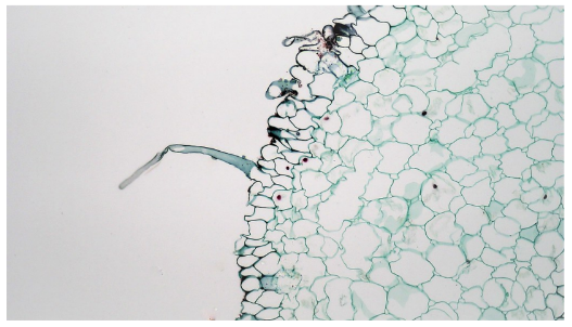
Fig. 2.3.4 Microscopic view of root hairs in a lily plant
The lower and upper parts of leaves are protected by the epidermis (as shown in Fig. 2.3.5). The lower epidermis of a leaf has guard cells. Guard cells are specialized structures that regulate the opening and closing of stomata. Stomata are slit-like structures on the lower epidermis of leaves which aids in the exchange of gases between plants and the environment.
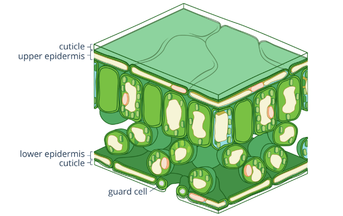
Fig. 2.3.5 Guard cells are specialized structures on the lower epidermis of leaves.
Ground Tissues
Cells that are neither dermal nor vascular are called ground tissues. They are considered as fillers of plants and form the bulk of plants. Ground tissues are usually found between dermal and vascular tissues. The ground tissues found on the exterior of vascular tissues are referred to as cortex. Meanwhile, pith is found at the center of the stem. These simple permanent tissues are generally responsible for photosynthesis, support, and storage. Parenchyma, collenchyma, and sclerenchyma are the kinds of ground tissues found in plants that differ in function and in location.
Parenchyma cells (shown in Fig. 2.3.6) are found in all parts of plants. Their structure is the least specialized among other ground tissues. Parenchyma cells have thin and flexible primary walls but lack secondary walls. These cells are mostly responsible for the synthesis and storage of plant food. Parenchyma cells in leaves contain chloroplasts and perform photosynthesis. In stems and in roots, parenchyma cells contain plastids that act as storage of starch, which is a complex sugar found in most plants. Although considered as permanent tissues, parenchyma cells can also divide and differentiate into specialized cells but under certain conditions, such as wound repairing.
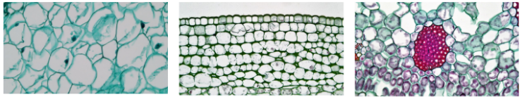
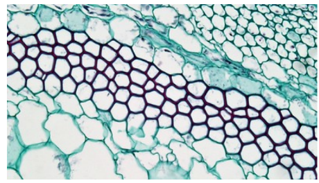
Fig. 2.3.6 Ground tissues parenchyma (left), collenchyma (center), and sclerenchyma (right) are generally responsible for storage, support, and photosynthesis. In these microscopic pictures, it is noticeable that they differ in structure in terms of primary and secondary walls.
Collenchyma is a type of ground tissue found in the young stems and petioles in plants. Unlike parenchyma, the primary walls of collenchyma are relatively thick although its thickness is uneven. Collenchyma lacks secondary walls and their primary walls do not contain lignin, which is a polymer providing rigidity. This thus provides a furnishing flexible support to immature parts of plants.
Collenchyma cells
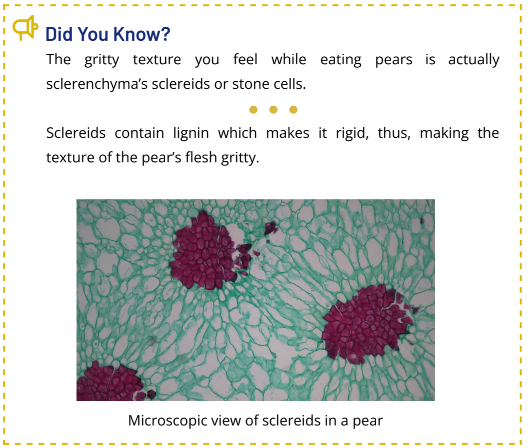
Sclerenchyma is a ground tissue with thick secondary walls. Their secondary walls are strengthened by lignin, thus, making it more rigid compared to collenchyma. Sclerenchyma provides support and rigidity to plants. It has two kinds, namely sclereids and fibers. Sclereids strengthen seed coats and are responsible for gritty-textured flesh of some fruits. On the other hand, fibers are used commercially as components of making rope and flax fibers.
Complex Permanent Tissues
Plants need a constant supply of food, water, and minerals for their survival. The distribution of food, water, and minerals are acted upon by xylem and phloem. Xylem is considered as water-conducting tissue since its main responsibility is to distribute water and minerals absorbed by the roots. On the other hand, phloem is considered as food-conducting vascular tissue since it distributes sucrose and other inorganic compounds throughout the plant’s body.
Xylem
Xylem (shown in Fig. 2.3.7) is composed of two types of conducting cells—tracheids and vessel elements. Tracheids are thin and elongated cells where water passes through. Tracheids, as well as vessel elements, have thin primary walls but thick secondary walls. Their secondary walls have pits that allow the transport of water from one cell to another. The thick secondary walls of tracheids serve as a rigid and strong wall against the tension of water transport. Moreover, vessel elements are thin-walled cells of xylem. Unlike tracheids, vessel elements have perforated plates that allow the transport of water through the vessels.
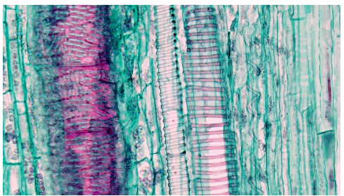
Fig. 2.3.7 Microscopic view of xylem in vascular plants
Phloem
Sugar and other organic nutrients are distributed throughout the plant’s body by phloem. Phloem is composed of sieve tubes consisting of sieve-tube elements (as shown in Fig.2.3.8). Sieve-tube elements are cells where nutrients, sucrose, and organic compounds are transported. These cells lack some of the essential cell parts like the nucleus and ribosomes, which makes the transport of substances effective. Furthermore, sieve-tube elements have sieve plates between their end walls. These sieve plates contain pores that regulate the flow of nutrients from one cell to another. Aside from sieve plates, sieve-tube elements also have companion cells, which is believed to be a helpful structure in transporting nutrients through the phloem.
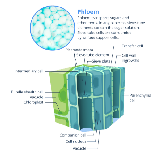
Fig. 2.3.8 Phloem is composed of sieve-tube elements that help in the transport of nutrients throughout the plant’s body.
Xylem and phloem are located along the stretch of the plant’s body. It extends from the roots to stems to leaves. Xylem and phloem form vascular bundles (shown in Fig. 2.3.9) which arrangement differs in roots and stems. In dicot and monocot roots, vascular bundles are arranged like a stellar. Meanwhile, in monocot and in dicot stems, vascular bundles are scattered and circular, respectively.
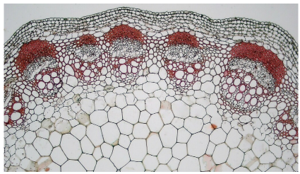
Fig. 2.3.9 Xylem and phloem form vascular bundles and are arranged in circular form in dicot stems. The picture shows a microscopic view of vascular bundles in a dicot stem.
Key Points
- Plants have two major tissues, namely meristematic and permanent tissues.
- Meristematic tissues give rise to permanent tissues which will differentiate into specialized cells with certain functions.
- Permanent tissues are generally responsible for photosynthesis, support, and transport of water and nutrients.
- Apical and lateral meristems are responsible for primary and secondary growth, respectively.
- Primary growth is an increase in plant’s height.
- Secondary growth is an increase in plant’s diameter and girth.
- Ground tissues are also called fundamental tissues because their functions are essential for the growth and development of plants.
- Parenchyma is responsible for storage and photosynthesis.
- Collenchyma and sclerenchyma provide support to the plant’s body.
Different types of plant cells
LESSON 2.4 Structures and Functions of Modified Cells
Specialized Cells and Cell Structures
Cells, as the basic unit of life, carry out different processes that help organisms to properly function. There are tiny structures inside our cells called organelles that help cells to carry out their specific functions. A typical cell has a nucleus, endoplasmic reticulum, Golgi apparatus, ribosomes, and mitochondria that are suspended in the cytoplasm. Cells are surrounded by a cell membrane. An additional cell wall is also present in cells of some organisms. However, there are certain cells that have different structures that cannot be found in a typical cell. Cells with these unique structures are called modified or specialized cells.
All specialized cells have undergone cell modification, a process in which cells develop special structures to carry out their specific functions. Modification in cells includes an increase in the number of organelles like mitochondria in the muscle cells or the loss of organelles like the nucleus in the red blood cells. Moreover, some specialized structures may be in the form of outgrowths like root hairs or numerous folds like microvilli which can increase the surface area for the absorption of nutrients. There are various specialized cells that can be found in animals and plants, and each type carries out specific functions that help in the development and survival of these organisms.
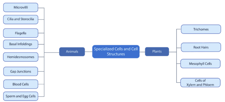
Specialized Cells and Cell Structures in Animals
Microvilli
Microvilli (singular, microviillus) are specialized structures on the surface of epithelial cells. These cellular extensions are made of actin microfilaments that serve as their structural core. A microvillus’ average diameter and length are 0.1 and 2 micrometers, respectively. They are found in the small intestines, kidneys, egg cells, and white blood cells.
In small intestines, microvilli are found on the surface of simple columnar epithelial cells (as shown in Fig 2.4.1). These increase the surface area for the absorption of nutrients and other essential substances from the gut cavity into the underlying tissues and blood vessels. Also, microvilli are found on the simple cuboidal epithelial cells in kidneys. Furthermore, microvilli are found on the surface of egg cells that help sperm cells to attach to it during fertilization. In white blood cells, microvilli serve as a motile structure which helps them migrate. Generally, microvilli do not only increase the area of surface absorption but also aids in the motility of cells.
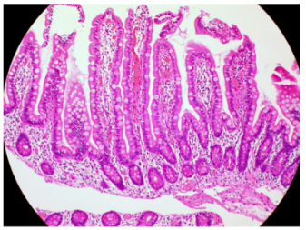
Fig 2.4.1 Microvilli in the small intestines
Cilia and Stereocilia
Cilia (singular, cilium) are tiny hair-like projections on the surface of epithelial cells. A cilium’s diameter is about 0.25 micrometers, and its length ranges from 2 to 20 micrometers; thus, it is bigger than a microvillus. A cilium’s structural core is made of nine pairs of microtubules on the outside ring with 2 microtubules on the central portion. Found in between the nine pairs of microtubules on the outside ring of a cilium is the dynein, a protein responsible for the motility of the cilium. Fig. 2.4.2. shows cilia in the bronchioles which are responsible for ensuring that the respiratory tract is free of dust, dirt, and pathogens. The peripheral nine pairs (with dynein between) and central pair of microtubules are evident in the figure.
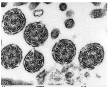
Fig. 2.4.2. Cilia (in cross-sectional view) found in bronchioles
Cilia are found on the respiratory tract (as shown in Fig. 2.4.3). These hair-like structures above the pseudostratified epithelia of the trachea prevent mucus (from goblet cells), bacteria, and dirt from entering the lungs. These particles and pathogens are either coughed out or moved to the mouth to be swallowed into the gastrointestinal tract. Cilia are mainly responsible for motility. In the respiratory tract, cilia brush off cells and substances like bacteria, mucus, and dirt that may infect and harm our lungs. Moreover, the cilia in fallopian tubes move the egg cell to the uterus. Cilia can also be found in some unicellular organisms like the Paramecium.
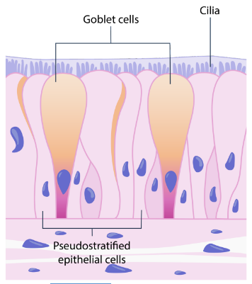
Fig. 2.4.3. Cilia above pseudostratified epithelia of the trachea
Cilia aid in the movement of these unicellular organisms in an aquatic medium.
Stereocilia (singular, stereocilium) are surface extensions of the epidermis which can be found in the epididymis of the testis and in the inner ear (shown in Fig. 2.4.4) of humans and animals. They are longer than microvilli with a length of 10–50 micrometers. The structural core of a stereocilium is made of actin microfilaments. Unlike microvilli and cilia, stereocilia do not function as a motile structure of cells, instead, they are mainly responsible for fluid absorption and detection of sound vibrations.
Stereocilia in the epididymis (which is a duct or passageway in the male reproductive system through which sperm cells pass) absorb the fluid that propels sperm cells from the seminiferous tubules. Initially, sperm cells are nonmotile when they are released from seminiferous tubules. In order to allow the sperm cells to become motile to be sent to the epididymis, a fluid is secreted. As sperm cells become motile, the stereocilia in the epididymis reabsorb this fluid. By contrast, stereocilia in the inner ear serve as sensors of vibrations. Aside from sensing vibrations, stereocilia also transform these vibrations into neural signals that will be interpreted by the brain.
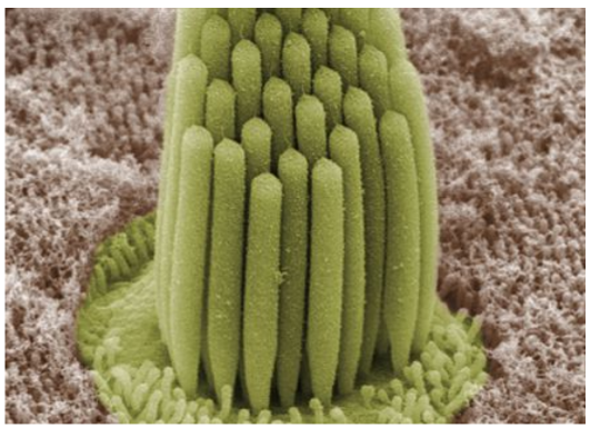
Figure 2.4.4. Stereocilia in a frog’s inner ear allow it to detect sounds from its environment. Through these, frogs have a mechanism to detect and escape from potential threats.
Flagella
The flagella (singular, flagellum) are tail-like structures that provide motility to cells. Flagella and cilia are structurally similar because both of their outer rings are composed of nine pairs of microtubules and another two pairs on their cores. Flagella are found in sperm cells which help them travel from the male reproductive tract to the egg cell in the fallopian tube. The flagellum is also found in some unicellular organisms like the Euglena (as shown in Fig. 2.4.5) and does the same function.
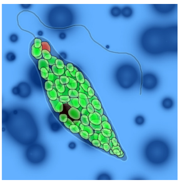
Fig 2.4.5. The flagellum (tail-like structure) helps unicellular organisms, such as the Euglena, to move from one location to another in an aquatic medium. Usually, this structure helps some photosynthetic unicellular organisms to move to locations with sufficient sunlight.
Basal Infoldings and Hemidesmosomes
Basal infoldings and hemidesmosomes (as shown in Fig 2.4.6) are both found at the basement membrane of epithelial cells. Epithelial cells sit on a basement membrane that serves as their foundation. Hemidesmosomes are the ones that anchor epithelial cells to their basement membrane. Epithelial cells found in the salivary glands and excretory duct contain basal infoldings in their basement membrane. These basal infoldings increase the surface area of these cells. Moreover, basal infoldings are responsible for ion and fluid transport. Numerous mitochondria are also found in the basal infoldings which provide energy, in the form of adenosine triphosphate, to be utilized during active transport.
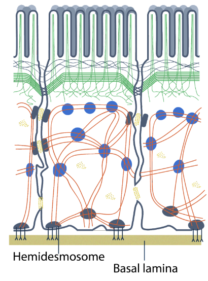
Fig 2.4.6. Hemidesmosomes are specialized protein structures that anchor the epithelial cells to their underlying basement membrane. They help the epithelial tissue provide protection and structural support to the underlying cells.
Cell Junctions
Cell junctions are specialized structures found on the lateral membrane of the cells. They are responsible for adhering cells to other cells or to the extracellular matrix. Tight junctions, adherens junctions, desmosomes, and gap junctions (shown in Fig 2.4.7) are cell junctions found in epithelial tissues. Tight junctions are found surrounding each cell and serve as impermeable structures that prevent leakage of substances when transmitted from one cell to another. Adherens junctions and desmosomes are specialized protein structures responsible for connecting adjacent cells. Adherens junctions are found below tight junctions, while desmosomes are found below adherens junctions. Furthermore, gap junctions serve as channels of ions, water, and other essential substances needed by cells.
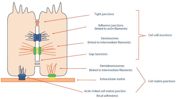
Fig 2.4.7. Cell junctions are found in epithelial cells and are mainly responsible for connecting adjacent cells. In addition, they may also facilitate the transport of certain substances between the adjacent cells of epithelial tissue.
Red and White Blood Cells
A typical animal cell contains one nucleus, numerous mitochondria, multiple Golgi bodies, millions of ribosomes, and an endoplasmic reticulum. However, some cells lack or have an excess of these structures like the red and white blood cells. Red blood cells or erythrocytes (as shown in Fig 2.4.8) are blood cells that lack nuclei and mitochondria. The absence of a nucleus and mitochondria in each red blood cell allows it to perform its work properly. Red blood cells are responsible for transporting oxygen throughout the body. The lack of nucleus in red blood cells gives more space for hemoglobin—a protein that carries oxygen; thus, more oxygen molecules can be transported. The biconcave shape of red blood cells is also a consequence of the absence of nuclei. This shape of red blood cells also contributes to the diffusion of substances. Red blood cells also lack mitochondria. They generate their energy through anaerobic respiration. If red blood cells have mitochondria, the oxygen molecules will just be consumed for aerobic respiration. Therefore, the transport of oxygen molecules will become less efficient.
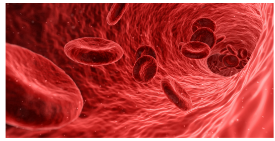
Fig 2.4.8. Red blood cells are biconcave to aid in the diffusion of gases from the air sacs of the lungs and into the oxygen-deprived tissues of the rest of the body.
White blood cells, also known as leukocytes, are specialized cells that are classified into granulocytes and agranulocytes. Granulocytes are white blood cells that contain granules in their cytoplasms which are observable under a light microscope. These cytoplasmic granules are enzymes that digest pathogens that can cause diseases in the body. Neutrophils, basophils, and eosinophils are types of granulocytes. The nuclei in granulocytes are multilobed unlike that of a typical cell’s nucleus (as shown in Fig. 2.4.9). By contrast, agranulocytes do not have distinct granules in their cytoplasm. Also, their nuclei are not lobed as compared to granulocytes. Lymphocytes and monocytes are types of agranulocytes. Generally, white blood cells act as defenders of our bodies by identifying and targeting disease-causing microorganisms and parasites and even developing cancer cells.
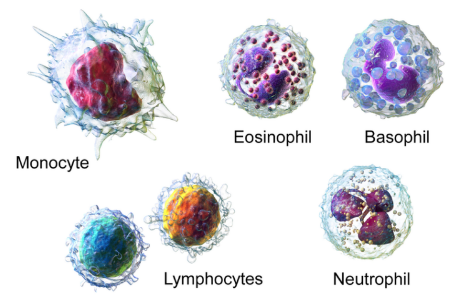
Fig. 2.4.9. While blood cells can be classified into granulocytes and agranulocytes.
Sperm and Egg Cells
Gametes or sex cells are considered as specialized cells because of their distinct structures that help them carry out their specific functions. Egg cells or ova (singular, ovum), with a diameter of 0.12 micrometers, are the largest cells in humans. Egg cells have two outer membrane layers namely zona pellucida and corona radiata (as shown in Fig.2.4.10). Zona pellucida is the inner layer of the egg’s outer membrane. It assists the sperm cell upon fusion with the egg cell. The outermost layer is the corona radiata which consists of three layers of cells derived from follicles. The two outer membrane layers of the egg cells prevent polyspermy—an event when an egg cell is fertilized by more than one sperm cell.
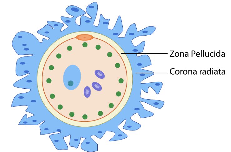
Fig.2.4.10. Egg cells are the largest cells in the human body and are necessary for sexual reproduction. They have two outer membrane layers, namely, zona pellucida and corona radiata which protect and nourish the egg cell.
Sperm cells, by contrast, are relatively small compared with the egg cells. Sperm cells are specialized cells that have a flagellum. The flagella in sperm cells help them move from the reproductive tract of males to that of females. The sperm cells have three distinct parts— head, midpiece, and flagellum (as shown in Fig. 2.4.11). The sperm’s head contains acrosome that helps them penetrate the egg cell’s outer membrane, while the midpiece contains numerous mitochondria that provide energy to the flagellum. The tail-like region in the figure is the flagellum itself which provides motility to the sperm cell.
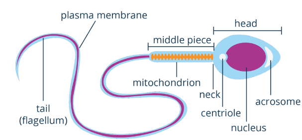
Fig. 2.4.11. Sperm cells travel from the testes to the female’s fallopian tube to facilitate an event called fertilization.
Sperm cells and egg cells are specialized because they are the only ones that can carry out the fertilization process. These gametes are produced by having half of the genetic material of the source parent organism. During the process of fertilization, the sperm and egg cells fuse, and these halves of the genetic material are combined to restore the normal condition in the offspring.
Specialized Cells and Cell Structures in Plants
Trichomes
The epidermis of plants has many outgrowths that vary in their functions. Some outgrowths increase the plant’s surface area for absorption, while others protect plants from insects and other herbivores and from too much sunlight. One group of epidermal outgrowths in plants include the trichomes. Trichomes are hair-like structures that developed from the plant’s epidermis and are mostly found on the leaves. They may be unicellular or multicellular. Their size and functions vary from one type of plant to another. Trichomes with glands on their tip release chemicals that prevent some insects from feeding on plants. But trichomes may also produce chemicals that some useful products are derived from, like mint fragrance. Trichomes may also help some plants to adapt to a hotter and drier environment by acting as a shade on leaves, as well as on stems. Trichomes found on insect-eating plants (shown in Fig. 2.4.12) serve as a trap as insects try to feed on them.

Fig. 2.4.12. Trichomes on insect-eating plants, such as Drosera hartmeyerorum or sundew, act as traps to insects that are trying to feed on them. Aside from protecting them from possible consumption, these glandular trichomes also aid in the digestion of captured insects.
Root Hairs
Plants need a constant supply of water to carry out their physiological functions. Water is essential to plants as it is one of the reactants that initiate photosynthesis. Roots are the ones responsible for absorbing water from the soil, and water is then distributed throughout the plant’s body. Increasing the rate of the absorption of water in roots is advantageous to plants. Root hairs (as shown in Fig. 2.4.13) are specialized cell structures that help plants to efficiently absorb water from the soil by increasing the surface area for absorption. Root hairs are tiny hair-like structures that originated from the epidermis of plants. These specialized structures die off after 2 to 3 weeks but are constantly replaced by new ones to maintain the efficiency of water absorption.
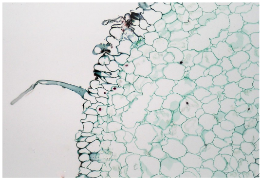
Fig. 2.4.13. Microscopic view of root hairs in a lily plant. These specialized structures in the roots of plants facilitate the absorption of water from the substrate.
Mesophyll Cells
Leaves are among the most important parts of a plant since they serve as major sites of photosynthesis. The composition and structures of the leaves are important features that enable them to produce food through photosynthesis. Photosynthesis happens in the mesophyll layer of the leaves located between the lower and upper epidermis. This layer is the specific site of photosynthesis as it contains a large number of chloroplasts. Mesophyll layer is composed of two types of parenchyma cells, namely palisade and spongy cells (as shown in Fig. 2.4.14). The palisade cells are elongated and cylindrical and form a palisade layer beneath the upper epidermis of the leaves. Because of their shape, palisade cells accommodate 70% of the plant’s chloroplasts. The arrangement and location of palisade cells allow them to obtain enough sunlight needed for photosynthesis. By contrast, spongy mesophyll cells are irregularly-shaped cells found beneath the palisade layer and above the lower epidermis of leaves, and form a spongy mesophyll layer. The term “spongy” is derived from the arrangement of their cells in this layer. Cells in this layer are loosely packed and have many spaces in between them. The spaces between the cells in the spongy layer allow more efficient gas exchange during photosynthesis.
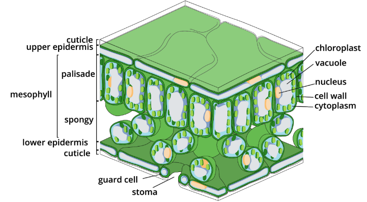
Fig. 2.4.14. Leaves have a mesophyll layer composed of palisade and spongy cells. The mesophyll layer is the primary site of photosynthesis in the leaves of a plant.
Cells of Xylem and Phloem
As discussed in the previous lesson, food, water, and nutrients are transported through a network of tubes inside the plant’s body that consists of xylem and phloem tissues (as shown in Fig. 2.4.15). Xylem and phloem are not themselves cells, but are instead composed of specialized cells that play a vital role in a plant’s growth and development. These are tube-like cells that transport water, nutrients, and food throughout the plant’s body. Xylem comprises cells that are specifically responsible for transporting water and nutrients obtained from the soil. By contrast, the phloem comprises cells that are responsible for transporting food, usually the by-products of photosynthesis.
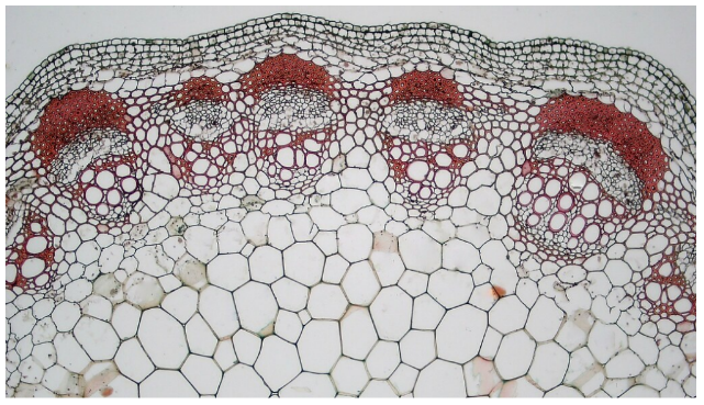
Fig. 2.4.15. Xylem and phloem tissues consist of specialized cells that are responsible for transporting essential substances, such as water, minerals, and food needed by the plants. The figure shows a bundle of cells (beneath the group of cells stained with red dye) called the vascular bundle. This consists of the cells of the xylem and phloem tissues.
Key Points
- Modified or specialized cells have developed structures that help them carry out their functions.
- Specialized cells in animals include the following:
- Microvilli are responsible for increasing the surface area for absorption.
- Cilia and stereocilia are responsible for movement and sensation, respectively.
- Flagella are responsible for locomotion or motility.
- Basal infoldings and hemidesmosomes are responsible for fluid transport and attachment, respectively.
- Cell junctions serve as connections between adjacent cells.
- Red and white blood cells are responsible for transporting oxygen and protecting the body from pathogens, respectively.
- Sperm and egg cells aid in the reproduction process.
- Specialized cells in plants include the following:
- Trichomes serve as protection of plants from extreme temperatures and insect or herbivore attacks.
- Root hairs increase the surface area for the absorption of water.
- Mesophyll cells serve as the sites of photosynthesis.
- Xylem and phloem consist of cells that are responsible for the transport of essential substances such as water and photosynthetic by-products.
Animals and plants have specialized cells and cell structures that ultimately contribute to the growth, development, and survival of the organism.