lecture 10: protein modification
chaperones
Many eukaryotic proteins need to be sorted to different intracellular destinations.
Cytosolic proteins
Most protein synthesis in a eukaryotic cells starts on free cytosolic ribosomes - except mitochondrial and plastid translation.
There is a common pool of ribosomal subunits in the cytosol that assemble on a suitable mRNA. This ribosome starts to produce a protein - once the ribosome has moved on there is the docking of another protein and another one etc. This is called a polysome which continue making protein until the end of translation where they disaggregate and reenter the pool of common ribosome subunits. Remember that mRNAs are pseudo circularised that makes this more efficient 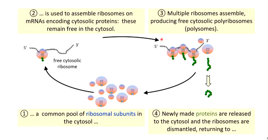
There is a problem with this simple description as the cytosol is more crowded with small molecules not shown. Macromolecular crowding favours aggregation of proteins. The effects of crowding are very large and estimates of reaction rates and equilibria made in test tubes may differ by orders of magnitude from those of the same reactions operating within cells. Many of these proteins are making inappropriate contact with other molecules. If hydrophobic regions start touching, you start to favour aggregation of proteins
Nascent proteins are also crowded. 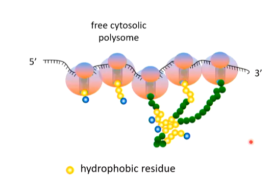
They are being produced in close proximity so hydrophobic patches interact which may result in aggregation - nascent proteins are in a non native state so are aggregation and protease proof. This means it is non functional
The proteins can misfold very easily. There is a class of cellular proteins (Chaperones) that ensure that the folding of other polypeptides occur correctly.
Hydrophobic patches are recognised by HSP40 co-chaperones on nascent proteins and masks them which keeps them soluble. An HSP40 delivers the protein to HSC70 and stimulates the ATPase activity of HSC70. ATP in HSC70 is degraded to ADP and HSC70 closes around the hydrophobic patches. Multiple HSC70s on a protein protecting the hydrophobic parts mean the hydrophilic parts can fold correctly. Upon nucleotide exchange - ADP replaced by ATP - HSC70 opens and releases the protein. This partly folded protein now snaps into its final conformation 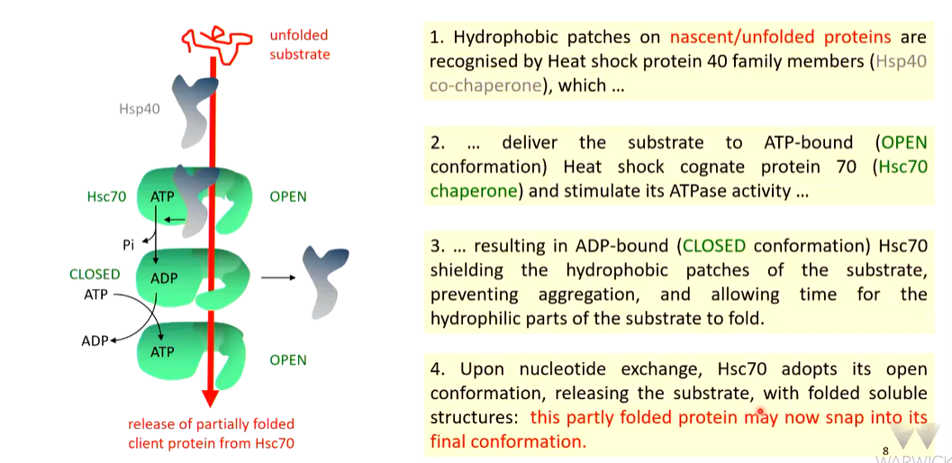
How to study chaperone interactions by in vitro recapitulation
The heat target protein is chosen so it is easy to denature by heat for 15 mins at 45 degrees. A lot of this protein becomes aggregated and insoluble so is spun out as a pellet. The soluble fraction is run on a gel amd this can be quantified by silver staining. Heating in the presence of HSP40 increases the soluble fraction on the gel. In this process, HSP40 becomes denatured by lasts long enough to rescue the target protein. Heating in the presence of HSC70 has a larger effect as almost half the protein is rescued from aggregation. Together, HSP40 and HSC 70 have a larger effect with half the protein soluble. Adding ATP obtains maximal solubilisation 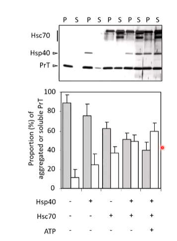
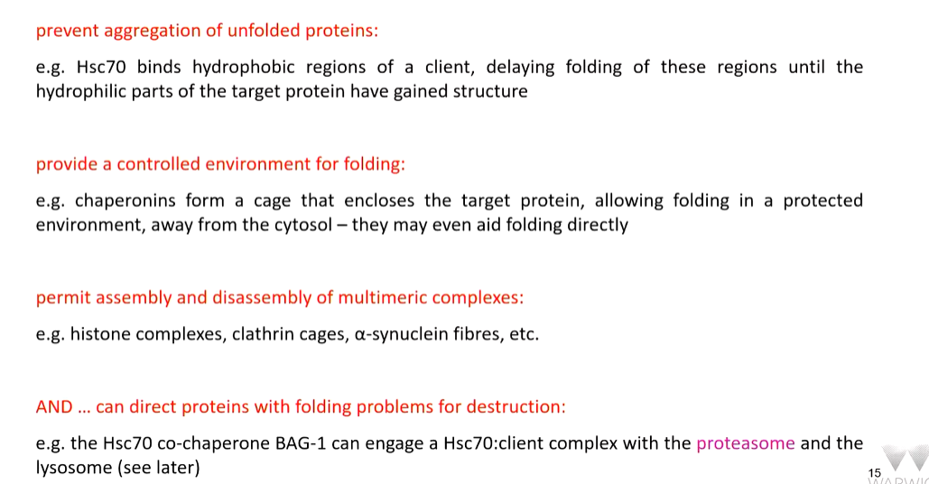
HSC 70 shields the hydrophobic regions and so reduces aggregation of nascent or unfolded proteins and acts as a holdase by holding the client in a soluble condition. This protein can enter different routes - productive or destructive routes.
In the productie route, the protein is released and if there is enough time for hydrophilic parts to fold, it will snap into its conformation. It may also be passed on to other chaperones for further folding and or assembly into multimeric complexes.
In the destructive route, they can be transported by HSC70 to a lysosome where it is autophagy or it can be passed to proteasomes for degradation
Both productive and non productive fates can proceed from an HSC70 bound state
Release from HSC70
This requires a nucleotide exchange factor (NEF) that removed ADP from the closed conformation of HSC 70. It inserts ATP into the nucleotide binding cleft that results in a change in the conformation that opens and released the part folded protein
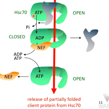
This tells you at HSC70 is also important but the co chaperones operate the chaperone. Co chaperones involve NEF such as BAG1,2 and HSPBP1 as well as HSP40 family membrane and CHIP
Further multimeric assembly
HSP90 can provide a platform for further protein folding. This is a dimer. It can take other partially folded proteins and combine them and allow the final folding to form a multimeric complex. iT CAN ALSO INCREASE THE FOLDING OF A PARTIALLY FOLDED PRODUCT
there are also cages that trap nascent proteins away from the cellular medium and are called chaperonins. There are either 14 16 or 18 subunits in rings that isolates the client away and allows correct folding. Small molecules need to fold away from the cytoskeleton and are produced and then captured by the caged. There is a hole in the cage between upper and lower chambers which communicate. The upper chamber closes and makes contact with the protein and limits its steric movement. It provides a sense of folding information. As the top ring closes, the bottom one opens and the fully folded protein falls out which allows this chamber open to another protein to enter 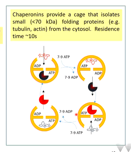
This is an expensive process - each round takes 7-9 ATPs.
The fate of a protein is not predetermined. From chaperone complexes, proteins can be folded, patt of a complex, inactivated etc and it is the prevailing concentrations of HSC70 and HSP90 that determines the proportion of an unfolded/misfolded protein that can gain a stable conformation and the proportion that is destroyed - co chaperones mae decisions by competing to release chaperone clients
There is an interaction competing network of co chaperones that
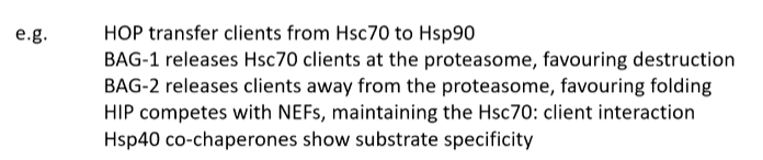 determines the fate of a chaperone client
Some HSC 70 co chaperones convert it into a disaggrease. Fibrils of Tau alpha syn and huntington is associated with neurodegenerative diseases and one way to treat this is by deaggregation. HSC 70 can be convinced to do this.
A-syn fibres have directionality andone bind can be seen by the binding of SNAJB1 (hsp40) binding to it. This recruits HSC70 binding at the targeted end. APG2 is a nucleotide exchange factor that makes HSC 70 that is bound to let go and fills the space with more HSC 70 - increases the concentration of bound HSC 70. As there is not much room for HSC70, they start to stretch and pull on the fibres that can rip fibrils apart 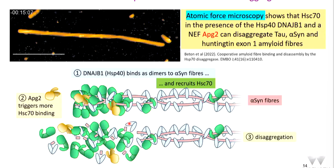
SUMMARY
engaging the proteasome
Architecture of the proteasome
Proteasomes have 4 rings at the centre comprised of 2 beta rings flanked by 2 alph rings. The catalytic activity is in the beta rings and have three proteolytic activities: chymotrypsin, trypsin and caspase like (peptidyl glutamyl-peptide hydrolysing). This is in the core of the proteasome and is surmounted on one or both ends by a 19s regulatory particle cap that regulates what comes in and what doesn’t. Two caps mean the protein is 30s and one means it is 26s.
How is a protein targeted to the proteasome
This is done by ubiquitination of the target. It is 76 aa found in eukaryotic cells with a characteristic fold. A protein requires multiple ubiquitin factors on a chain to provide a degradation signal. Typically this is 4 or more and elsewhere mono ubiquitination sends things to lysosomes
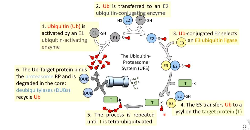
The ubiquitin is captured by an E1 ubiquitin-activating enzyme which is kept in a reduced state but has Ub added after the addition of Ub. There are 9 E1 enzymes with different affinities for different substrates
The activated Ub is transferred to an E2 ubiquitin conjugating enzyme of which there are about 30 in mammalian cells. 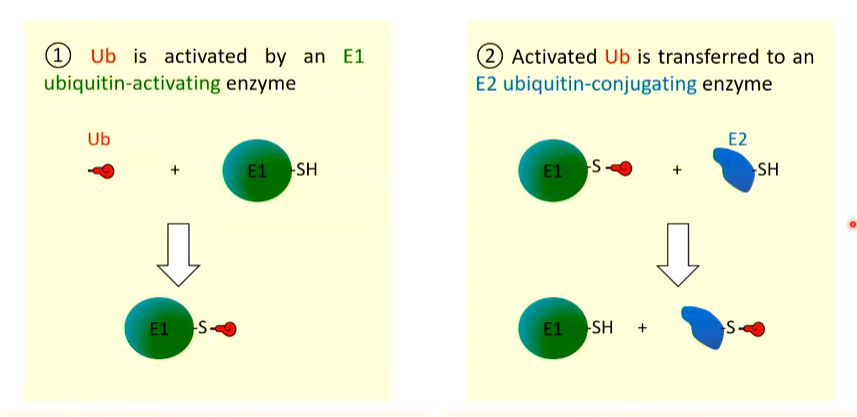
The E2-Ub selects appropriate E3 Ub ligases and associates with it. There 100s of E3 ligases in mammalian cells.
The E3-E2-Ub complex is targeted to the Hsc 70 which is holding the misfolded protein. E3 transfers the Ub to the protein and marks it for the destructive pathway. 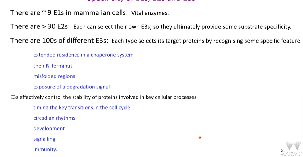
Ub is normally added covalently to the side chain of an available Lys residues on the target molecule. The process is repeated using side chain lys residues in Ub until there is a chain of at least 4. This is a degradation signal and polyub proteins can be bound by the proteasomal 19S RP
Targeting the proteasome
The polyUb proteins bind the cap of the proteasome (regulatory particle). The RP uses ATP to generate energy to unfold the target protein and feed it to the 20S core - DeUblases remove Ub molecules and return them to the common pool. The protein is unfolded by the ATPase activity of the cap. The protein is unfolded by the proteolytic activity in the beta core protein and is degraded which are ejected. Sometimes, these ejected proteins have cellular functions
UPS
twist
Not every unit of the proteasome has a function and is also a fail-safe mechanism that can bind proteins. It can act as a chaperone to make decisions about the state of the protein that can re fold proteins to their native conformation.
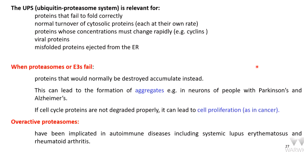
When UPS fail
Other modifications
Proteolytic cleavage
This is to activate a protein where the effector protease of apoptosis are stored in an inactive condition. Activation is by proteolytic cleavage and subunit rearrangement 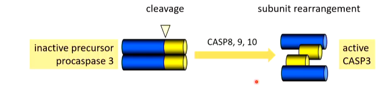
Addition of lipids to permit membrane targeting
Many regulatory proteins are modified by the addition of lipids. RABs are doubly prenylated - either a 15C faresyl or 20C geranylgeranyl. When the prenyl groups are used by GDI (GDP dissociation inhibitor), it is inactive. If a membrane has to be reorganised, RABs coordinated this and so can be activated by nucleotide exchange
ADP is removed and replaced with GTP which disrupts the interaction with GDI that exposes hydrophilic lipids that embed in the nearest membrane - typically a vesicle as RAB is activated at the edge. RAB can now coordinate the movement of the vesicle across the membrane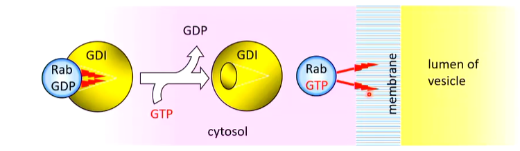
Phosphorylation
The addition or removal of phosphates can alter the activity of a protein - they can activate or inactivate eg control of CDK activation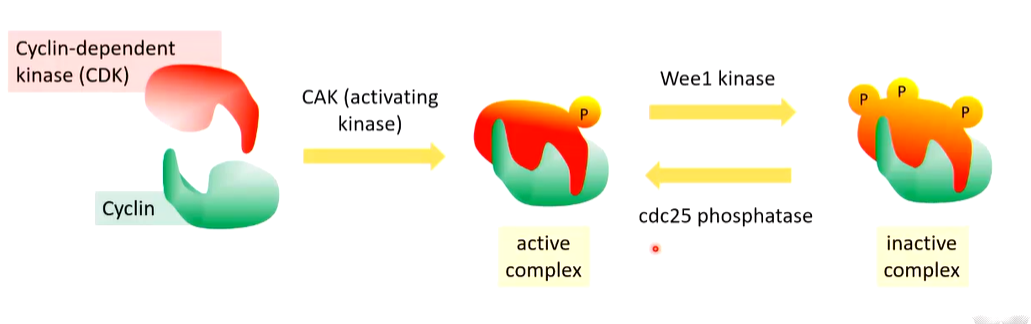
Wee1 inactivates almost immediately.
Phosphorylation is studied in vitro recapitulation.
Mek is phosphorylated that activates and phosphorylated ERK. There is an inhibitor of this (U0126) that when treated and probed for in cells for cycling B it is found to reduce the phosphorylation of cyclin B.
SCH772984 acts on phosphorylated erk and also reduces this and so cyclin B1 cannot enter the nucleus. 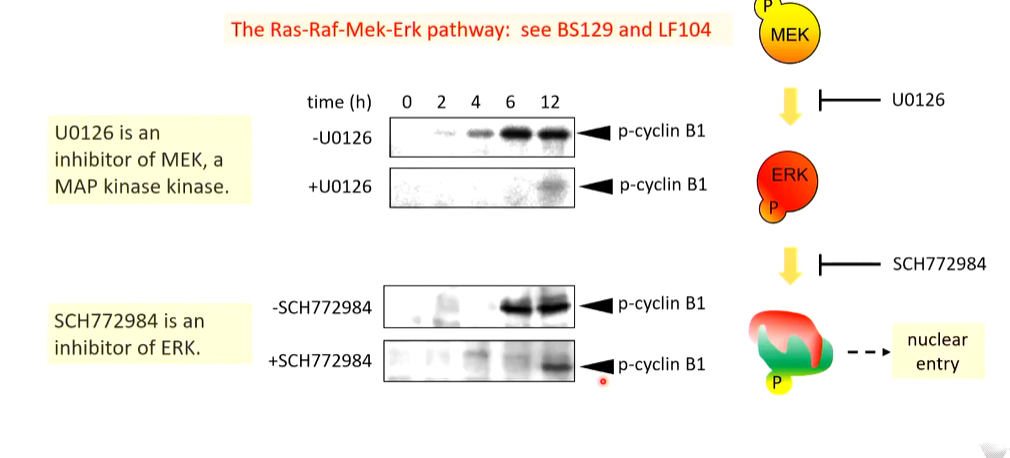
Phosphorylation can be complex
P53 is modified by phosphorylation (24 phos sites but not at the same time). Different stressors activate different kinases with different specificity that phosphorylates specific sites. When phosphorylated, it forms a tetramer - each phosphorylation means it folds in a different way - DNA binding domains displayed in different orientations depending on the stress the cell is encountering. P53 binds dna and trans activates anti stress responses that are tailored to the stress the cells is receiving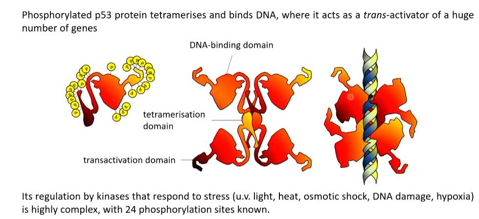
Other
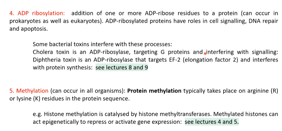 4 - diphtheria toxin.