3.3.2 Cells
Structure and function of Eukaryotic cells
Cell surface membrane
Structure:
main components are phospholipids (glycerol + phosphate head, 2 fatty acids as tail) and proteins
phospholipids form a bilayer (hydrophobic tails point towards each other and hydrophilic heads on outside)
proteins embedded between phospholipids (intrinsic proteins extend through bilayer, extrinsic proteins exits on outer layer)
cholesterol within bilayer stabilises and reduces permeability of membrane
Function:
forms a barrier between cells and surroundings
strong = offers structural support
flexible = allows cell to grow and divide
self sealing - cell can divide without bursting
selectively permeable - controls passage of material in and out of cells
Nucleus
Structure:
nucleolus produces RNA used to make ribosomes
endoplasmic reticulum on surface with nuclear pores between them
chromatin - form of DNA when cell not in mitosis
Function:
control cell via DNA and protein synthesis
contain genetic material
synthesise RNA and ribosomes
Mitochondria
Structure:
double membrane forms mitochondrial envelope
inner membrane folded into cristae (increased surface area for enzymes in respiration)
Function:
location of anaerobic respiration
outer membrane controls exit/entry
Chloroplasts
Structure:
Thylakoids contains chlorophyll
Stacks of thylakoids = granum
Function:
grana provide large surface area for chlorophyll
stroma contains enzymes for stage 2 photosynthesis
absorb light energy for photosynthesis
Golgi apparatus/vesicles
Structure:
compact stack of membranes
consists of cisternae
Function:
assembles, modifies and packages lipids
forms lysosomes
Lysosomes
Structure:
contain lysozymes
Function:
store hydrolytic enzymes
digest and recycle worn out organelles
hydrolyse ingested bacteria
autolysis of cell after death
Ribosomes
Structure:
larger 80S ribosomes in cytoplasm and RER
smaller 70S ribosomes in mitochondria
composed of 2 subunits
Function:
protein synthesis (location of condensation reactions that join amino acids)
Rough/smooth endoplasmic reticulum
Structure:
Rough ER = ribosomes attached
Smooth ER = no ribosomes
Function:
transport system
RER = transport of proteins
SER = synthesis, storage, modification and transport of lipids and carbohydrates
Cell wall
Structure:
made from cellulose
plasmodesmata between plant cells
Function:
cell walls allow plant cells to become full of water without bursting
cell wall is fully permeable
plasmodesmata allow water and minerals to pass through cells easier
provides strength and structure for cell/plant
Cell vacuole
Function:
holds solutions
Structure and function of Prokaryotic cells
Prokaryotic cells are much smaller than Eukaryotic cells
Cytoplasm
Lacks membrane bound organelles
Ribosomes
smaller 70S ribosomes
Free DNA
circular chromosome not enclosed in nucleus and not associated with proteins
Cell wall
contains murein (glycoprotein)
Plasmids
small circular loops of DNA
Cell capsule
Flagella
Microscopy
formula: magnification = size of image/size of real object
resolution = ability to distinguish between two objects that are close together and see greater detail. Determined by wavelength
magnification = increase in apparent size of object
Light microscope
Uses lenses
Lower resolution due to longer wavelength = less detailed
lower magnification = can only see cells not organelles
living specimen on slide to see active processes e.g. mitosis
Produces colour images
Electron microscope
Transmission EM - used to study fine internal structure of cells
Scanning EM - used to study surface structure of cells and to obtain 3D images
In this type of microscope specimens are dead because they are in vacuum (need oxygen) and sections must be extremely thin to allow electrons to pass through
Uses magnets
Higher resolution due to shorter wavelength = more detailed
higher magnification = can see smaller organelles
Only black and white images produced
Risk of artefacts - visible details that aren’t part of specimen being observed e.g. finger prints
Requires more complex preparation
Cell fractionation
Cell fractionation = isolating organelles from cells for autolysis to obtain knowledge about their function
Organelles separated due to differences in density
Homogenisation:
chopped tissues homogenised in an isotonic, buffered, cold solution
COLD = slows metabolic activity and prevents autolysis
ISOTONIC = salt concentration same as cell so water will not enter/leave cell via osmosis so organelles will not burst/shrivel and can function as usual
BUFFERED = maintains specific pH which prevents damage to enzymes and other proteins in organelles (no denaturing)
homogenate transferred into centrifuge tubes
Centrifugation:
first sample centrifuged at low speed for short period of time
cell debris collects in pellet at the base below supernatant, which contains organelles
supernatant centrifuged at high speed for long time
separated by densities - heavier on bottom
Densities: nucleus, chloroplast, mitochondria, lysosomes, ER, ribosomes
Cell replication
Mitosis
Interphase - cell carries out normal functions:
Growth 1 - increase in rate of synthesis
Synthesis - DNA is replicated
Growth 2 - energy stores increase
MITOSIS:
Prophase
chromosomes condense and become visible as 2 sister chromatids joined by a centromere
nucleoulus disappears
centrioles migrate to poles of cell
nuclear envelope starts to disappear
Metaphase
chromosomes line up along equator of cell
spindle fibres attach to centromere
nuclear envelope disappears
Anaphase
spindle fibres contract and pull sisters chromatids to opposite poles of cell
poles move further apart - lengthening cell
each chromatid now called daughter chromosome
Telophase
daughter chromosomes form groups at opposite poles and become surrounded by new nuclear envelopes
spindle disappears
nucleoli reform
Cytokinesis - cytoplasm divides to form 2 daughter cells
Mitosis is a controlled process
Uncontrolled cell division can lead to the formation of tumours and cancers
Many cancer treatments are directed at controlling the rate of cell division e.g. slowing down cell cycle, stopping spindle fibres attaching to centromere, inhibiting DNA replication
Binary fission
Occurs in prokaryotic cells:
replication of the circular DNA and of plasmids
division of the cytoplasm to produce two daughter cells, each with a single copy of the circular DNA and a variable number of copies of plasmids
Viral replication
virus attachment proteins bind to complementary receptor proteins on the surface of a host cell
the virus then injects its DNA or RNA into the host cell
the host cell then uses its nucleic acid and protein-building machinery (ribosomes) to produce new viral particles
host cell bursts and new viral particles released
Transport across cell membranes
Plasma membrane structure
Fluid mosaic:
fluid because individual molecules can move relative to each other
mosaic because its made up of phospholipids and proteins
Diffusion
The net movement of molecules from a region of high concentration to lower concentration down a concentration gradient (passive process)
Factors affecting diffusion:
size of concentration gradient
thickness of exchange surface (diffusion pathway)
areas over which diffusion takes place (surface area)
temperature
Facilitated diffusion:
carrier proteins move large molecules in or out of cell down a concentration gradient
molecules bind to specific carrier protein
carrier undergoes conformational shape change
molecule released on other side
passive process
Active transport
Movement of molecules in/out of a cell from region of low concentration to high concentration against a concentration gradient using energy in the form of ATP and carrier proteins
specific solute binds to carrier protein on one side of membrane
hydrolysis of ATP into ADP and Pi causes conformational change in shape of carrier protein
solute molecule is consequently translocated accross membrane against gradient and released
Co-transport
A form of active transport which uses ATP to transport 2 distinct molecules across plasma membrane using carrier proteins
sodium ions actively transported out of cell by potassium pump
higher concentration of sodium in intestinal lumen compared to inside cell
sodium diffuse down their concentration gradient through the co transport protein and carry glucose or amino acids with them
glucose or amino acids pass into blood by facilitated diffusion
Osmosis
Net movement of water from a high water potential to a low water potential through a selectively permeable membrane down a water potential gradient (passive)
Water travels through the membrane via aquaporins (channel proteins)
Water diffusing into cell = hypotonic
water diffusing out of cell = hypertonic
Immune system
Antigens
Antigen = any part of organism or substance (usually a protein) that is recognised as foreign. This stimulates an immune response causing production of antibodies
Antigens recognise:
pathogens
toxins
abnormal body cells (cancerous)
foreign material
Phagocytosis
phagocyte attracted by a substance
pathogen engulfed and enclosed in vesicle (phagosome)
vesicle fuses with lysosome to form phagolysosome
lysosome contains enzymes (lysozymes)
pathogen digested/hydrolysed
molecules leave by exocytosis
Humoral immunity
macrophages engulf pathogen and become antigen presenting cells
T helper cells bind through their complimentary receptor to the antigen and are activated
Surface antigens taken up by B cells which also display them on surface
Specific T helper cells attach to same antigen on the B cells
Specific B cells are activated by T helper cells and divide by mitosis to form clones of same B cell
Plasma cells (secrete antibodies) or memory B cells formed
Antibodies
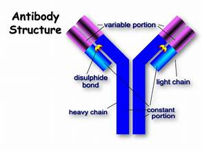
4 polypeptide chains - quaternary protein structure
Complimentary to antigens to form antigen antibody complex
Function of antibodies:
AGGLUTINATION - leading to destruction of pathogen
Mark pathogens for destruction (by macrophages)
Monoclonal antibodies:
antibodies produced by a single clone of B cells so they all have the same tertiary structure - can only bind and will all recognise 1 antigen
used to target cancer cells
ELISA test:
sample added and antigen stick to well
mAB that is specific to antigen added
excess washed away to remove any unbound antibody
2nd antibody that binds to 1st antibody added - has an enzyme attached to it
excess washed away
colourless substrate of enzyme is added and will change colour of solution - amount of antigen present is relative to the intensity of colour that develops
Vaccines
Herd immunity: immunising a sufficiently large number of people to protect an entire population from the spread of a particular disease
Problems with vaccines:
pathogen may mutate
some may not get vaccinated
some many variations of antigens that it is impossible to develop vaccines for them all
HIV
antibiotics are ineffective against HIV as viruses rely on host cells
Anti-retroviral therapy (drugs)