Human Anatomy & Physiology Overview
2. Anatomy and Physiology
2.1 Definitions
Anatomy: Study of the structure of body parts and their relationships to one another.
Physiology: Study of the function of body parts; how body parts work to carry out life-sustaining activities.
2.2 Reference Values
Reference Male: A healthy, young male weighing approximately 155 lbs (70 kg).
Reference Female: A healthy, young female weighing approximately 125 lbs (57 kg).
2.3 Anatomical Variability
Humans exhibit slight variations in both external and internal anatomy.
Over 90% of anatomical structures match textbook descriptions.
Variations may include:
Nerves or blood vessels being somewhat out of place.
Small muscles may be absent.
Extreme variations are rare and usually incompatible with life.
2.4 Sex and Gender
Sex: Set of biological attributes based on chromosomes, gene expression, and hormone action; reflected in reproductive anatomy and physiology (male or female).
Gender: Psychosocial construct comprising behaviors, expressions, and identities (e.g., man, woman, transgender, non-binary).
3. Topics of Anatomy
3.1 Subdivisions of Anatomy
Gross (Macroscopic) Anatomy: Study of large body structures visible to the naked eye.
Regional Anatomy: Examines all structures in a specific area of the body.
System Anatomy: Focuses on one system (e.g., cardiovascular, nervous).
Surface Anatomy: Looks at internal structures in relation to overlying skin.
Microscopic Anatomy: Studies structures too small to be seen with the naked eye.
Cytology: Study of cells.
Histology: Study of tissues.
Developmental Anatomy: Traced through life span.
Embryology: Study of developmental changes occurring before birth.
3.2 Studying Anatomy
Essential skills for studying anatomy:
Observe
Manipulate
Palpate: Feeling organs with hands.
Auscultate: Listening to organs using a stethoscope.
Medical Imaging Technology: Tools to view inside the body without surgery (e.g., X-ray, MRI, CT, ultrasound).
4. Topics of Physiology
4.1 Subdivisions of Physiology
Based on organ systems, including:
Renal Physiology: Functions of the kidney.
Neurophysiology: Functions of the nervous system.
Cardiovascular Physiology: Functions of the heart and blood vessels.
Often focuses on cellular or molecular levels; explores chemical reactions in cells.
Understanding basic chemical and physical principles (e.g., electrical currents, pressure, lever systems) is required.
5. Complementarity of Structure and Function
Anatomy and physiology are inseparable; structure dictates function.
Known as the Principle of Complementarity of Structure and Function.
6. Levels of Structural Organization
Chemical Level: Atoms and molecules.
Cellular Level: Cells and organelles.
Tissue Level: Groups of similar cells.
Organ Level: Two or more types of tissues.
Organ System Level: Organs that work closely together.
Organismal Level: All organ systems combined to form the whole organism.
7. The Body’s Organ Systems and Their Major Functions
11 Organ Systems:
Integumentary System: Protects internal structures.
Skeletal System: Supports and protects the body.
Muscular System: Enables movement.
Nervous System: Responds to stimuli.
Endocrine System: Hormonal regulation.
Cardiovascular System: Transports blood.
Lymphatic System: Immune response.
Respiratory System: Gas exchange.
Digestive System: Nutrient breakdown and absorption.
Urinary System: Waste removal.
Reproductive System: Produces offspring.
8. Homeostasis and Its Control
8.1 Homeostasis Defined
Homeostasis: Maintenance of stable internal conditions in response to external changes; a dynamic equilibrium.
Law of Mass Balance: Steady state where total substance intake equals its output.
8.2 Homeostatic Control Mechanisms
The body must monitor and regulate homeostasis using:
Receptors: Monitors environment and responds to stimuli.
Control Center: Determines set point and appropriate response.
Effectors: Provides means to respond (muscles or glands).
8.3 Negative and Positive Feedback
Negative Feedback: Reduces or shuts off the original stimulus; promotes stability (e.g., body temperature regulation).
Positive Feedback: Enhances original stimulus; useful in processes like childbirth.
Feedforward Response: Anticipates changes in the internal environment.
8.4 Homeostatic Imbalance
Refers to disturbances in homeostasis leading to disease or aging; inefficient control systems may lead to positive feedback taking over destructive systems (e.g., heart failure).
9. Anatomical Terms
9.1 Orientation and Directional Terms
Anatomical Position: Standard position of the body, facing forward with arms at the side.
Directional Terms: Describe the location of one body structure relative to another.
Examples include:
Superior: Above.
Inferior: Below.
Anterior: Front.
Posterior: Back.
Medial: Toward the midline.
Lateral: Away from the midline.
Intermediate: Between medial and lateral structures.
Proximal: Near to the origin of body part.
Distal: Farther from the origin.
Superficial: On the surface.
Deep: Away from the surface.
9.2 Regional Terms
Major divisions of the body:
Axial Part: Main axis (head, neck, trunk).
Appendicular Part: Limbs (arms, legs).
9.3 Body Planes and Sections
Body Plane: Flat surfaces dividing the body parts:
Sagittal Plane: Divides body into right and left parts.
Frontal Plane: Divides body into anterior and posterior parts.
Transverse Plane: Divides body into superior and inferior parts.
Oblique Section: Cuts made at angles other than right angles.
10. Cavities of the Body
10.1 Body Cavities
Internal cavities enclosed to protect organs.
Dorsal Body Cavity: Supports nervous system.
Cranial Cavity: Encases the brain.
Vertebral Cavity: Encases the spinal cord.
Ventral Body Cavity: Contains visceral organs.
Divided into:
Thoracic Cavity: Houses lungs and heart.
Abdominopelvic Cavity: Contains digestive organs and reproductive organs.
10.2 Serous Membranes
Serosa (Serous Membrane): Covers surfaces in ventral body cavity.
Parietal Serosa: Lines cavity walls.
Visceral Serosa: Covers organs.
Cavity Between the Two: Filled with lubricating serous fluid.
11. Summary
Essential concepts in human anatomy and physiology are organized hierarchically: from cells to body systems, with a focus on structure, function, homeostasis, and anatomical terminology. Understanding these is crucial for further studies in health and medical sciences.
The Cell Cycle
The cell cycle is a series of phases that a cell goes through as it grows, duplicates its DNA, and divides into two daughter cells. It is fundamental to growth, development, and tissue repair in multicellular organisms.
1. Phases of the Cell Cycle
The cell cycle is typically divided into two main stages: interphase and mitotic phase (M phase).
1.1 Interphase
Duration: The longest phase, accounting for about 90% of the cell cycle.
Subphases: Interphase consists of three subphases:
G1 Phase (Gap 1):
The cell grows in size, synthesizes proteins, and produces organelles.
The cell checks the environment for favorable conditions to continue the cycle (nutrients, growth factors).
Cells may exit the cycle and enter a resting state known as G0 phase if conditions are not favorable.
S Phase (Synthesis):
DNA replication occurs, resulting in two copies of each chromosome, known as sister chromatids.
Genetic material is duplicated, ensuring that each daughter cell will receive an identical set of chromosomes.
G2 Phase (Gap 2):
Further growth occurs, and the cell prepares for mitosis (M phase).
Organelles are duplicated, and additional proteins are synthesized.
The cell checks for DNA damage and ensures that all DNA is replicated accurately before proceeding.
1.2 M Phase (Mitotic Phase)
Duration: Typically short, lasting only a few hours.
Process: Mitosis is divided into several stages:
Prophase:
Chromatin condenses into visible chromosomes.
The nuclear membrane begins to break down.
The mitotic spindle forms and begins to capture chromosomes.
Metaphase:
Chromosomes line up in the center of the cell along the metaphase plate.
Spindle fibers attach to the centromere of each sister chromatid.
Anaphase:
Sister chromatids are pulled apart and move toward opposite poles of the cell.
The separation ensures that each daughter cell will receive an identical set of chromosomes.
Telophase:
Chromosomes reach the poles of the cell and begin to de-condense back into chromatin.
The nuclear membrane re-forms around each set of chromosomes.
The cell prepares to divide into two.
Cytokinesis: Immediately following mitosis, this process divides the cytoplasm of the parent cell into two daughter cells, completing the cell cycle.
2. Regulation of the Cell Cycle
Regulation of the cell cycle is critical for normal cell function and involves:
Cyclins and Cyclin-dependent Kinases (CDKs): Proteins that regulate progress through the cell cycle.
Checkpoints: Specific points in the cell cycle where the cell evaluates whether to proceed with division, based on factors such as DNA integrity and cell health:
G1 Checkpoint: Determines whether the cell will divide, enter G0, or proceed to S phase.
G2 Checkpoint: Ensures that DNA replication is complete and damage-free before entering mitosis.
M Checkpoint: Ensures that all chromosomes are correctly attached to the spindle before anaphase begins.
3. Importance of the Cell Cycle
The cell cycle is crucial for:
Growth and development of organisms.
Cellular repair mechanisms.
Maintaining tissue homeostasis.
Proper functioning of immune cells in response to pathogens. Furthermore, dysregulation can lead to uncontrolled cell proliferation, contributing to cancer development.
Four Abdominopelvic Quadrants (Clinical Use)
The quadrants are formed by one vertical and one horizontal line crossing at the umbilicus.
Right Upper Quadrant (RUQ)
Liver (right lobe)
Gallbladder
Duodenum
Head of pancreas
Right kidney & adrenal gland
Hepatic flexure of colon
Clinical note: Pain here is often associated with gallbladder disease.
Left Upper Quadrant (LUQ)
Stomach
Spleen
Left lobe of liver
Body & tail of pancreas
Left kidney & adrenal gland
Splenic flexure of colon
Clinical note: Trauma here may injure the spleen.
Right Lower Quadrant (RLQ)
Cecum
Appendix
Portions of small intestine
Right ureter
Right ovary & uterine tube (female)
Right spermatic cord (male)
Clinical note: Appendicitis pain classically localizes here.
Left Lower Quadrant (LLQ)
Descending & sigmoid colon
Portions of small intestine
Left ureter
Left ovary & uterine tube (female)
Left spermatic cord (male)
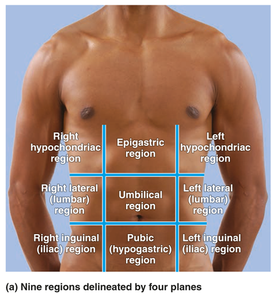
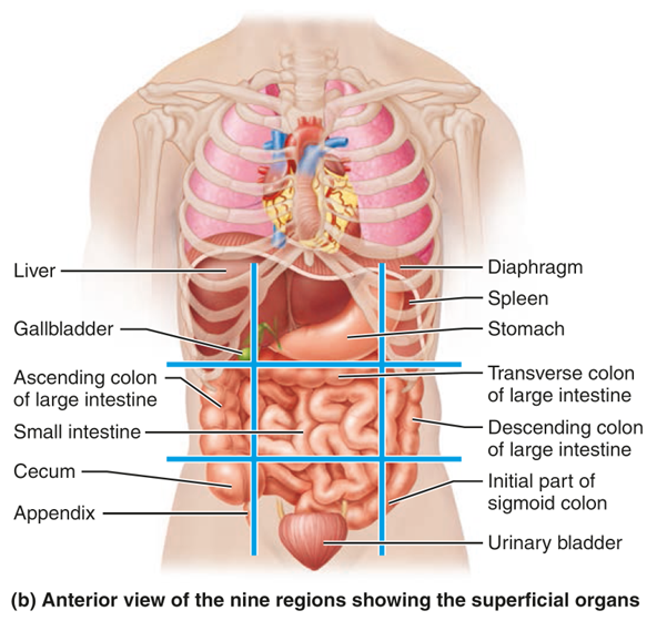
Nine Abdominopelvic Regions (Anatomical Use)
These regions are formed by two vertical and two horizontal lines.
Right Hypochondriac Region
Right lobe of liver
Gallbladder
Upper right kidney
Portions of small intestine
Key idea: Located beneath the ribs ("hypo" = below, "chondr" = cartilage).
Epigastric Region
Stomach
Left lobe of liver
Pancreas
Duodenum
Key idea: Often associated with stomach-related pain or indigestion.
Left Hypochondriac Region
Spleen
Fundus of stomach
Upper left kidney
Tail of pancreas
Right Lateral (Lumbar) Region
Ascending colon
Right kidney
Portions of small intestine
Umbilical Region
Small intestine (jejunum & ileum)
Transverse colon
Key idea: Center of the abdomen at the navel.
Left Lateral (Lumbar) Region
Descending colon
Left kidney
Portions of small intestine
Right Iliac (Inguinal) Region
Cecum
Appendix
Portions of small intestine
Clinical note: Appendicitis pain may begin near the umbilicus and migrate here.
Hypogastric (Pubic) Region
Urinary bladder
Portions of small intestine
Uterus (female)
Prostate (male)
Left Iliac (Inguinal) Region
Sigmoid colon
Portions of small intestine