Abdomen V
The first paired branches of the abdominal aorta are the ________.
A) inferior phrenic arteries
B) superior mesenteric arteries
C) gonadal arteries
D) renal arteries
E) common iliac arteries
2) Which vessel(s) originates between the superior and inferior mesenteric arteries?
A) splenic artery
B) gonadal arteries
C) lumbar arteries
D) descending geniculate artery
E) common hepatic artery
3) Which vessels supply the urinary bladder?
A) celiac trunk vessels
B) suprarenal arteries
C) gonadal arteries
D) sacral arteries
E) internal iliac arteries
4) The common iliac arteries divide to form a branch that enters the pelvic cavity and a branch called the ________ that proceeds to the lower limb.
A) inferior branch
B) femoral artery
C) inguinal artery
D) external iliac artery
E) sacral artery
5) Which of the following increases the surface area for digestion and absorption in the mucosa of the small intestine?
A) taenia coli
B) rugae
C) omenta
D) microvilli
E) lacteals
6) The region of the stomach superior to the gastroesophageal junction is which of the following?
A) cardia
B) pylorus
C) fundus
D) greater curvature
E) body
Which of the following is the deepest layer of the muscularis externa of the stomach?
A) oblique layer of smooth muscle
B) longitudinal layer of smooth muscle
C) muscularis mucosae
D) circular layer of smooth muscle
E) adventitia
8) Which vessel directly supplies the fundus of the stomach?
A) right gastric artery
B) splenic artery
C) gastroepiploic artery
D) common hepatic artery
E) left gastric artery
9) The fundus is the region of the stomach inferior to the junction between the stomach and the esophagus, also known as the gastroesophageal junction. TRUE
10) The lining of the small intestine bears a series of transverse folds called ________, which are a permanent feature of the intestinal lining.
A) taenia coli
B) plicae circulares
C) intestinal villi
D) rugae
E) haustra
11) Which type of epithelium lines the renal pelvis and ureters?
A) cuboidal
B) columnar
C) transitional
D) squamous
E) glandular
The cells of the zona fasciculata form cords that radiate outward like a sunburst from the innermost zona reticularis, and produce steroid hormones collectively known as mineralocorticoids. FALSE
13) Chromaffin cells, which are large, rounded cells of the medulla of the suprarenal gland, resemble the neurons in sympathetic ganglia and are innervated by preganglionic sympathetic fibers. TRUE
14) The ________ is/are the largest structure(s) visible in a cross section at the level of vertebra T10.
A) right lobe of the liver
B) left lung, inferior lobe
C) cardia of the stomach
D) longissimus thoracis muscles
E) latissimus dorsi muscles
15) The ________ form the prominent bony features located bilaterally in the lower abdomen.
A) costal margins
B) anterior superior iliac spines
C) tendinous inscriptions of the rectus abdominis muscles
D) pubic symphyses
E) inguinal ligaments
16) Flexion of the spinal column and depression of the ribs are actions accomplished by the ________ group of muscles in the abdominal wall.
A) inferior serratus posterior
B) diaphragm
C) rectus abdominis
D) internal intercostals
E) transversus abdominis
Which layer of the urethra is thick and elastic?
A) adventitia
B) lamina propria
C) submucosa
D) serosa
E) muscularis externa
18) An obstruction in the glomerulus would increase the blood pressure in the ________.
A) afferent arteriole
B) renal artery
C) efferent arteriole
D) intralobular artery
E) lobular vein
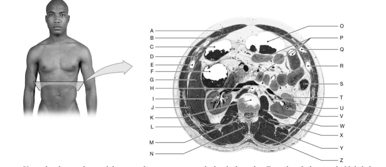
Identify the structure(s) indicated by Label A.
A) Transversus abdominis muscle
B) Intercostal muscles
C) Rectus abdominis muscle
D) Ileum
E) External oblique muscle
20) Identify the structure(s) indicated by Label B.
A) Rectus abdominis muscle
B) Transversus abdominis muscle
C) Ileum
D) External oblique muscle
E) Internal oblique muscle
21) Identify the structure(s) indicated by Label D.
A) Intercostal muscles
B) Rib 9
C) Jejunum
D) Transversus abdominis muscle
E) Psoas thoracis muscle
22) Identify the structure(s) indicated by Label F.
A) Cecum
B) Transverse colon
C) Ileum
D) Jejunum
E) Ascending colon
23) Identify the structure(s) indicated by Label G.
A) Kidney
B) Right lobe of liver
C) Spleen
D) Diaphragm
E) Intercostal muscles
24) Identify the structure(s) indicated by Label I.
A) Renal artery
B) Renal pelvis of right kidney
C) Psoas thoracis muscle
D) Right lobe of liver
E) Psoas major muscle
25) Identify the structure(s) indicated by Label K.
A) Sacrum
B) Spinal cord
C) Ileum
D) Intervertebral disc
E) Vertebral foramen
Identify the structure(s) indicated by Label L.
A) Iliocostalis lumborum muscle
B) Longissimus thoracis muscle
C) Psoas major muscle
D) Latissimus dorsi muscle
E) Gluteus medius muscle
27) Identify the structure(s) indicated by Label M.
A) Spinal cord
B) Abdominal aorta
C) Ascending colon
D) Thoracic aorta
E) Intervertebral disc
28) Identify the structure(s) indicated by Label O.
A) Cecum
B) Ileum
C) Jejunum
D) Descending colon
E) Transverse colon
29) Identify the structure(s) indicated by Label S.
A) Ascending colon
B) Inferior vena cava
C) Descending colon
D) Ileum
E) Abdominal aorta
30) Identify the structure(s) indicated by Label V.
A) Jejunum
B) Quadratus lumborum muscle
C) Left kidney
D) Psoas major muscle
E) Iliacus muscle
Identify the structure(s) indicated by Label W.
A) Iliacus muscle
B) Psoas major muscle
C) Iliocostalis muscle
D) Psoas thoracis muscle
E) Spinalis thoracis muscle
32) Identify the structure(s) indicated by Label X.
A) Longissimus thoracis muscle
B) Spinalis thoracis muscle
C) Psoas major muscle
D) Quadratus lumborum muscle
E) Iliocostalis lumborum muscle
33) Identify the structure(s) indicated by Label Y.
A) Iliocostalis lumborum muscle
B) Latissimus dorsi lumborum
C) Longissimus thoracis muscle
D) Iliacus muscle
E) Psoas thoracis muscle
34) Identify the structure(s) indicated by Label Z.
A) Trapezius muscle
B) Latissimus dorsi muscle
C) Iliocostalis lumborum muscle
D) Spinalis thoracis muscle
E) Longissimus thoracis muscle
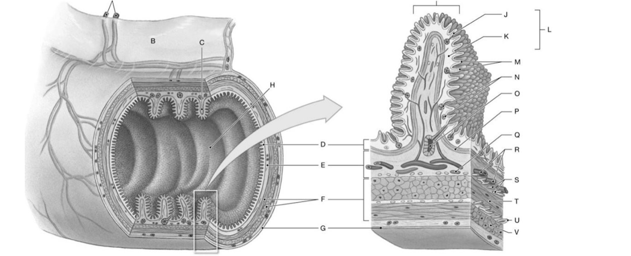
Identify the structure(s) indicated by Label B.
A) Mucosa
B) Mesentery
C) Submucosa
D) Muscularis externa
E) Plica
36) Identify the structure(s) indicated by Label C.
A) Plica
B) Muscularis externa
C) Mesentery
D) Mucosa
E) Submucosa
37) Identify the structure(s) indicated by Label D.
A) Muscularis externa
B) Plica
C) Submucosa
D) Mesentery
E) Mucosa
38) Identify the structure(s) indicated by Label E.
A) Serosa
B) Adventitia
C) Mucosa
D) Submucosa
E) Muscularis externa
39) Identify the structure(s) indicated by Label F.
A) Adventitia
B) Mucosa
C) Muscularis externa
D) Serosa
E) Submucosa
Identify the structure(s) indicated by Label G.
A) Serosa
B) Myenteric plexus
C) Mucosa
D) Submucosa
E) Muscularis externa
41) Identify the structure(s) indicated by Label I.
A) Muscularis externa
B) Taenia coli
C) Submucosa
D) Rugae
E) Omentum
42) Identify the structure(s) indicated by Label L.
A) Submucosa
B) Muscularis externa
C) Serosa
D) Adventitia
E) Mucosa
43) Identify the structure(s) indicated by Label M.
A) Mucosal gland
B) Circular muscle layer
C) Lymphatic vessel
D) Muscularis mucosae
E) Villi
44) Identify the structure(s) indicated by Label O.
A) Lymphatic vessel
B) Submucosal gland
C) Muscularis mucosae
D) Villi
E) Circular muscle layer