Lec 12: Cortical auditory evoked potentials (CAEP)
Introduction to Auditory Evoked Potentials (AEP)
Definition: Auditory evoked potentials are electrical activities within the auditory system evoked by an auditory stimulus. These neural responses are time-locked to specific auditory events.
Timeline and Generators:
Auditory Brainstem Response (ABR): Occurs very early, within the first after stimulus initiation. Generators are located in the brainstem.
Cortical Auditory Evoked Potential (CAEP): A much later response occurring as early as and extending into the to range. Generators are located higher up in the central nervous system, specifically in the auditory cortex.
Complexity: As the signal moves from the auditory nerve up the brainstem to the cortex, the pathway becomes more complex with increased connections, interconnections, and contributing centers. Unlike the "one train line" of the lower brainstem, the auditory cortex involves significant cognitive and interactive components.
Amplitude: Cortical responses have a much larger amplitude than ABR responses because the neural regions are larger and the electrodes are placed closer to the generators.
Classification of Potentials: Exogenous vs. Endogenous
Exogenous Potentials:
Primarily determined by the physical characteristics of the stimulus.
Recorded regardless of the subject's attention (e.g., ABR can be recorded while the patient is asleep).
Endogenous Potentials:
Highly dependent on the stimulus context (e.g., change in stimulus) and the subject’s internal state (e.g., attention, cognition, alertness).
If the patient is asleep, habituated, or not focused, the trace may be poor or absent.
Neural Generators and Waveform Components
Components: The standard cortical waveform consists of three primary peaks/troughs:
P1: The first positive peak.
N1: The first negative trough (most significant for visual identification).
P2: The second positive peak.
Identification: Clinicians look for presence versus absence and the general shape. Exact millisecond latency shifts are less critical than in ABR, though latency does increase as stimulus intensity approaches the hearing threshold.
Stimulus Parameters for Cortical Testing
Stimulus Type: Can use clicks, tone bursts, noise bursts, or speech stimuli (e.g., specific syllables or phonemes).
Rise and Fall Time: Requires a longer rise and fall time than earlier responses, typically between and .
Intensity: Higher intensity produces larger amplitudes. Latency remains fairly stable at supra-threshold levels but shifts/increases significantly only as we get close to the actual hearing threshold.
Inter-Stimulus Interval (ISI):
The amplitude increases as the ISI increases up to about , where it begins to plateau.
ABR uses fast rates ( to ), but CAEP requires very slow presentation (intervals of several seconds).
Sweeps: Over-averaging is counterproductive. While ABR may require thousands of sweeps, CAEP typically only requires to per average. Exceeding sweeps often results in a flattened response due to habituation.
Tonotopic Organization: Lower frequency sounds are processed in cortical regions closer to the scalp surface, often resulting in cleaner traces compared to high frequencies.
Patient Characteristics and Requirements
State of Alertness: Sleep is the "enemy" of cortical testing. Recordings performed during REM sleep result in significantly smaller or absent traces. Patients must be awake and attending to something (e.g., watching a silent movie with subtitles or reading a book).
Age and Maturation:
Adults: Show clean, large, mature traces.
Infants/Children: Traces are often messier and smaller in amplitude because the auditory cortex and associated pathways are not yet fully mature. Latency decreases and amplitude increases as the brain matures.
Habituation: The brain naturally tunes out boring, repetitive stimuli. This is a protective mechanism but hinders testing. Clinicians mitigate this by keeping runs short ( to sweeps) and changing the frequency or ear tested unpredictably.
Refractoriness: A biological process where neuronal excitability decreases because the brain gets "tired."
Clinical Implementation and Electrode Setup
Montage:
Low Center Forehead (Ground): Similar to ABR.
Center of the Coronal Line (Vertex/Cz): The active electrode is placed at the top of the head to be near the auditory cortex.
Earlobes or Mastoids: Reference electrodes.
Impedance: Goal is less than . Most importantly, impedances across all electrodes should be balanced (similar values). Coronal placement requires parting the hair and using exfoliating paste or conductive gel.
Equipment Scale: CAEP responses are much larger (measured in microvolts) than ABR (measured in sub-microvolts). Setting the wrong scale on the equipment can lead to false diagnoses of profound deafness if the clinician is looking for a tiny ABR trace on a large cortical scale.
Clinical Application: Cortical Evoked Response Audiometry (CERA/SARA)
Purpose: To objectively estimate hearing thresholds when behavioral testing is unreliable. It typically gets within to of actual behavioral thresholds.
Medico-Legal/WorkCover Context: Frequently used in cases regarding noise-induced hearing loss compensation. In these scenarios, a behavioral audiogram may be exaggerated (non-organic hearing loss). Courts require an objective definitive measure.
Search Technique: Descend in steps; once the response is lost, ascend in or steps.
Case Study: Jeffrey (54 years old):
History: Transport industry worker claiming noise-induced loss. Behavioral results were inconsistent (speech at a at but inconsistent PTA; OAEs present ).
SARA Results: Showed thresholds at to , proving no worse than a mild to moderate loss, contradicting his behavioral claim of severe loss.
Case Study: Ronald (Intellectual Disability):
Goal: Confirm if current hearing aids were sufficient as communication skills had declined.
Challenge: Could only sit still for intervals; distracted with AFLW (Carlton team) on an iPad.
Result: Corticals at and matched previous hearing levels, confirming that a change in hearing was not the cause of his decline.
Clinical Application: HearLab (Infant Testing)
HearLab System: Used for objective cortical testing in awake infants, often in the free field (loudspeakers).
Speech Phonemes: Specifically utilizes "m", "g", "t", and "s" to cover the speech frequency range.
Statistical Evaluation: The software uses a -value (typically p < 0.05) to objectively determine if a response is present, rather than relying solely on visual inspection.
Aided Cortical Testing: Evaluates the efficacy of a hearing aid fitting about six weeks post-fitting. If an infant does not show cortical responses to soft speech phonemes (), the clinician increases the hearing aid gain or considers cochlear implant (CI) referral.
Case Study: Eleanor (5 weeks old): Showed no response to soft "m" phonemes; hearing aid low-frequency gain was increased, and subsequent visit showed improved access to sounds.
Case Study: Xu (4.5 months old): Diagnosed with profound loss. Aided HearLab showed no response even after gain adjustments, providing objective evidence for cochlear implant referral.
Other Clinical Applications
Auditory Processing Disorder (APD): Used to track the maturation of the auditory pathway. APD children often show "younger"/immature cortical responses compared to age-matched controls. Maturation (decreased latency, increased amplitude) can be tracked after training programs.
P300 (Cognitive Potential):
Occurs roughly post-stimulus.
Requires an "oddball" paradigm: a "standard" stimulus and a "deviant" stimulus. The brain's response to the change is measured.
Used in research for Alzheimer's, dementia, aphasia, and traumatic brain injury.
Emerging Research:
Hyperacusis: Investigating larger-than-normal amplitudes to loud tones.
Tinnitus: Exploring objective pitch matching and identifying sites of generation.
Troubleshooting Guidelines
No Response: Check if the stimulus is actually playing and verify electrode connections.
Small Response: The patient might be dozing off or habituating to the stimulus.
Noisy Trace: The patient may be tense or jaw-clenching. Re-instruct them to relax and check for tangled electrode leads or mobile phone interference.
Questions & Discussion
Question: What kind of scenario in clinic might you consider corticals?
Response: Closely associated with non-organic presentations where behavioral tests fail, and also used extensively in APD clinics.
Question: Why not use ASSR instead of SARA for adults with disabilities?
Response: SARA/CAEP in adults is more reliable and gets closer to actual behavioral thresholds ( vs. common elevations in ASSR). ASSR requires the patient to be asleep and quiet, whereas CAEP is better for awake adults.
Question: Does a report showing non-organic results get used against someone in court?
Response: It's less about "slapping their wrist" for being naughty and more about using formulas to determine true entitlement and accurate compensation based on the objective objective SARA numbers.
Question: Why use speech sounds in infants?
Response: It is more relevant to language development and potentially more interesting to the child than pure tones.
Cortical Auditory Evoked Potentials (CAEPs)
Learning Outcomes
Describe the neural generators and principles to record obligatory CAEP.
Describe the requirements to prepare patients for CAEP testing.
Be able to label straightforward traces and analyse/interpret the results .
Be able to identify stimulus, recording, and subject factors that affect obligatory CAEP interpretation.
Recommend trouble shooting techniques to improve poor responses.
Describe different clinical applications of obligatory CAEP in complex audiological cases.
Be familiar with discriminative CAEPs.
Apply knowledge of cortical auditory evoked potentials to accurately diagnose hearing disorders and evaluate treatment outcomes in both children and adults.
Overview
CAEPs are another type of auditory evoked potential used by audiologists.
They were previously mentioned in relation to:
Non-organic hearing loss presentations.
Cases where behavioural testing cannot provide reliable thresholds.
Auditory Processing Disorder (APD) assessment.
CAEPs are often considered when:
Behavioural testing is unreliable.
Objective confirmation of hearing thresholds is required.
APD investigations are being conducted.
Auditory Evoked Potentials
Definition
Auditory evoked potentials are:
Neural activity within the auditory system.
Evoked by an auditory stimulus.
Time-locked to an auditory event.
Examples of stimuli:
Clicks
Tones
Speech sounds
General principle:
Greater stimulus intensity produces larger responses.
Position of CAEPs Along the Auditory Pathway
ABR vs CAEP
Auditory Brainstem Response (ABR)
Occurs within approximately the first 10 ms after stimulus onset.
Generated by the brainstem.
Cortical Responses
Generated much higher in the auditory pathway.
Begin around 50 ms after stimulus onset.
Later components occur around:
100 ms
200 ms
As neural activity is recorded from higher levels of the auditory system:
Complexity increases
Different cortical regions and systems interact with each other
Attention and cognition begin to influence responses.

Characteristics of CAEPs
Larger Amplitude
Compared with ABRs:
CAEPs have much larger amplitudes.
Electrodes are physically closer to the neural generators.
Responses are easier to see above the noise floor.
Cognitive Influence
As responses originate from higher auditory centres:
Cognition contributes.
Attention contributes.
State of arousal contributes.
These factors are much less important for ABRs.
Exogenous vs Endogenous Potentials
Exogenous Potentials
A product of the stimulus characteristic
Can be recorded regardless of whether the patient attends to the stimulus.
Example:
ABR
Endogenous Potentials
Less dependent on stimulus characteristics
Highly dependent on:
Stimulus context
Changes in the stimulus
Patient state
Attention
Responses may be reduced if the patient is:
Sleeping
Not paying attention
Habituated to the stimulus
CAEPs have important endogenous influences.
Generators of CAEPs
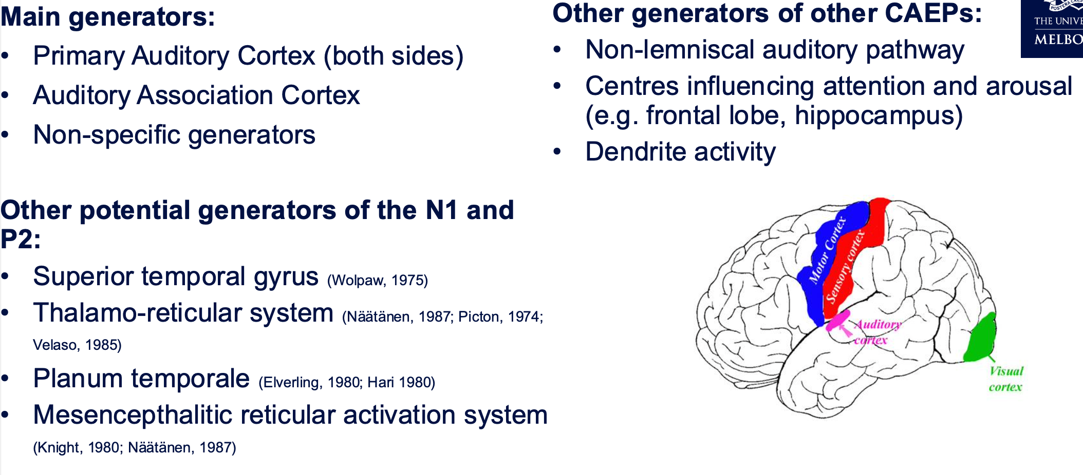
The cortical response is generated by many regions within the brain.
Key point:
There are numerous contributors.
Not expected to memorise all anatomical names
The important concept is that many brain regions influence the recorded waveform
Adult CAEP: onset response
P1-N1-P2 complex

Components
The classic waveform consists of:
P1
First positive peak.
N1
First negative peak.
P2
Second positive peak.
Naming
P = positive peak.
N = negative peak.
The waveform is therefore:
P1 → N1 → P2
Clinical Focus
Unlike ABR:
Precise latency measurement is less important.
Clinicians are mainly interested in:
Presence vs absence of a response.
Amplitude is much larger than ABR.
Terminology
CAEPs may be referred to by several names.
• Cortical Event-Related Potential (CERP)
• Auditory Late potential (ALP)
• Slow Vertex Potential (SVP)
• P1-N1-P2
• N1-P2
Stimulus Factors Affecting CAEPs
Stimulus Type
Any transient onset stimulus may be used:
Clicks
Tone bursts
Noise bursts
Speech syllables
Longer rise/fall times are required compared with ABR:
Approximately 10–20 ms rise time.
Responses are broadly similar across different stimulus types.
Other Possible Stimuli
The stimulus can also be:
A gap in a tone
A gap in noise
An omission within a stimulus train
A change within a continuous sound
Choice depends on the clinical application.
Clinical Context for Threshold Estimation
A major clinical use is:
Objective Threshold Estimation
Particularly when behavioural thresholds cannot be trusted.
Common situations:
Medico-legal cases
WorkCover cases
Non-organic hearing loss presentations
Purpose:
Produce an objective audiogram.
Confirm true hearing status.
The courts often require objective evidence because behavioural audiograms may be exaggerated.
Inter-Stimulus Interval (ISI)
Effect on Response
As ISI increases:
N1P1 amplitude increases.
Response improves up to approximately 5 seconds.
Improvement then plateaus.
Recommended ISI
Approximately:
4–5 seconds
Comparison with ABR
ABR uses much faster rates:
Paediatric ABR ≈ 33 Hz
Adult ABR ≈ 11 Hz
CAEP testing is therefore much slower.
Test Duration
Obtaining a full objective audiogram using CAEPs can take:
Approximately 3 hours
Reasons:
Slow presentation rates.
Need for multiple frequencies.
Need for replication.
Tonotopic Organisation
Frequency Effects
Lower-frequency sounds:
Produce greater spread of activity (basilar membrane)
Are processed closer to the scalp (auditory cortex)
→ Produce larger amplitude responses
Higher-frequency sounds:
Produce responses that are generally more difficult to record.
Responses can still be obtained at high frequencies, but lower frequencies are usually easier to interpret.
Intensity Effects
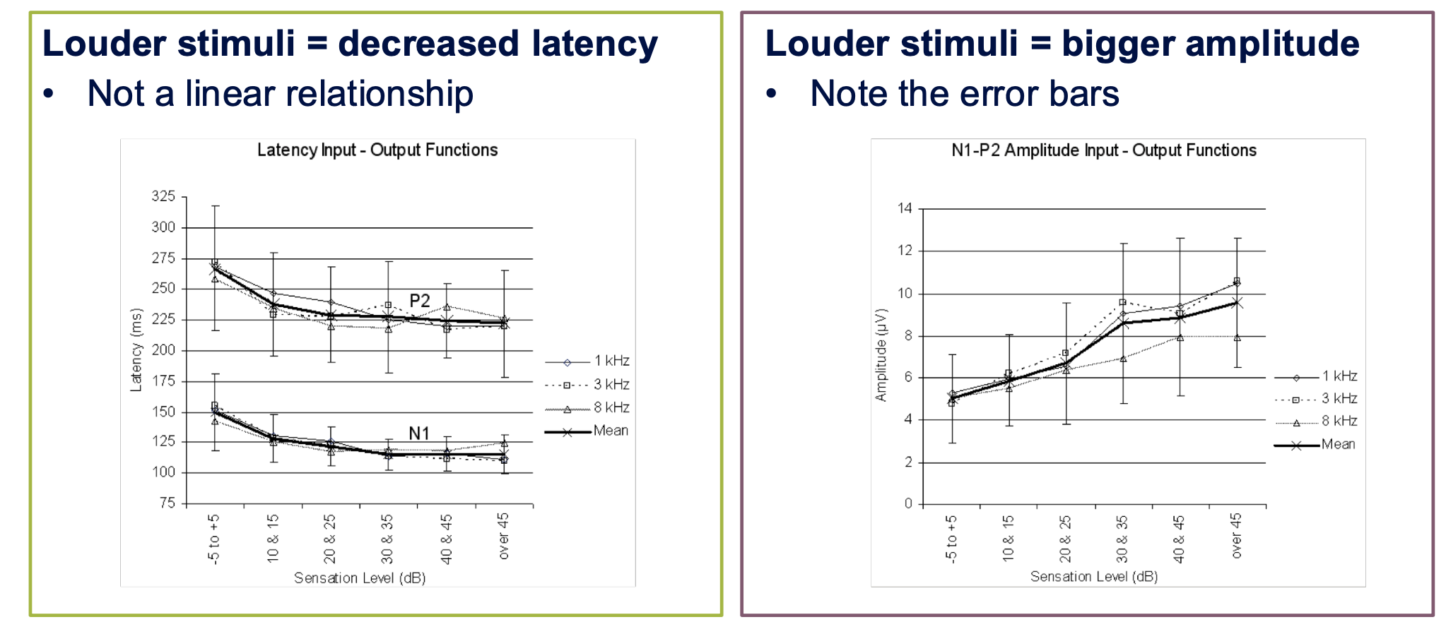
Latency
Louder stimuli:
Produce shorter latencies.
Unlike ABR:
Relationship is not strongly linear.
Latency remains relatively stable at high levels.
Latency increases noticeably only near threshold.
Amplitude
Louder stimuli:
Produce larger amplitudes.
Softer stimuli:
Produce smaller amplitudes.
Sweeps and Averaging
Important Difference from ABR
ABR:
More sweeps generally improve waveform quality.
CAEP:
Over-averaging is counterproductive
Why?
Habituation occurs.
Attention decreases.
Neural responsiveness reduces.
Recommended Number
Stimuli above about 20 dBSL usually produce a clear response after 10
still need to replicate responses
Patient Factors Affecting CAEPs
Attention
Attention is critical.
Poor attention can reduce response quality.
Relaxation
Patients should:
Sit still.
Relax.
Avoid muscle tension.
Jaw tension can create artefact.
Sleep
Unlike ABR:
Sleep is a major problem.
Patients should remain:
Awake
Alert
Strategies:
Bright room
Movie without sound
Reading material
Games
Avoid:
Dim lighting
Situations that encourage sleep.
Maturation (Age Effects)
Latency
With increasing age:
Latency decreases.
Amplitude
With increasing age:
Amplitude increases.
Children
Responses are:
Smaller
Less mature
More variable
Adults
Responses are:
Larger
Cleaner
Easier to interpret
This developmental effect makes CAEPs useful for:
Monitoring auditory development.
Monitoring outcomes following intervention.
Habituation
Definition
Reduction in neuronal responsiveness following repeated stimulation.
The brain naturally learns to ignore repetitive, unimportant stimuli.
Consequences
Reduced amplitude.
Poorer recordings.
Minimising Habituation
Use:
Short runs (~10 sweeps)
Longer ISIs
Unpredictable stimuli
Strategies:
Alternate ears
Alternate frequencies
Vary presentation order
This prevents the brain from predicting upcoming stimuli.
Refractoriness
Definition:
Decrease in neuronal excitability due to fatigue.
Effect:
Reduced response amplitude.
Different from habituation, which is related to repeated stimulation and reduced attention.
Effects of Sedation, Alcohol and Medication
Responses may be affected by:
Sedation
Sleeping tablets
Alcohol
Particularly relevant in:
WorkCover
Medico-legal
Non-organic hearing loss investigations
Patients may attempt to influence results.
Sleep and sedation substantially reduce CAEP responses.
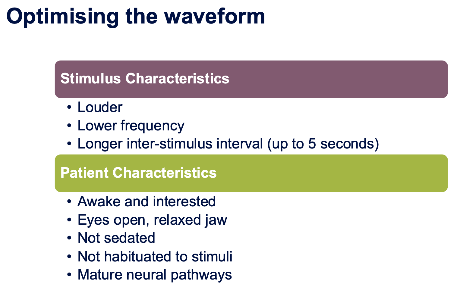
Optimising Waveforms
Stimulus Factors
Use:
Louder stimuli
Lower frequencies
Longer ISIs
Patient Factors
Ensure:
Awake
Alert
Eyes open
Relaxed jaw
No sedation
Minimal habituation
Older patients generally produce cleaner responses.
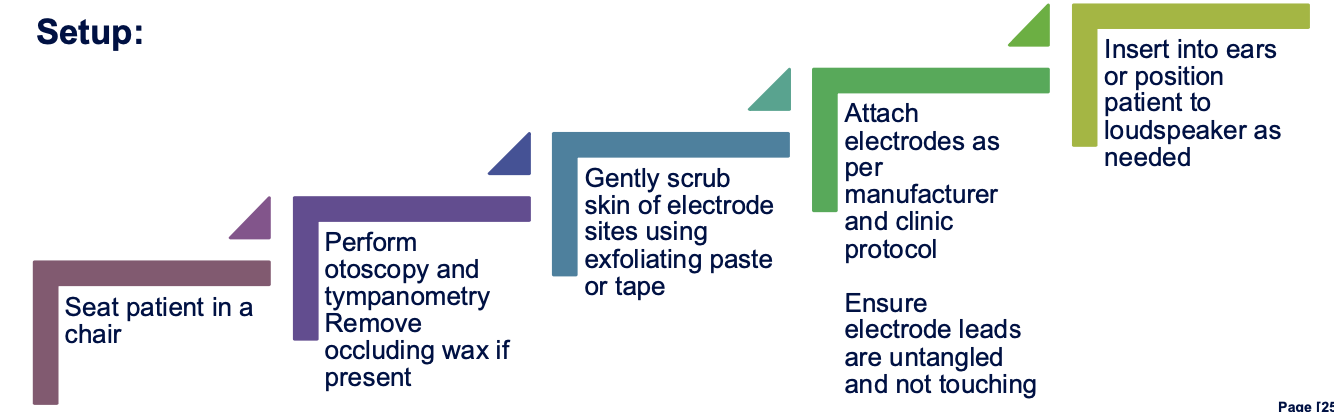
Electrode Placement

Montage
Electrodes are placed:
Low forehead
Vertex (top of head; coronal line)
Earlobes or mastoids
Practical Challenge
Hair makes obtaining low impedance difficult.
Strategies:
Part the hair
Use exfoliating paste
Use impedance gel
Patients are often advised they will need to wash their hair afterwards.
Impedance
Target:
Less than 10 kΩ
Compared with ABR:
ABR target is usually <5 kΩ.
Most important principle:
Electrode impedances should be balanced.
Balanced moderate impedances are preferable to one very high impedance electrode.
Patient Instructions
We're measuring your brain's electrical activity in response to sounds played through
your ears. The test is automatic, so you don't need to do anything. Testing takes a
while, so feel free to read a magazine or use this iPad to pass the time.
Patients should be told:
They will hear sounds.
They do not need to respond.
The test is automatic.
The test may take a long time.
Provide activities to maintain alertness:
Books
Movies with subtitles
Nintendo Switch
Colouring activities
iPads
The goal is to keep the patient:
Quiet
Comfortable
Awake
Engaged
Clinical Applications
Main Applications
1. Cortical Evoked Response Audiometry (CERA)
Technique to objectively estimate hearing thresholds
Used for:
Objective audiograms
Non-organic hearing loss
Medico-legal assessment
Patients unable to complete behavioural testing
2. HearLab
Used for:
Infants
Hearing aid verification
Cochlear implant candidacy assessment
3. Auditory Processing Disorder Assessment
Used to:
Assess maturation of auditory pathways.
Monitor changes following intervention.
Other applications were mentioned but described as more specialised.
CERA Threshold Search
Features:
Excellent frequency specificity (250-8000 Hz)
Good validity
Threshold estimates often within 5–15 dB of behavioural threshold
Typical procedure:
Descend in 20 dB steps.
Ascend in 10 dB steps if response is lost.
Replicate responses.
Indicators of threshold:
Response becomes smaller.
Latency increases.
Response eventually disappears.
Scale Differences: ABR vs CAEP
Important clinical warning:
CAEP amplitudes are much larger than ABR amplitudes.
If the wrong display scale is used:
An ABR may appear flat.
Normal hearing may be misinterpreted as profound hearing loss.
Checking the display scale is one of the first troubleshooting steps.
Troubleshooting
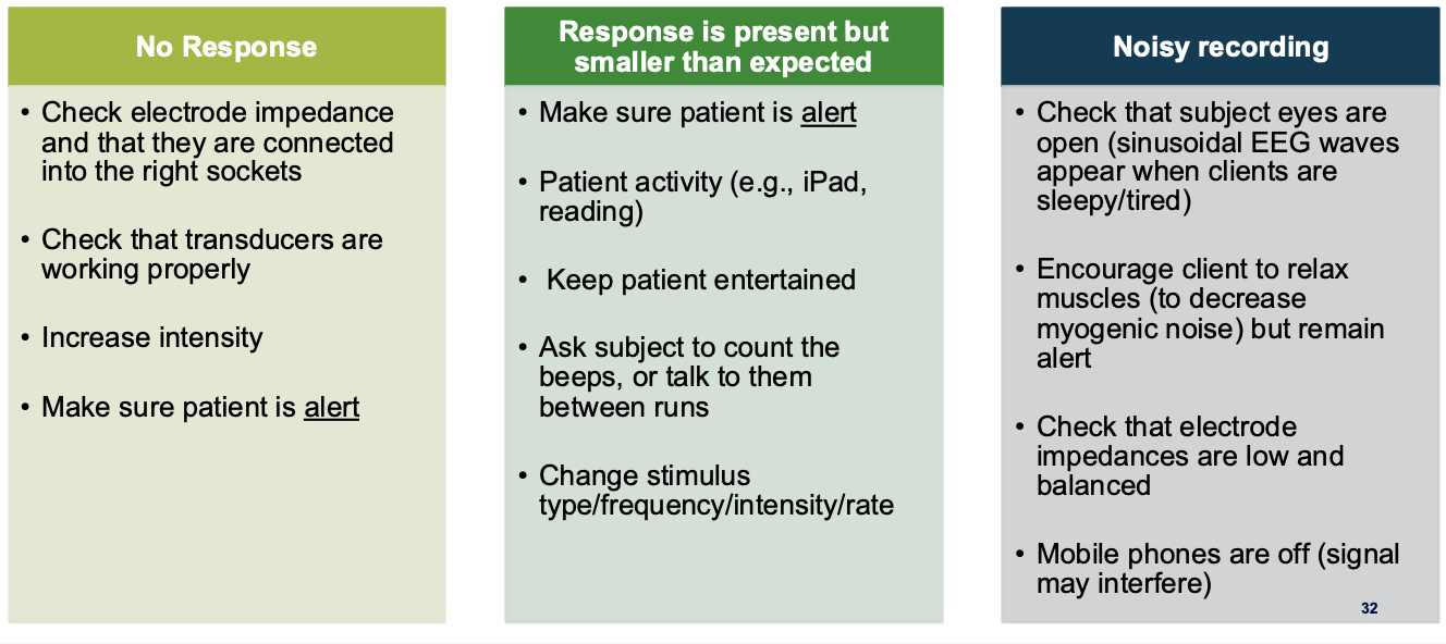
No Response
Check:
Stimulus presentation
Electrode placement
Small Response
Consider:
Patient attention
Drowsiness
Habituation
Noisy Response
Consider:
Muscle tension
Jaw movement
Chewing gum
Poor electrode impedance
Tangled leads
Mobile phone interference
Reinstruct and relax the patient if necessary.
Cortical Auditory Evoked Potentials (CAEPs / “SARA”) – Detailed Lecture Notes
Based strictly on the provided lecture transcript only.
1. Adult Clinical Application: Non-Organic / Unreliable Behavioural Audiograms
Case 1: Jeffrey (54 years old)
History
Referred by a medical panel for SARA testing.
Had a hearing test previously.
Fitted with hearing aids 6 months earlier at another clinic.
Reports hearing aids are annoying because he hears “too much”.
No ENT concerns.
Constant bilateral buzzing/ringing tinnitus for 2 years.
Annoying but not affecting sleep.
Worked in the transport industry.
Significant occupational noise exposure.
No hearing protection provided.
Seeking compensation related to workplace noise exposure.
Occasionally experiences lightheadedness when standing.
Otherwise generally well.
Behavioural Audiogram Findings
Inconsistency 1: Speech Results vs Audiogram
Observed:
Speech scores close to 100% at 50 dB.
Speech scores also close to normal at 30 dB.
Interpretation:
If speech recognition is close to 100% at 30 dB:
Estimated hearing thresholds around 1–2 kHz should be approximately normal.
This is inconsistent with an audiogram suggesting severe hearing loss.
Inconsistency 2: Acoustic Reflexes
Why were reflexes considered reassuring?
Reflexes present around 80 dB.
Based on minimum sensation level calculations:
Patient should at least be hearing around 65 dB.
Therefore:
Reflex findings do not support the severe behavioural hearing loss shown on the audiogram.
Why Was Bone Conduction Not Performed?
Reason:
Air-conduction responses were not reliable.
If AC responses are unreliable:
Bone conduction will not provide useful additional information.
Better use of clinical time:
Use alternative techniques.
Perform objective tests.
Clinical reasoning:
No point obtaining BC thresholds if AC thresholds themselves cannot be trusted.
Inconsistency 3: Multiple Different Thresholds at Same Frequency
Example:
Right AC threshold at 1 kHz varied depending on technique:
80 dB
60 dB
35 dB
Interpretation:
A reliable audiogram should not show this degree of variation.
Therefore:
Audiogram is unreliable.
Bone conduction would not be helpful.
Inconsistency 4: OAEs
Findings:
OAEs present from 1–4 kHz.
Interpretation:
OAEs suggest hearing is no worse than mild–moderate loss.
This conflicts with severe behavioural thresholds.
2. SARA Testing Interpretation
General Principles
Reading the Trace
Clinician labels:
Frequency tested.
Presentation level.
Example:
At 1 kHz:
Response obtained at 40 dB.
Response obtained at 20 dB.
Slight latency increase at lower level.
Interpretation:
Increased latency near threshold is expected.
Presence of repeatable waveform indicates response.
Estimated Hearing Status from SARA
Approximate findings:
Around 20–30 dB at 1–2 kHz.
Around 40–50 dB at 4 kHz.
Interpretation:
Mild to moderate hearing loss.
Important fact:
CAEP/SARA thresholds are generally within 5–15 dB of true behavioural threshold.
Therefore:
Much closer to true hearing levels than many other objective tests.
Identifying a Response
Key Features
Look for:
Expected latency region.
P1
N1
P2
Repeatability between traces.
Important teaching point:
Negativity (N1 region) often aligns most clearly.
Peaks may not align perfectly.
Repeatable negativity is often easier to identify.
Using Louder Responses as Templates
A stronger response at a higher level:
Helps identify weaker responses near threshold.
Clinical caution:
A borderline response might not be accepted if a louder template was unavailable.
3. Latency Behaviour in Cortical Responses
Relationship Between Level and Latency
Unlike some other tests:
Latency remains relatively stable at louder levels.
Latency only increases when approaching threshold.
Therefore:
Similar latency across several loud levels.
Delayed latency near threshold.
Clinical value:
Helps determine when threshold is being approached.
4. Reporting Non-Organic Presentations
Example wording:
Behavioural responses:
Moderate to severe bilateral hearing loss.
However:
Variable responses obtained.
Ascending technique.
Speech testing.
Objective tests.
All suggest behavioural responses are suprathreshold.
Reporting SARA Results
Example reporting style:
For thresholds obtained at 20 dB:
Report as ≤20 dB.
Reason:
Testing not performed below 20 dB.
True threshold may actually be better.
Conservative interpretation:
No worse than a mild-to-moderate hearing loss from 500 Hz–4 kHz bilaterally.
5. Hearing Aid Implications
Why Were the Hearing Aids Too Loud?
Likely explanation:
Previous clinic accepted exaggerated behavioural thresholds.
Hearing aids fitted according to exaggerated loss.
Result:
Excessive amplification.
Hearing aids perceived as too loud.
Clinical Consequences
Failure to detect non-organic presentation may result in:
Inappropriate hearing aid fitting.
Discomfort.
Potentially unsafe amplification.
Important Clinical Point
Patients with non-organic presentations often:
Still have genuine hearing loss.
Exaggeration may occur on top of real hearing loss.
Lecturer's comment:
Sometimes patients are desperate for help.
Exaggeration may be partly subconscious.
Not always deliberate deception.
6. Adult Clinical Application: Intellectual Disability Case
History
Patient:
Long-term hearing aid user through Hearing Australia.
Previously diagnosed with moderate-severe hearing loss.
Intellectual disability.
Recent decline in:
Responsiveness
Attention
Communication
Question:
Has hearing deteriorated?
Problem:
Reliable behavioural audiogram could not be obtained.
Testing Challenges
Patient:
Could only sit still for ~15 minutes.
Management:
Used AFLW football videos on an iPad to maintain cooperation.
Testing priority:
Answer the referral question efficiently.
Results
Only obtained:
1 kHz
4 kHz
One ear
This limited information was enough.
Hearing Australia later confirmed:
No significant hearing change.
Clinical conclusion:
Hearing was not the cause of the patient's deterioration.
Why Not ASSR?
Lecturer explanation:
ASSR:
Smaller amplitude.
More elevated relative to behavioural threshold.
Requires quietness/sleep.
For this adult:
Cortical responses provided a better estimate of hearing status.
7. HearLab in Infants
Purpose
HearLab:
Objective cortical testing in awake young infants.
Uses speech stimuli.
Conducted in free field.
Correlates well with:
Infant perception of sound.
Main Uses
1. Hearing Assessment
Assess access to speech sounds.
Can help re-estimate audiogram.
2. Hearing Aid Verification
Usually ~6 weeks after fitting.
Determines whether amplification is adequate.
3. Cochlear Implant Assessment
Determine whether hearing aids provide sufficient speech access.
Supports referral decisions.
4. Auditory Neuropathy
Helpful before fitting.
More detail taught later in course.
8. HearLab Speech Stimuli
Speech sounds:
/m/
/g/
Other speech phonemes spanning frequency range.
Characteristics:
/m/ = lower frequency
Higher-frequency phonemes represent upper frequency region.
Statistical Detection
Software evaluates:
Repeatability.
Consistency.
Shape matching.
Statistical significance.
Criterion:
p < 0.05
If achieved:
Response considered present.
Software is more objective than visual interpretation alone.
Why Use Speech Stimuli?
Reason:
Goal is speech and language development.
More clinically meaningful than:
Pure tones alone.
Question:
Does the child have access to speech sounds needed for language development?
9. Infant Case: Eleanor
Background
5 weeks old.
Recently fitted with hearing aids.
HearLab performed at Visit 1 and Visit 2.
Visit 1 Interpretation
Findings:
Poor access to low-frequency sounds.
Poor access to soft mid-frequency speech sounds.
Management:
Increase low-frequency gain.
Increase mid-frequency gain.
Visit 2
Results:
More responses present.
Better access to soft speech sounds.
Outcome:
Improved hearing aid performance.
Parent feedback:
Felt relieved.
Confidence in hearing aids restored.
10. Infant Case: Zu
History
Profound SNHL diagnosed at 3 months.
Bilateral hearing aids fitted at 4 months.
HearLab performed at 4.5 months.
Visit 1
Results:
No clear responses.
No access to speech sounds despite hearing aids.
Management:
Hearing aids adjusted.
Visit 2
Results:
Still inadequate speech access.
Interpretation:
Hearing aids unable to provide required amplification.
Management:
Refer for cochlear implantation.
Importance of HearLab
Provides objective evidence that:
Hearing aids are insufficient.
Helps families:
Make informed cochlear implant decisions.
11. Auditory Processing Disorder (APD)
Clinical Use
Not used for threshold estimation.
Instead:
Present suprathreshold stimuli.
Examine maturation of auditory pathways.
Research Findings
Compared:
APD children
Age-matched controls
Findings:
APD group had:
Later latency.
Smaller amplitude.
Responses resembling younger auditory systems.
Interpretation:
Auditory pathways appear less mature.
Typical APD Protocol
Often:
500 Hz
4 kHz
Presented around:
80–85 dB
May repeat after auditory training.
Improvement indicators:
Reduced latency.
Increased amplitude.
Limitation
Need:
Better normative data.
Currently:
Used as one component of an assessment battery.
Not a standalone diagnostic tool.
12. P300
What Is It?
Measure of:
Attention
Recognition of change
Occurs:
~300 ms after stimulus.
Indicates:
High-level cognitive processing.
Broad brain involvement.
Stimuli
Can use:
Auditory
Visual
Other sensory stimuli
Not limited to hearing.
Method
Two stimulus categories:
Standard Stimulus
Frequent/common stimulus.
Deviant Stimulus
Rare/different stimulus.
Brain response to deviant stimulus:
Much larger.
Responses are compared:
Standard vs deviant.
Difference reveals P300.
Requirements
P300 only occurs if:
Brain recognises the difference.
Examples:
Different frequencies.
Different colours.
Different flashing patterns.
Attention is required.
If attention is elsewhere:
P300 may not occur.
Applications
More common in:
Neuropsychology
Used for:
Cognitive dysfunction
Alzheimer's disease
Dementia
Aphasia
Cognitive tracking over time
Also used in research:
Auditory perceptual skills.
13. Final Summary of Clinical Applications
Established Clinical Uses
Adult hearing threshold estimation.
Infant hearing threshold estimation.
Aided hearing threshold estimation.
Hearing aid verification.
Cochlear implant candidacy support.
Emerging Applications
Auditory Processing Disorder.
Cognitive tracking.
Neuropsychological assessment.
Traumatic brain injury.
Research Applications
Hyperacusis
Observed:
Larger amplitude responses to loud sounds.
Potential use:
Understanding mechanisms.
Evaluating management effectiveness.
Tinnitus
Research exploring:
Objective pitch matching.
Site of generation.
Kahoot Exam Pearls
Cortical Response Timing
Much later than ABR.
Approximately 50–200 ms response window.
Inter-Stimulus Interval (ISI)
Increasing ISI increases N1-P2 amplitude.
Effect plateaus around 5 ms.
Aim for approximately 5 ms ISI.
Patient Preparation
Recommended:
Keep patient alert.
Not recommended:
Dimly lit room.
(Unlike ABR, where sleepiness is desirable.)
Tonotopic Organisation
Lower frequencies are processed closer to the scalp.
Therefore produce larger cortical responses.
Over-Averaging
Problem:
Causes habituation.
Result:
Can be counterproductive in cortical recordings.
HearLab
Purpose
Objective cortical testing in awake infants.
Used to:
Estimate hearing.
Verify hearing aid fittings.
Assess cochlear implant candidacy.
Evaluate auditory neuropathy cases.
Procedure
Child is:
Seated approximately 1.5 m from the loudspeaker.
Kept awake and engaged.
Possible distractors:
Toys
Cartoons (without sound)
Tablets
Sensory toys
Avoid:
Noisy toys
Videos with sound.
Speech Stimuli Used in HearLab
Speech sounds:
/m/
/g/
/t/
These broadly represent different frequency regions of speech.
Purpose:
Determine whether amplified speech sounds are reaching the auditory cortex.
Emerging and Research Applications
Mentioned areas include:
Hyperacusis research
Larger cortical responses to louder tones.
Tinnitus research
Objective pitch matching.
Investigating site of generation.
Currently these remain primarily research applications.