Week 5: Cariology
Caries is the ongoing cycle of mineral loss and replacement in teeth. When we eat, bacteria use carbohydrates to produce acids that cause demineralisation. Saliva then neutralises the acidity, allowing lost minerals to be reincorporated into the tooth surface (remineralisation). This balance of loss and repair occurs constantly in everyone’s mouth.
Process:
caries = demineralization/weakening of tooth
weakening of enamel causes a caries lesion
this leads to cavitation
Health Determinanats:
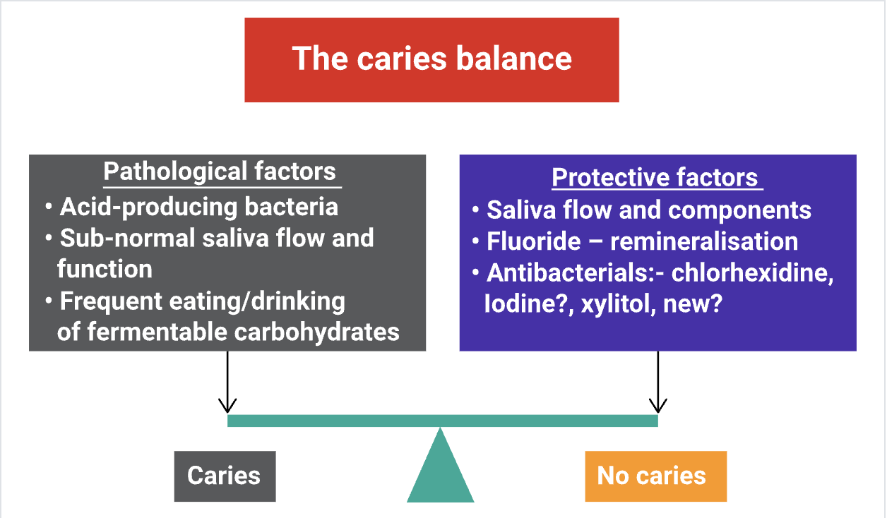
pathogenic factors cause demineralization
protective factors cause remineralization
Protective Measures:
fluoride promotes remineralization + inhibits demineralization
antibacterials (chlorhexidine, iodine, xylitol) affect oral bacteria
Definitions:
Caries is not the term for a lesion, it is a disease process
Caries process is an interaction between the biofilm (plaque) and the tooth surface and subsurface.
Caries lesion is the manifestation of the stage of the process at one point in time. Caries lesions range from white spot (incipient) lesions (ICDAS 1 and 2) to breakdown of enamel (ICDAS 3) or initial cavitation to the destruction of dentine (ICDAS 4, 5 and 6).
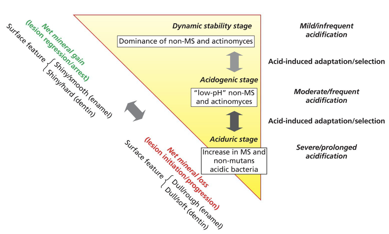
Caries progression occurs when demineralisation and remineralisation are out of balance, leading to net mineral loss. Remineralisation can arrest or reverse the progression of the disease and can lead to changes in tooth structure.[6] So, the caries progression is an active process, where caries is progressing from very early to more advanced stages of mineral loss. If we were to measure mineral loss, we could see a lowering of net minerals in the two surfaces as the lesion progresses over time.
Biofilm + Oral Microbiome
Three critical factors contribute to dental caries:
the biofilm or the plaque that grows on teeth and other hard structures
the diet, what we put into our mouths in terms of food and drinks
the health or state of the oral environment
Salivary Microbiome
Saliva countains 10^7-10^8 microorganisms/mL in adults
eating/drinking will immediately eliminate the major portion of micoorganisms by swallowing
1 min rinsing w chlorhexidine reduces microorganism levels from 10^8 to 10^6
bacteria on oral surfaces grows using nutrition from saliva
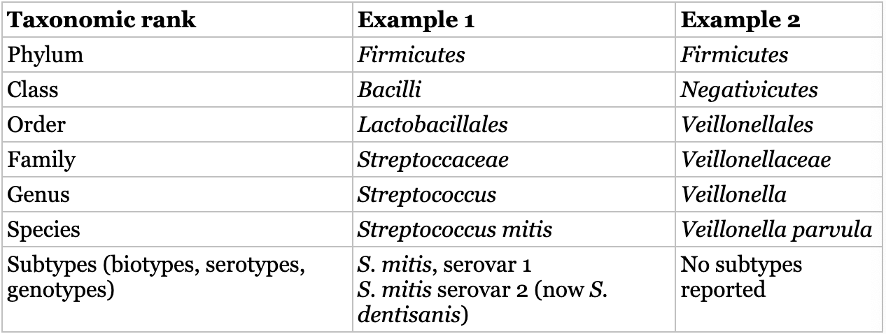
“The oral microbiome is dominated by the phylum Firmicutes but also include a high proportion of Actinoacteria, proteobacteria, bacteroidetes, fusobacteria, and spirochaetes, which stands for 96% of total oral bacteria”
Functions of oral microbiome in caries
dental caries is the result of the metabolic activities of bacteria growing in microbial communities on teeth (a.k.a. dental plaque)
therefore, plaque on tooth surface is the prerequisite for caries lesions to develop
Caries-Pomoting Characteristics
Acidogenicity
the ability to rapidly transport fermentable sugars when in competition with other plaque bacteria
conversion of sugars to acid (lactic acid, acetic acid, formic acid)
Acidurance
ability to maintain sugar metabolism under extreme environmental conditions (low pH)
Extracellular Polysaccharide Production
the ability to produce extracellular polysaccharides including glucans and fructans (contribute to biofilm matrix
Root + Dentin Caries
the caries process in dentin + root cementum differs from enamel caries bc of the organic content of these tissues
Biofilms + Formation of Cariogenic Dental Plaque
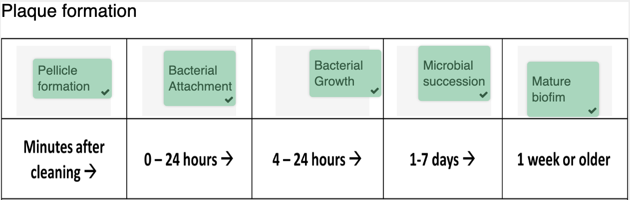
salivary proteins form a lubricating layer which forms the salivary pellicle
salivary pellicle forms on all surfaces
it also provides binding sites for bacteria
epithelial cells continuously shed which prevents bacteria build-up
teeth have non-shedding surfaces which allows for build-up
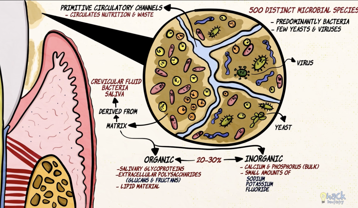
Biofilm on teeth
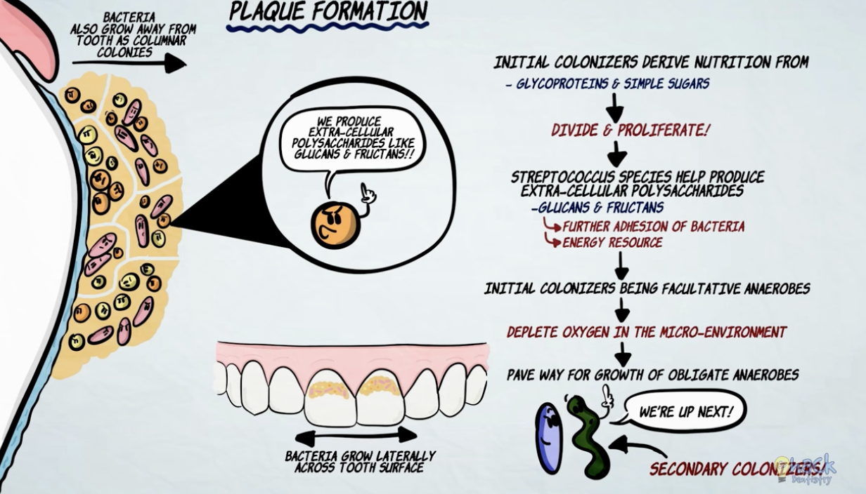
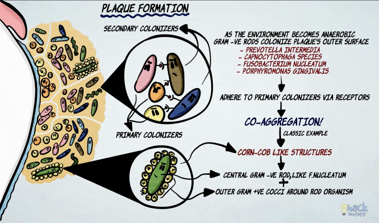
bacteria thrive on soft deposits/biofilm on teeth
soft deposit = dental plaque
yellowish white substance
made up of bacterial colonies + exfoliated epithelial cells + salivary proteins
accumulates in the gingival 1/3
rest of the tooth gets rubbed off during mastication
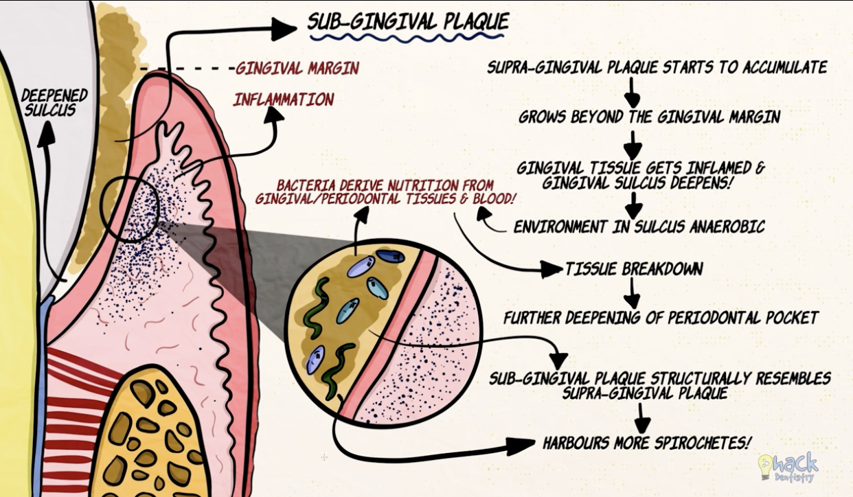
sub gingival can either be tooth associated or sulcular tissue associated
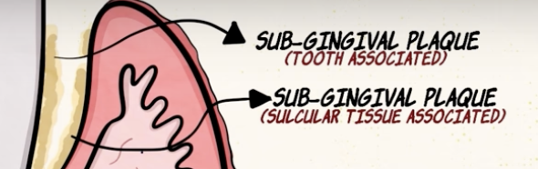
supra gingival plaque + tooth associated sub gingival plaque contributes to calculus
sulcular tissue associated sub gingival plaque causes periodontitis
Plaque Hypothesis
Specific Plaque Hypothesis:
specific types of bacteria are responsible for progression
we could eliminate certain species through vaccination however not everyone that has the bacteria progresses to cavitation
these statements are problematic because people have the micro-organisms and not the disease
Non-specific Plaque Hypothesis
you do not need a particular level of plaque for disease progression
progression requires overall bacteria and a specific threshold level
many individuals get new cavities, despite having good plaque control
Ecological Plaque Hypothesis
We all have these particular species of bacteria in our mouths, but if we keep everything ecologically under control (balanced), there will be no disease progression.
Streptococcus Mutans:
a.k.a. primary coloniser
start up the process + lead to cavitation
-Why is Streptococcus Mutans so successful?
S. mutans creates a water-insoluble glucan from sucrose that glues them to the tooth surface
makes plaque thicker
xylitol interferes with ability of microorganisms to stick to teeth
s. mutans produces lots of lactic acid (causes demineralization)
s. mutans makes intracellular polysaccharides ehich supports acid production without dietary substrates (s. mutans can continue to made acid without food)
s. mutans are aciduric (thrives in low pH)
Diet:
Vipeholm study. In 1954, researchers in Scandinavia, led by a guy called Gustafsson = the more frequently the patients were exposed to sugars, the higher the levels of caries.
something was sticky and could be retained on teeth for longer. In that case, it will increase the risk of dental caries,
Carbohydrates!
Fermentable carbohydrates allow bacteria to produce acids and those little glues dextrans and levans that enable bacteria to aggregate on tooth surfaces and stick on quite well
complex carbohydrates and starches are less cariogenic, as they are not completely digested in the mouth
Carbohydrates are dangerous because they have a low molecular weight, meaning once they are in the mouth, they can diffuse very rapidly into the dental plaque
Sugars!
Intrinsic sugars = inside the cell structure of particular unprocessed foods, including whole fruits and vegetables. These sugars are not considered to be cariogenic.
But if that sugar is released from the cells of fruits through processing, it is considered cariogenic.
Extrinsic sugars are found outside of the cells of the food and drink, and there are two different types.
Milk extrinsic sugars include lactose; these are found in dairy products such as milk and milk products (milk, cheese, yogurt).
Non-milk extrinsic sugars (NMES) are found in table sugar, confectionery, soft drinks, biscuits, honey and fruit juice; these are considered cariogenic. Many people consider honey and fruit juice healthy because they are natural. But unfortunately, there is nothing natural about fruit juice as its structure changes when it transforms from whole fruit into a reduced form.
Understanding Food Labels + Reducing sugar Intake
sugars are listed under carbohydrates
1 tsp = 4 grams
WHO recommends no more than 25g/6 tsp of free sugar in a day
Acid + Tooth Decay
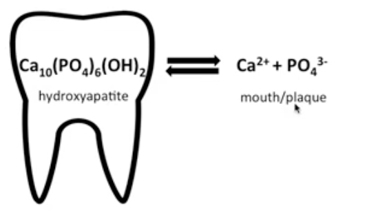
bacteria digests sugar and produce lactic acid via fermentation
the H+ ions from acid reacts w PO43- to form phosphoric acid
shifts reaction to right
pulls phosphate out of the tooth which weakens enamel
Demineralistion is the leeching of minerals from teeth
white spot is the first clinical sign of demineralisation
Stephan Curve = pH of tooth after acidic foods
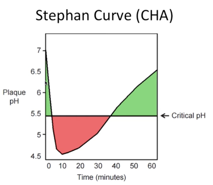
Patients with normal saliva function, the plaque remains acidic for 20 to 30 minutes, so when you talk to your patients about how many times they are exposed to sugars, you need to treat each exposure as a 20–30-minute period
Overall, repeated and frequent sugar consumption will keep plaque at low pH, thus favouring net loss of minerals from the tooth
Usually, teeth are continually exposed to saliva, which is super-saturated with calcium and phosphate ions, and these are the ions that will remineralise early lesions, especially in the presence of fluoride