Chapter 1: Whole Brain Imaging
__Chapter’s purpose__
- modern brain imaging technology has the ability to produce detailed images of the human brain without physically penetrating the skull.
- explain what whole-brain imaging technology is and provide insight into how experiments are designed and interpreted.
Two categories of whole-brain imaging technology: structural and functional
- ==Structural==: produce images of the structure/anatomy of the brain
- ==Functional==: display images of the physiological processes that underlie neural activity
{{Structural Brain Imaging Techniques{{
are used:
- to correlate neural activity in specific anatomical regions with behavioral or cognitive functions
- Activation of hippocampus when recalling first childhood memory
- To measure anatomical changes over time
- Brain mass decreases with aging or certain diseases
- In clinical neuroscience and neurology to diagnose diseases
- Tumors and vascular disorders
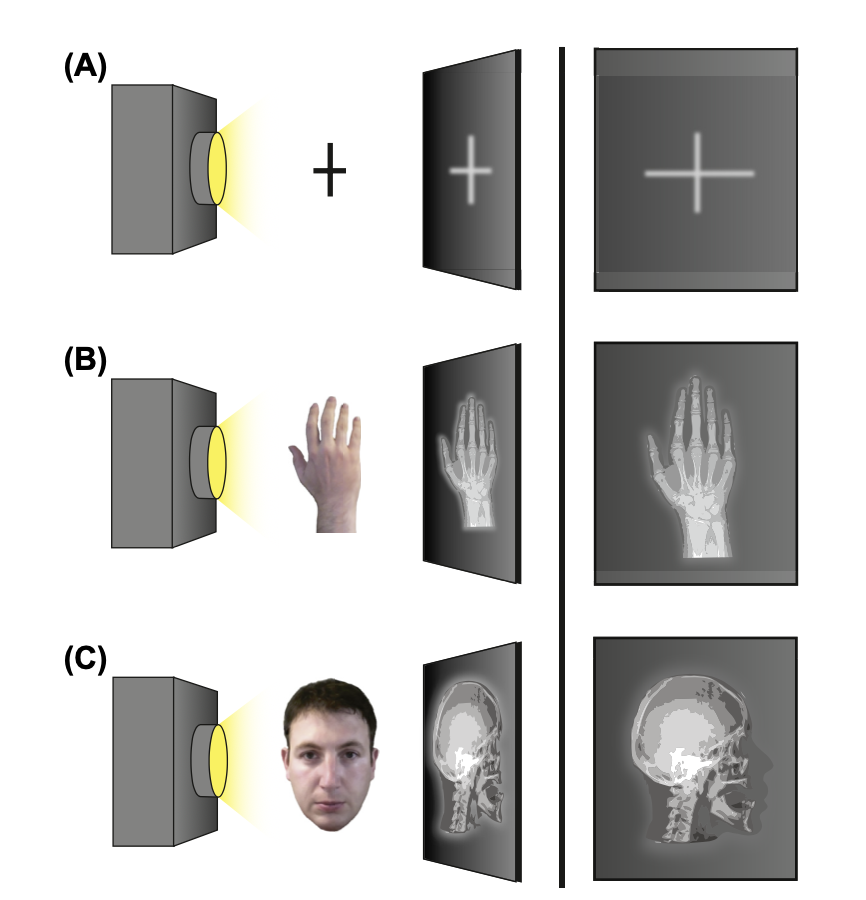
==X-rays (procedure)==
- (A) X-ray beams pass through an object onto a photographic plate. The portions of the x-ray that don't get absorbed create an image
- (B) X-ray images are created when there is enough contrast between skin and muscle (soft) and bone (hard).
- However, (C) X-ray images of the brain can't be made because x-rays can’t detect the contrast between various soft tissues within the nervous system
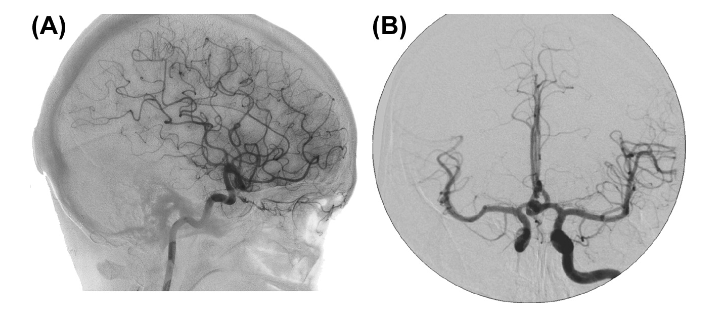
==Cerebral Angiography (aka an angiogram)==
An X-ray of the blood vessels.
It’s used to look for changes in the blood vessels, such as:
- Ballooning of a blood vessel (aneurysm)
- Narrowing of a blood vessel (stenosis)
- Blockages
General process:
- a catheter (a tube inserted into the bladder) is inserted into an artery
- usually in the groin
- the contrast dye is injected, followed by a series of X-rays
- These x-ray images show the arteries, veins, capillaries, and blood flow in the brain
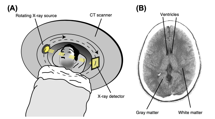
==Computerized Tomography (“CT Scan”)==
a series of X-ray images taken from different angles around a body and uses computer processing to create cross-sectional images (slices) of the bones, blood vessels and soft tissues inside the body
they’re used to:
- Diagnose muscle and bone disorders
- bone tumors and fractures
- Detect and monitor diseases and conditions
- cancer, heart disease, lung nodules and liver masses
- Detect internal injuries and bleeding
CT scanners are faster, cheaper to operate, and less prone to motion artifacts than MRI scanners
General process:
- CT scanners are shaped like a large doughnut standing on its side. patients lie on a narrow, motorized table that slides into the scanner.
- patient slides through the “doughnut,” causing detectors and an X-ray tube to rotate around the patient, taking several x-ray images during each rotation.
==MRI==
- An MRI uses a magnetic field and computer-generated radio waves to create a detailed image of one’s body's organs and tissues
- a noninvasive way to examine your organs, tissues and skeletal system
- they’re used to help diagnose:
- Aneurysms of cerebral vessels
- Multiple sclerosis
- Stroke
- Tumors
- Brain injury from trauma
- General process:
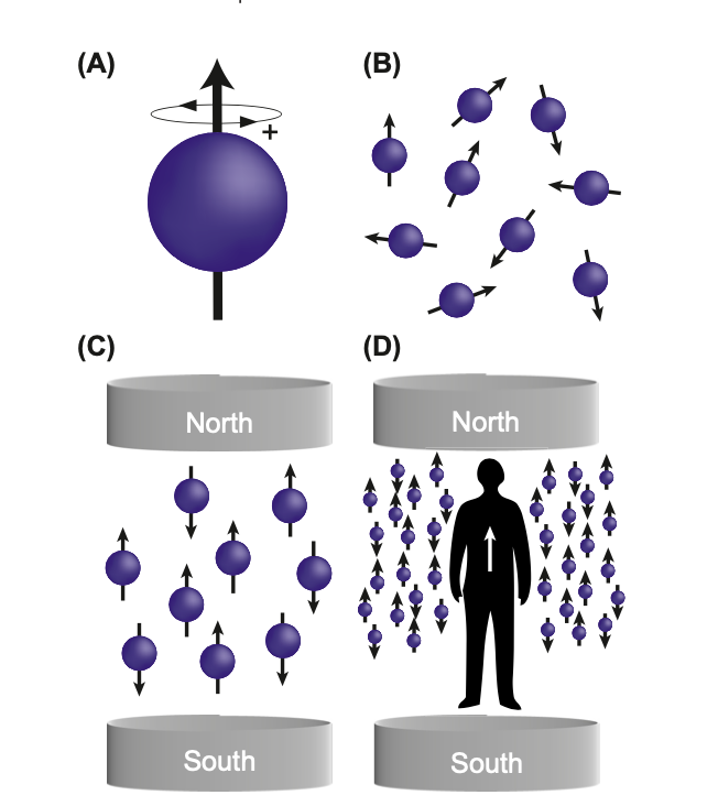
- powerful magnets are used to produce a strong magnetic field
- the magnetic field forces the protons in the body to align with that field
- a radiofrequency current is then pulsed through the patient, stimulating the protons. The protons spin out of equilibrium, straining against the pull of the magnetic field.
- When the radiofrequency field is turned off, the MRI sensors are able to detect the energy released from the protons realigning with the magnetic field.
- To obtain an image, the patient is placed inside a large magnet and must remain very still during the imaging process. The faster the protons realign, the brighter the image \n
- To obtain an image, the patient is placed inside a large magnet and must remain very still during the imaging process. The faster the protons realign, the brighter the image \n
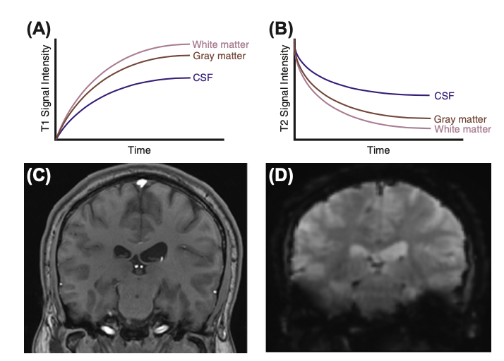
different substances exhibit different T1 and T2 values
- : white matter’s signal will be greater than CSF’s; white matter will be bright and CSF will be dark
- : CSF’s signal will be greater than the white matter’s; CSF will be bright and white matter will be dark
{{Functional Brain Imaging Techniques{{
are used:
- to measure neural activity in the central nervous system
- to correlate the activation of specific brain regions with a particular stimulus, emotional state, or behavioral task
- fear → amygdala
- for neuroscientists to study the neural basis of cognition, emotion, sensation, and behavior in humans
==FMRI==
- detects signals from excited hydrogen protons in a magnetic field
- addresses , the protein in the blood that carries oxygen to cells.
- researchers can examine changes in the oxygenation state of hemoglobin over time
- : the signal measured in fMRI's that relates to the concentration of deoxyhemoglobin in the blood
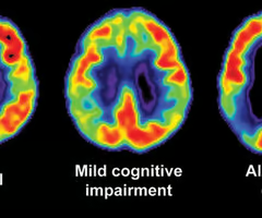
==Positron Emission Tomography (PET)==
shows the metabolic or biochemical function of tissues and organs.
uses radioactive drug () to show both normal and abnormal metabolic activity
- : a radioactive form of glucose commonly used as a tracer in PET scans
they’re used to:
- help identify a variety of conditions, including cancer, heart disease and brain disorders
General process:
- A small amount of radioactive glucose (a sugar) is injected into a patient’s vein. the scanner then rotates around the body and takes a picture of where the glucose is being used in the brain.
- Malignant tumor cells show up brighter in the picture because they are more active and take up more glucose than normal cells do
==SPECT==
uses a radioactive substance and a special camera to create 3D pictures
can show how your organs are functioning
- how well blood is flowing to a heart
they help determine the parts of the brain are being affected by:2
- Clogged blood vessels.
- Seizure disorders.
- Parkinson's disease
General process:
involve two steps:
- : the substance is injected intravenous infusion. The tracer dose is very small, being absorbed by the more active tissues in the body
- EX: part of brain causing a seizure retains more of the radioactive tracer
- : The SPECT machine is a large circular device containing a camera that detects the radioactive tracer. During the scan, a patient lies on a table while the SPECT machine rotates around them. The machine takes pictures of internal organs and other structures. These pictures are then sent to a computer that uses the information to create 3D images of your body.
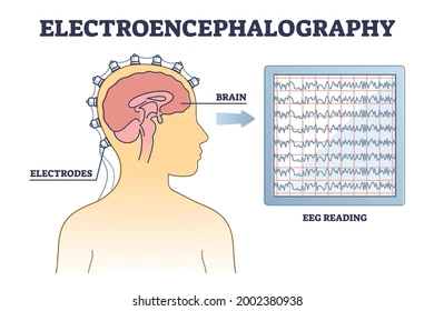
==Electroencephalogram (EEG)==
An electroencephalogram (EEG) is a recording of
- measured in
they’re used:
- to help identify the cause of certain symptoms or find out more about a diagnosed condition
- (primarily) to detect and investigate epilepsy, a condition that causes repeated seizures.
- to investigate other problems, such as dementia, brain tumors, concussions, and sleep disorders (sleep apnea)
General process:
- Sensors called electrodes are attached to the head (usually with glue or paste) and connected to an EEG recording machine. Patients are either seated up or laid down flat.
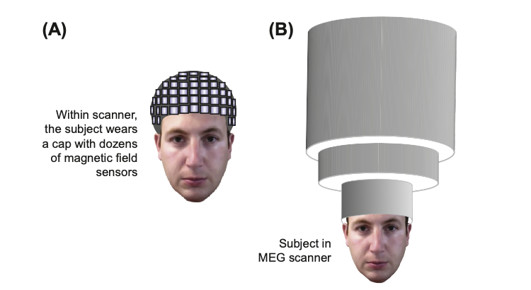
==Magnetoencephalography (MEG)==
measuring a magnetic field generated by the electrical activity of neurons.
usually combined with a magnetic resonance imaging
- hence the name .
MEG is used to identify or map:
- the functional areas of the brain, including centers of sensory, motor, language, and memory activities
- the precise location of the source of epileptic seizures
General process:
- completed in a special room that is shielded from outside magnetic and electric noise. A helmet-shaped container with tiny magnetic sensors lined on the inside is placed on the patient’s head.
- A computer workstation detects and records the signals from the helmet.
- patient either lies still or completes a series of tasks, such as listening to a series of words or looking at pictures while the activity is being recorded.
- After the data are obtained, an analysis of the recording is conducted to help determine where specific activities in the brain are located.
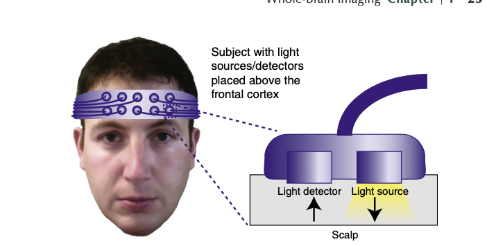
==Optical imaging==
produce images of neural activity by measuring changes in blood flow and metabolism from the surface of the brain
reduces patient exposure to harmful radiation by using non-ionizing radiation
- visible, ultraviolet, and infrared light.
- can be used for repeated procedures because it’s so safe
useful for measuring multiple properties of soft tissue
- soft tissues absorb and scatter light in many ways, so early markers of abnormal functioning of organs and tissues can be detected using this technique
General process:
- Light is shined onto the surface of the brain
- A portion of this light reflects off the brain and detected by multiple
- : an optical sensor that measures a specific substance with a chemical transducer
- Changes in neural activity produce changes in the amount of light that is absorbed and reflected by the brain
- optical imaging can indirectly detect these changes in neural activity
- These techniques can be either invasive (the skull is opened to reveal the surface of the brain) or noninvasive.
- These techniques can be either invasive (the skull is opened to reveal the surface of the brain) or noninvasive.
{{{{
- Researchers use fMRI to study attention, learning, memory, executive function, language, emotion, and higher-order sensory processing, such as art appreciation.
- humans or similar animal models are used
- ex: fMRI provides evidence that humans' visual pathways are anatomically comparable to those of other mammals.
- fMRIs are “unique” compared to other techniques used in neuroscience, so only a few dedicated researchers who it regularly
==Planning the Experiment==
- Cost is very important
- Equipment costs millions of dollars, not including the necessary maintenance, utilities of the lab, subjects, “stimuli” supplies, etc.
- fMRIs are very noisy (for the entire experiment)
- Difficult for researchers studying sleep, attention, or the auditory system
- they examine the effect of an independent variable on a dependent variable.
- independent variables: stimuli, tasks, and even differences between the subjects, such as age, gender, or disease state.
- Dependent variables:
- fMRI = signal intensity for a particular part of the brain.
- PET or SPECT = .
| Advantages | Disadvantages | |
|---|---|---|
| Blocked | - more statistical power for detecting subtle differences across different conditions- tend to be simpler to implement and analyze- good for examining cognitive state change | - predictability of stimuli may confound results- information about the time course of the activation response is lost within a block- not applicable to certain tasks (ex: novelty = antecedent of attention, emotion, memory, and behavior) |
| Event-Related | - reduces confounds of predictable stimulus order, because stimuli can be presented randomly - can sort trials after the experiment according to specific behavioral outcomes- useful for examining temporal characteristics of responses - flexible analysis strategies | - more complex design and analysis than blocked design- lower signal-to-noise ratio than blocked - must perform longer scan runs to compensate for the loss in statistical power |
| Mixed | can compared short term transient activity with long term sustained activity | - most complicated analysis |
- Pilot experiments: necessary tests that demonstrate the efficacy of stimulus delivery in producing an appropriate human response
- Because MRIs are so expensive
- a lab must be given permission to use humans as research subjects by an Institutional Review Board (IRB).
- : committee of about 10 individuals (physicians, research faculty, nurses, minister, graduate students, or lawyers) who review studies that use human subjects and determine if the study meets ethical, safety, and scientific obligations
- : regulates the protection, security, and confidentiality of private health information.
==Conducting the Experiment==
- Usually 10–20 subjects are necessary to obtain data and make an appropriate conclusion.
- Recruiting human subjects can be easy or difficult depending on the experiment
- is difficult when subjects must represent a specific population
- Ex: individuals with PTSD, elderly subjects, or individuals diagnosed with depression
- Researchers may need to recruit participants from other organizations
- secondary schools, nursing homes, hospitals, or universities
- Coordinating with clinical neurologists may be beneficial to identify people with diverse mental health issues.
- Prior to the experiment, the investigator must inform the subject about the nature of the experiment (just enough for the information not to affect the results).
- The subject must sign a release form indicating that they are willing to participate in the study and that any data gathered can be used for publication
- the subject must also complete a brief survey about his or her personal health to ensure that there are no confounding variables
- history of mental health disease, cardiovascular disorders, or heavy use of alcohol or other drugs.
- During the experiment, the investigator ensures that the subject is as comfortable as possible inside the scanner and that there are no (head) movements
- Each participant takes part in 1-3 sessions, which is the time allotted for the execution of the experiment.
- The brain is repeatedly scanned throughout each run in a session to test the hypothesis.
- : The smallest functional segment of brain tissue that can be examined for changes in signal strength over time.
- 4 dimensions: the voxel's location in space is defined in three dimensions (x, y, z), while its signal intensity is determined in the fourth dimension.
- an fMRI experiment can generate huge quantities of data because of this
==Manipulating Neural Activity during an Experiment==
- it is very difficult to influence the human brain while studying the nervous system
- Can’t just make someone nervous to exhibit the desired (neural) results
- Techniques applied on animals are not applicable to humans because we deem them unethical
- Lesions, stimulating microelectrodes or overexpressing genes
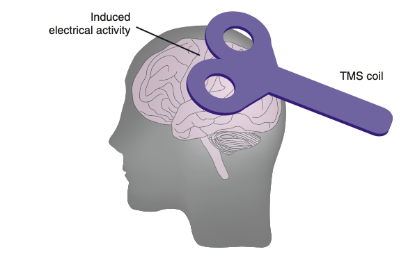
- two technologies can modulate neural activity in humans
- activates or inactivates human brain regions reversibly.
- where low-intensity ultrasound (US) is delivered to nervous system tissue