Visual Perception
Definition (#f7aeae)
Important (#edcae9)
Extra (#fffe9d)
Perception:

Perception: The set of processes by which we recognise, organise and make sense of the sensations we receive from environmental stimuli.
Desensation: There is a transduction at your receptors.
Perception to cognition:
Perception questions relate to identity and form patterns and movement.
Cognition occurs when this information is used to determine further goals.
Image → retina → brain (nerve impulses) → image: perception of a coherent image.
Ex: Apple
Is that red thing an apple? - Perception
Is that edible, can we buy it? - Cognition
Perceptual constancies:
Occurs when our perception of an object remains the same even when our proximal sensation of the distal object changes.
Perception remains stable despite changing inputs.
2 main consistencies:
Size
Shape
Size consistency:
Size constancy: The perception that an object maintains the same size despite changes in the size of the proximal stimulus.
The size of an image on the retina depends directly on the distance of that object from the eye.
The same object at 2 different distances projects different-size images on the retina.
The projection into the retina is different because of the distance it’s in.
Is this inborn:
It is inborn but the accuracy differs by age.
Different ages were told to judge the size of different coins, 50+ and 7-10 were able to guess the size accurately, the rest underestimaed the size.
Sensory adaptiation:
Receptor cells adapt to constant stimulation by not firing until there is a change in stimulation.
Through sensory adaptation, we may stop detecting the presence of a stimulus.
The Ganzfeld effect:
Phenomenon of perception caused by exposure to an unstructured uniform stimulation field.
Sensory organs are designed to detect changes in the environment rather than constance.
Stimulus remains unchanged → receptors slow down/stop signals → prevent nervous system from sensory overload.
Ex: At night, within minutes the brain blocks out color except grays, Due to lack of information we ‘see’ shadows in the dark.


Why perceptual illusions:
The existence of perceptual illusions suggests that what we sense (in our sensory organs) is not necessarily what we perceive (in our minds).
Our minds must take the available sensory information and manipulate that information to create mental representations of objects, properties, and spatial relationships within our environments.
Visual system:
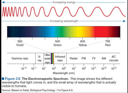
Humans can perceive only a small range of the wavelengths that are from 380-750 nanometres.
The precondition for vision is the existence of light.
Vision:
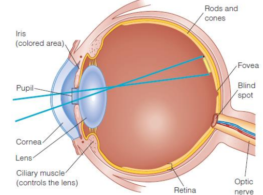
Parts:
Cornea: Clear protective outer layer, acts as the eyes primary fixed lens to bend incoming light.
Pupil: Black opening in centre of the eye.
Iris: Multi-colored muscle surrounding the pupil, constricts or dilates to control how much light enters the eye.
Lens: Clear structure behind the pupil, it dynamically flips and sharpens the incoming image.
Ciliary muscles: Muscular ring attached to the lens, changes the lens shape to focus on near/far objects.
Retina: The light sensitive inner lining at the back of the eye, acts as a projection screen.
Rods & cones: The photoreceptor cells imbedded in the retina. Rods detect light, darkness and motion, cones detect color and fine details.
Fovea: A small pit in the retina containing the highest concentration of cones, provides a sharper cental vision.
Optic nerve: The biological cable bundle at the back & carries all the electrical impulses directly to the brain’s visual cortex.
Blind spot: Specific point where the optic nerve exits, contains no rods or cones and creates a gap in vision.

Each eye contains roughly 120 millions rods and 8 millions cones.
Rods and cones differ not only in shape but also in their compositions, locations and responses to light
Shape:
Rods are elongated & cylindrical.
Cones are tapered and cone-shaped.
Location:
Rods sit in the periphery.
Cons are packed tightly in the central retina.
Light response/function:
Rods work more to night vision, highly sensitive to light, they function in dim settings but cannot process color, give us black & white perpiheral vision.
Cones are day vision and require bright light to activate, they handle fine details, sharp focus and vibrant color visions.
3 cone types: Based on wave length that they can absorb
Short wavelength (S cones): Sensitive to blue light.
Medium wavelength (M cones): Sensitive to green light.
Large wavelength (L cones): Sensitive to red lights
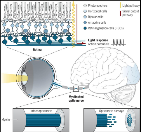
2 stream Hypothesis: Once light travels down the optic nerve, they split into 2 processing pathways.
Dorsal stream: The where and how and goes to the parietal lobe.
Primary visual: Goes to the cortex through the ventral stream and that goes to the temporal lobe.
Ventral stream:
Decides ‘what is this object I am seeing’, extends down into the temporal lobe and handles conscious perception, object identification and recognition.
Responsible for creating mental representations.
Dorsal stream:
Extends up into the parietal lobe, handles unconscious motor roles and spatial awareness, determines where objects are in space and guides physcial actions in real time.
Calculates the size, shape, distance of an object.
Sections of the visual cortices:

(Low order thinking question for labelling the diagram, or what does visual cortices do)
Visual cortices: Show how visual signals travels from the eyes to specialised mapping zones in the occipital lobe, it breaks down the distinction between primary entry zone (striate cortex) vs the advanced processing zone (extriate cortex).
Process:
Light enters LGN.
LGN: Lateral geniculate nucleus. The central relay station in the thalamus receiving raw signals from the optic nerve.
Visual radiation: The biological fibre pathway that carries data from the LGN to the back of the brain.
Striate cortex:
v1: Primary visual cortex, it catalogues all incoming inputs, collects primary inputs (landing zone).
Extrastriate cortex:
v2, vp: Relay networks that splits & distribute signals to higher order processing sensors (EC).
v3: Specialise zone, analyses the form and shapes of moving object.
v3a: Specialised zone mapping global coherent motion across the field of view.
v4: Specialised zone for processing color and complex geometric forms.
v5: Motion. Specialised zone for processing the directions and speed of visual motion.
Lateral occipital (LO): Crucial for object recogition and boundary detection.
v7, v8: Advanced areas processing complex spatial configuration and nuanced color configuration (hues).
Sagittal section: Shows the medial phase of the occipital lobe. (all above is in occipital lobe).
2 pathways of visual processing:

What:
Ventral stream.
Decodes the properties of an object.
Recognises shape, color, texture & identity.
Answers the question, ‘What am I looking at?'
Where:
Dorsal stream
Looking at spatial relationship and motion.
Answers the question. ‘Where is it and how do I grab it?'

Patient D.F. suffered bilateral carbon monoxide poisoning. Severe form of Visual Agnosia.
The arrows and color coded labels, show us areas of severe localized tissue destruction along widespread coritcal thinning.
Corticolization: The evolutionary migration and transfer of brain functions from primitive, subcortical areas (brainstem) to the cerebral cortex. Grants higher species the ability to perform complex cognitive processes like abstract thinking, and voluntary motor control
+40 shows not much corticolization.
Green arrowhead: Substantial localised tissue disruption in the ventral stream.
Yellow arrowheads: Smaller contration lesion in the upper left hemisphere.
Small red arrowheads: While Focal damage disrupted specific networks, oxygen deprivation caused global brain volume loss. Shows widened fissures where the brain tissue has shrunk away.
As the lateral occipital cortex was destroyed → cannot consciously recognise/describe shape, sizes or orientation.
But for her dorsal stream, it wasn’t impacted. Could gauge depth and spatial orientation.
Perception approaches:
How do we make sense of the world.
Bottom-up theories: Approaches in which perception starts with the stimuli whose appearance you take in through your eye. You look at a kitten, and perception happens where light information is transported to your brain.
Therefore, they are data-driven (i.e., stimulus-driven) theory.

Top-down theories: Perception is driven by high level cognitive processes, existing knowledge, and the prior expectations that influence perception.
Instead of raw stimulus data, uses cognitive context to intepret info.
Uses stored memory to fill in information.
Bottom- up theories:
4 main theories:
Direct perception:
How do you know the letter A when you see it?
Why does it not look like the letter H?
Gilson’s (1976): Theory of Direct Perception
How do we connect what we perceive to what we have stored in our minds?
We do not need higher cognitive processes or anything else to mediate between our sensory experiences and our perceptions.
Existing beliefs or higher-level inferential thought processes are not necessary for perception. (contextual knowledge is more helpful)
Direct perception may also play a role in interpersonal situations when we try to make sense of others’ emotions and intentions.
We can almost instantaneously recognize facial expression as a whole emotion.
Template theories:
Our minds have templates (highly detailed models of patterns we recognize).
We recognize a pattern by comparing it with our set of templates.
We then choose the exact template that perfectly matches what we observe.
Issues:
Fail to explain some aspects of the perception of letters.
We identify 2 different letters (A and H) from only one physical form.
Are we to believe that we have mental templates for each possible size, orientation, and form of a letter?
Feature-matching theories:
We attempt to match features of a pattern to features stored in memory, rather than to match a whole pattern to a template (Stankiewicz).
One such feature-matching model has been called Pandemonium.
In this model, metaphorical “demons” with specific duties receive and analyze the features of a stimulus (Selfridge).

Recognition by components theory:
Seeing with the Help of Geons (geometrical icons) Biederman (1987).
These shapes include objects such as bricks, cylinders, wedges, cones, and their curved axis counterparts.
We quickly recognize objects by observing the edges of them and then decomposing the objects into geons.

Top-down theories:
In constructive perception, the perceiver builds (constructs) a cognitive understanding (perception) of a stimulus.
The perceiver uses sensory information as the foundation for the structure to build the perception.
This viewpoint also is known as intelligent perception because it states that higher-order thinking plays an important role in perception.
Ex:
When you see a car approaching you on the street, its image on your retina gets bigger as the car comes closer.
And yet, you perceive the car to stay the same size. This suggests that high-level constructive processes are at work during perception.
Gestalt approach to form perception:
The Gestalt approach to form perception useful for understanding how we perceive groups of objects or even parts of objects to form integral wholes.
Gestalt does not see things as a separate being, he sees everything as a whole.
Principles include:
Figure-ground perception
Proximity
Similarity
Continuity
Symmetry
Figure-ground perception | When perceiving a visual field, some objects seem prominent and others aspects of the field recede into the background. |
Proximity | When we perceive an assortment of objects, we tend to see objects that are close to each other as forming a group. |
Similarity | We tend to group objects on the basis of their similarity. |
Continuity | We tend to perceive smoothly flowing or continuous forms rather than disrupted or discontinuous ones. |
Closure | We tend to perceptually close up or complete objects that are not complete. |
Symmetry | We tend to perceive objects as forming mirror images about their center. |


Shape Consistency:
Shape consistency: Perception that an object maintains the same shape despite changes in the shape of the proximal stimulus.
An object perceived shape remains the same despite changes in it’s orientation and hence in the shape of its retinal image.

Depth perception:
Depth: The distance from a surface, usually using your own body as a reference surface when speaking in terms of depth perception.
Generally depth cues are either monocular or binocular.
Monocular depth cues:
Cues for depth Perception | Appears closer | Appears farther away |
Texture gradients | Larger grains, farther apart | Smaller grains, closer together |
Relative size | Bigger | Smaller |
Interpostion | Partially obscures other object | Is partially obscured by other object |
Linear perspective | Apparently parallel lines seem to diverge as they move away from the horizon | Apparently parallel lines seem to converge as they approach the horizon |
Aerial perspective | Images seem crispier, more clearly delineated | Images seem fuzzier, less clearly delineated |
Location in the picture plane | Above the horizon, objects are higher in the picture plane; below the horizon, objects are lower in the picture plane | Above the horizon, objects are lower in the picture plane; below the horizon, objects are higher in the picture plane |
Motion parallax | Objects approaching get larger at an ever-increasing speed (i.e big & moving quickly closer) | Objects departing get smaller at an ever-decreasing speed (i.e small & moving slowly farther away) |
Binocular depth cues:
Cues for depth perception | Appears closer | Appears farther |
Binocular convergence | Eyes feel tug inward towards nose | Eyes relax outward toward ears |
Binocular disparity | Huge discrepancy between image seen by left eye and image seen by right eye | Miniscule discrepancy between image seen by left eye and image seen by right eye |
Perceptual deficits:
The what-how hypothesus is best supported by evidence of processing deficits.
Deficits can impair people’s ability to recognize what they see and can impair people’s ability to reach for what they see.
Visual agnosia:
People who suffer from an agnosia have trouble perceiving sensory information.
Agnosias often are caused by damage to the border of the temporal and occipital lobes or restricted oxygen flow to areas of the brain, sometimes as a result of traumatic brain injury.
They can perceive the colors and shapes of objects and persons, but they cannot recognize what the objects are.
Prosopagnosia:
Results in a severely impaired ability to recognize human faces.
A person with prosopagnosia might not recognize her or his own face in the mirror. Associated with damage to the right temporal lobe of the brain.
Ataxia:
Difficult knowing the how.
Optic ataxia: Impaired ability to use the visual system to guide movement.
People with this deficit have trouble reaching for things.
Processing failure in the posterior parietal cortex, where sensorimotor information is processed.
Immediate movements are executed through dorsal stream processing, whereas delayed movements make use of the ventral system, including the occipitotemporal and temporoparietal areas.
Color blindness:
Lesions to the ventromedial occipital and temporal lobes.
Least common is rod monochromacy, also called achromacy - no color vision at all.
In dichromacy (most common), only two of the mechanisms for color perception work, and one is malfunctioning.
The most common is red-green color blindness, difficulty in distinguishing red from green.
The other types of color blindness are:
Deuteranopia (trouble seeing greens with symptoms similar to protanopia).
Tritanopia (confusion of blues and greens, and yellows that disappear or appear as light shades of reds).