3.2 cerebral cortex
Describe the Layered Structure of the Cerebral Cortex
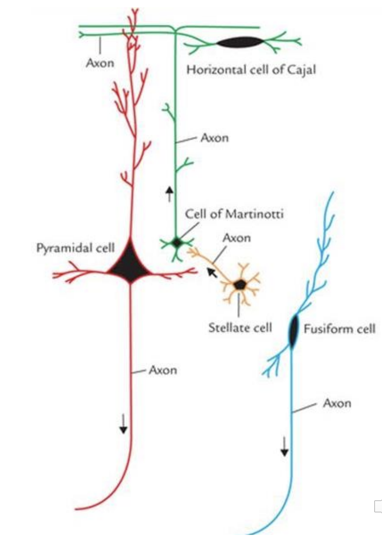
Pyramidal Cells: main output neuron; make up majority of cells in cortex
Granular (Stellate) Cells: small star shapes, short, locally projecting processes
Fusiform Cells: found in deepest layer, dendrites project to cortical surface, axons usually project to the thalamus
Horizontal Cells: rare, synapse locally
Layer I - Molecular Layer: Neither afferent nor efferent. Acellular with horizontal cells; contains many dendrites and axons.
Layer II - External Granular Layer: Afferent. Dense layer of small pyramidal and granular (stellate) cells.
Layer III - External Pyramidal Layer: Afferent. Medium pyramidal cells and loosely packed granular (stellate) cells.
Layer IV - Internal Granular Layer: Primary afferent. Densely packed granular (stellate) cells. Receives input from thalamus. Thick in sensory cortex but small/missing in motor cortex.
Layer V - Internal Pyramidal Layer: Primary efferent. Large pyramidal cells (Betz); primary motor output layer projecting to brainstem and spinal cord.
Layer VI - Multiform Layer: Efferent. Contains diverse cell types. Integrates inputs from the brainstem and sends projections back to thalamus.

Compare and Contrast the Composition and Structure of Grey Matter and White Matter of the Cerebral Cortex
Grey Matter:
Composed primarily of unmyelinated neuronal cell bodies (soma), dendrites, and synapses.
Forms the outer layer of the brain, processing information through cortical columns.
Measures approximately in thickness.
White Matter:
Composed of myelinated axons.
Located deep to the cortical ribbon; serves as long-range communication pathways between brain regions.
Axons are bundled into tracts classified as intrahemispheric (within) or interhemispheric (between). Associative (connect gyrus to gyrus), projection (connect crebral cortex with subcortical nuclei), or commissural fibres (connect hemispheres).
List and Locate the Four Lobes of the Brain
Frontal Lobe: Anterior to the central sulcus; responsible for motor control, executive function, and speech production.
Parietal Lobe: Posterior to the central sulcus; processes somatosensory information and visuospatial awareness.
Temporal Lobe: Inferior to the lateral (Sylvian) fissure; involved in auditory perception, language comprehension, and memory.
Occipital Lobe: At the posterior pole of the brain; dedicated to primary visual processing.
Identify and Describe the Functions of the Major Gyri Within Each Lobe
Frontal Lobe Gyri:
Precentral Gyrus (BA: 4): Primary motor cortex controlling voluntary muscle movement.
Pre-motor cortex (BA: 6): Programming and organisation of the postural adjustments to perform skilled movement. Medial surface is supplementary motor area, involved in planning and coordinating complex motor actions.
Inferior Frontal Gyrus (BA: 44): Houses Broca's area, critical for speech production.
Parietal Lobe Gyri:
Postcentral Gyrus (BA: 1, 2, 3): Primary somatosensory cortex for processing tactile information.
Superior Parietal Lobule (BA: 5): Stereognosis (ability to identify object through touch), recognition of objects/body parts and orderly performance of motor tasks
Inferior Parietal Lobule (BA: 7): Language and cognition
Temporal Lobe Gyri:
Superior Temporal Gyrus: Primary auditory cortex and emotional perception
Middle Temporal Gyrus: Language comprehension and semantic memory
Inferior Temporal Gyrus: Object recognition.
Occipital Lobe Gyri:
Cuneus and Lingual Gyrus (BA: 17, 18 and 19): Involved in visual processing and integration of visual information.