Organisation
Animal tissues, organs, and organ systems
The digestive system breaks down complex food substances and provides a large surface area for maximum absorption of food.
Region | Function |
Mouth | Begins digestion of carbohydrates |
Stomach | Begins digestion of proteins, small molecules are absorbed |
Small intestine | Completes digestion of carbs and proteins, begins digestion of lipids |
Large intestine | Absorption of water |
The small intestine is folded and has projections called villi, these villi have their own projections called microvilli. Villi increase the surface area of the small intestine
Enzymes are biological catalysts so they speed up reactions. They are proteins that are folded into a 3D structure, each enzyme has a region called an active site
The substrate fits into the active site
How do enzymes work
Substate binds to the active site - the active site is complementary to the substrate
Enzyme catalyses the breakdown of the substrate - the fit between the enzyme and substrate is called lock and key
Products are released from active site - the enzyme molecule is unchanged
Production and function of bile
The heart and blood vessels
Blood
Coronary heart disease
Lifestyle and non-communicable diseases
Cancer
Plant tissues organs and systems
Factors affecting enzymes
Temperature - At low temperatures the particles have less kinetic energy and so there are fewer successful collisions
If the temperature gets too high then the enzyme will denature meaning the active site has changed shape
pH - Changing th
e pH can also change the active site of the enzyme
Enzymes involved in digestion
Enzyme | Location | Function |
Carbohydrase | Mouth/intestine | Breaks down carbs |
Amylase | Mouth | Breaks down carbs |
Protease | Stomach/ intestine | Breaks down proteins |
Lipase | Intestine | Breaks down lipids |
The heart and blood vessels
The heart is an organ that pumps blood around the body in a double circulatory system.
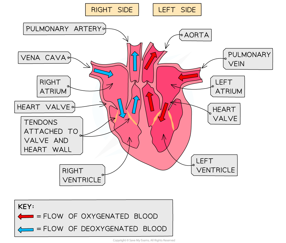
The right ventricle pumps blood to the lungs where gas exchange takes place. The left ventricle pumps blood around the rest of the body.
Blood enters the heart from the body through the vena cava where it flows into the right atrium and then the right ventricle, it is pushed towards the lungs through the pulmonary artery. When blood comes back to the heart it enters through the pulmonary vein into the left atrium and then the left ventricle and it leaves to go to the rest of the body aorta.
The natural resting heart rate is controlled by a group of cells located in the right atrium that act as a pacemaker. Artificial pacemakers can be used to correct irregularities in the heart rate.
Vein | Artery | Capillaries |
Blood to the heart | Blood away from the heart | Connect arteries and veins |
Carries deoxygenated blood | Carries oxygenated blood | One cell thick |
Low pressure | Thick muscular and elastic walls | |
Thin walls | Lumen carries blood | |
Wide lumen |