June 2 Cartilage Tissue: Types, Structure, and Growth
Overview of Cartilage Tissue
Definition: Cartilage is a specialized type of connective tissue characterized by being tough and durable.
Extracellular Matrix (ECM): Features high concentrations of glycosaminoglycans (GAGs) and proteoglycans.
Proteinoglycan is sulfated, so sulfated GAGs attach to a protein core. This composition provides cartilage with its unique properties, such as resilience and flexibility, making it essential for various functions within the skeletal system.
NO Vascularity: Entirely avascular; it possesses no blood supply.
Nutrients reach the tissue via diffusion from the perichondrium (a layer of connective tissue).
Cartilage is avascular and receives nutrients by diffusion. Most cartilage obtains nutrients from blood vessels in the perichondrium. Exceptions are articular cartilage and epiphyseal cartilage, which lack a perichondrium and receive nutrients by diffusion from neighboring tissues (articular cartilage specifically from synovial fluid)
NO Innervation: There is no neural supply or innervation within cartilage tissue.
Perichondrium → mainly Type I collagen (because it is connective tissue)
Hyaline cartilage → mainly Type II collagen
Elastic cartilage → Type II collagen plus elastic fibers
Elastic fibers :elastin + fibrillin, not Type II. ✅
Fibrocartilage → mostly Type I collagen (with some Type II)
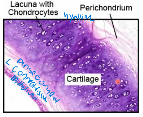
Perichondrium
Structure: A dense connective tissue layer surrounding most cartilage that contains blood vessels and the most important cells in there are the fibroblasts, which produce the EXM in other words, proteins and ground substances.
THE PER. IS NOT CARTILAGE TISSUE BUT DENSE IRR. TISSUE IN THE SURFACE OF THE CARILAGIN TISSUE.
Function: Acts as an interface between cartilage and surrounding tissues, providing vascular supply and a small neural component.
Specific Layers (in active growth):
Outer Fibrous Layer: Composed of Type I collagen and fibroblasts.
Inner Cellular Layer: Contains mesenchymal stem cells, which differentiate into chondroblasts (and subsequently chondrocytes).
Exceptions (Locations lacking perichondrium):
Articular cartilage (hyaline).
Epiphyseal cartilage (hyaline).
Fibrocartilage.
Surface / Surrounding Tissue
↓
Perichondrium
(Dense Irregular Connective Tissue)
(Type I collagen)
↓
Outer Fibrous Layer
- Fibroblasts
- Type I collagen fibers
↓
Inner Chondrogenic Layer
- Mesenchymal cells
↓
Chondroblasts
- Secrete cartilage ECM
- Produce Type II collagen
↓
Cartilage Matrix (ECM)
- Type II collagen
- Proteoglycans
- Water
Chondroblasts(secrete cartilage matrix)
↓
Hyaline cartilage matrix forms
↓
Chondroblasts become trapped in matrix
↓
Chondrocytes in Lacunae
- Maintain cartilage ECM
Structural-Functional Aspects
Mechanical Stress Resistance: Cartilage can bear mechanical stresses without permanent distortion due to the specific consistency of the ECM.
Framework Support: It forms a structural framework for soft tissues, including the respiratory tract, ears, and nose.
Joint Function:
Provides cushioning and sliding regions within joints.
Maintains high resiliency and creates smooth, lubricated surfaces.
Facilitates bone movements within the joint complex.
Developmental Role: It guides the development of long bones before and after birth by acting as a cartilaginous model in fetuses and infants.
Shock Absorption: Acts as a shock absorber due to its high content of bound water.
General Composition and Molecular Structure
Extracellular Matrix (ECM):
Constitutes: more than of the tissue volume.
The rest are cells like..:
Chondrocytes: Mature cartilage cells responsible for maintaining the cartilage matrix by producing and maintaining the ECM components, but are embedded within the matrix itself. They live in spaces called lacunae within the cartilage matrix.
Location: Are towards the interior, forming the cartilaginous tissues and providing structural support and resilience to the tissue.
This is where the perichondrium is able to provide additional support and nutrients to the chondrocytes, ensuring their health and the overall integrity of the cartilage tissue.
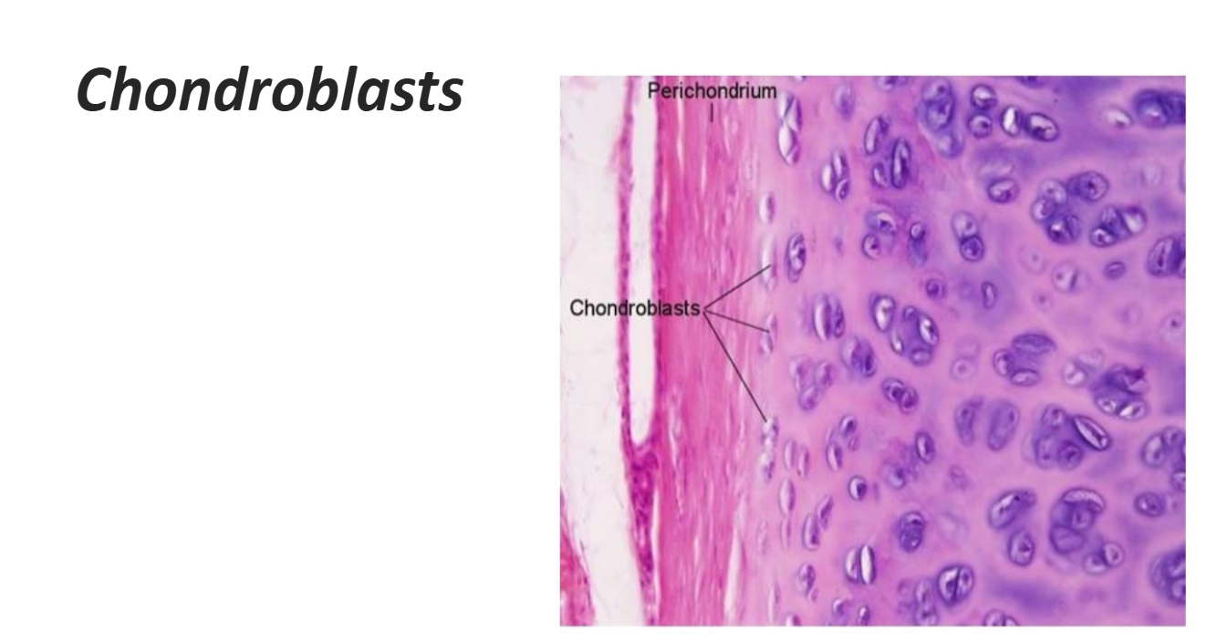
Chondroblasts: Once a chondroblast becomes enclosed by the matrix it secreted and becomes a chondrocyte, it resides in a lacuna and continues to synthesize and maintain components of the extracellular matrix (ECM).
Chondroblasts are immature cartilage-forming cells.
They are typically located near the periphery of growing cartilage, just beneath the perichondrium.Located: towards the surface, not THE/IN but TOWARDS the surfaces
They secrete cartilage matrix.
Once a chondroblast becomes surrounded by the matrix it produced, it differentiates into a chondrocyte and occupies a lacuna.
Components: include Type II collagen, GAGs (Hyaluronan and sulfated GAGs), and Proteoglycans.
Molecular arrangement: Type II collagen fibrils interact with proteoglycans. These proteoglycans consist of a core protein with side chains of chondroitin sulfate. Link proteins attach these to hyaluronan.
Binding: Type II collagen and proteoglycan components are unified by electrostatic bonds.
Molecular Comp. of ECM
Collagen: Represents of the organic matter (mostly barely discernible Type II collagen).
Hydration: to of the ECM in fresh hyaline cartilage is water bound to proteoglycans.
Chondronectin: A multiadhesive glycoprotein that mediates the adherence of chondrocytes to ECM components.
Acts like a "glue" that connects cartilage cells to the surrounding matrix. Chondronectin = cartilage cell attachment protein
Aggrecan: A specific proteoglycan with side chains of chondroitin sulfate and keratan sulfate.
Bound covantley to long polymer of hyaluronan
proteoglycan complexes bound to type ii collagen 60%-80% of ECM in frsh hyaline cartilage: water bound to proteoglycans
Keratan Sulfate and Chondroitin Sulfate
Types of glycosaminoglycans (GAGs) that are part of cartilage proteoglycans.Found in the cartilage extracellular matrix.
Functions:
Attract and retain water.
Form a gel-like matrix around collagen fibers.
Help cartilage resist compression.
Provide resilience and elasticity.
Contribute to the cartilage's structural integrity.
Keratan sulfate + Chondroitin sulfate = attract water → cushion cartilage → resist compression.
Keratin sulfates and chondroitin sulfates serve as crucial proteoglycans found in cartilage, contributing to its ability to withstand compressive forces and maintain structural integrity. These proteoglycans attract water, creating a gel-like matrix that supports the tissue's resilience and elasticity.
Cellular Components:
Chondroblasts: Located peripherally, they synthesize components of the ECM.
Chondrocytes: Derived from chondroblasts, they reside within lacunae. Their role is to synthesize and maintain ECM components. They often undergo anaerobic glycolysis.
Lacunae are small spaces or cavities within the cartilage matrix.
They are occupied by chondrocytes (mature cartilage cells).
Proteoglycan Aggregate in cartilage
Hyaluronan (Hyaluronic Acid)
A long, non-sulfated glycosaminoglycan (GAG).
Forms the central backbone of the aggregate.
Proteoglycan Monomer
Consists of:
A core protein
Sulfated GAGs attached to it, mainly:
Chondroitin sulfate
Keratan sulfate
Link Protein
Binds the proteoglycan monomer to the hyaluronan backbone.
Stabilizes the entire proteoglycan aggregate.
Hyaluronan (backbone)
|
Link Protein
|
Core Protein
/ \
Chondroitin Sulfate
Keratan Sulfate
Ionic attraction: between charged groups on proteoglycans and Type II collagen further stabilizes the structure of cartilage, enhancing its resilience under mechanical stress. This elaborate molecular architecture allows cartilage to withstand compressive forces, contributing to its function as a cushion in joints.
Proteoglycans (GAGs) = NEGATIVE
They have lots of sulfate (–SO₃⁻) and carboxyl (–COO⁻) groups
So they are strongly negatively charged
This is why cartilage attracts water 💧 (important for cushioning)
Type II collagen = RELATIVELY POSITIVE / less negative
Collagen is a protein, with some positively charged amino acids (lysine, arginine) exposed
So it can participate in ionic interactions
BUT it’s not strongly “positive” like a salt ion — just relatively positive compared to GAGs
So the interaction is:
Proteoglycans (–) ⟷ Type II collagen (more + regions)
→ electrostatic attraction stabilizes the matrix
Specialized Areas of the Extracellular Matrix
Capsular matrix
✓Densely staining matrix
✓Surrounds the cell
✓Highest concentration of proteoglycans
✓Type VI collagen surrounds each condrocyte and therefore in the lacuna
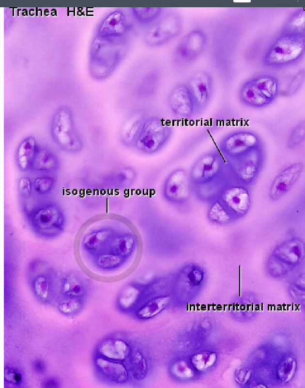
Territorial matrix
✓Intense basophilia
✓More GAGs than collagen (type II)
✓Surrounds isogenous groups
✓More basicophilic than inter.
territorial matrix is actually the dark matrix in between the isogenous groups, which contains higher concentrations of glycosaminoglycans (GAGs) and less collagen fibrils, providing the cartilage with resilience and support.
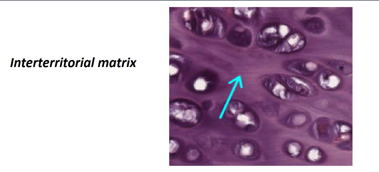
Interterritorial matrix
✓Weaker basophilia
✓Less GAGs
✓Areas between isogenous groups of chondrocytes
More lighter
Chondrocyte Morphology and Metabolism
Chondroblasts: Young chondrocytes located peripherally with an elliptic shape; their long axes are parallel to the surface.
Chondrocytes reside deeper in the cartilage. A single chondroblast can undergo mitosis to form an isogenous group of up to cells. Synthesis of ECM separates these cells into individual lacunae.
Chondrocytes are nuclues is not in the center
Active Chondrocyte (Light Microscopy): Shows cytoplasmic basophilia (due to RER protein synthesis), clear areas (Golgi apparatus), and an euchromatic nucleus.
Active Chondrocyte (TEM): Features an abundant RER, large Golgi apparatus, secretory granules/vesicles, an eccentric euchromatic nucleus, and a cytoskeleton comprising intermediate filaments, microtubules, and actin filaments.
Regulatory Factors: Somatomedins (insulin-like growth factors) produced by the liver stimulate chondrocyte activity.
Chondrogenesis and Cartilage Growth
Classification of Cartilage Types
Hyaline Cartilage
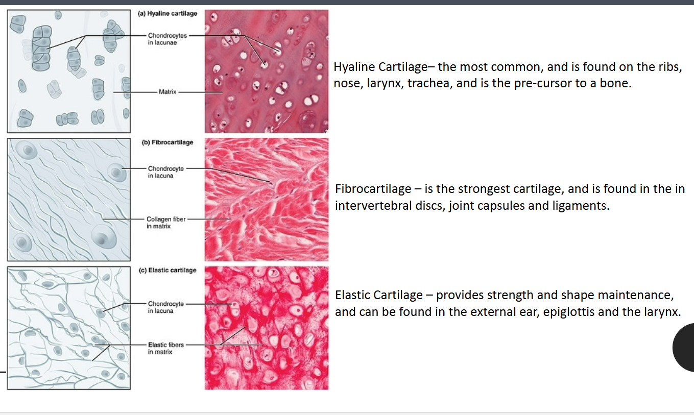
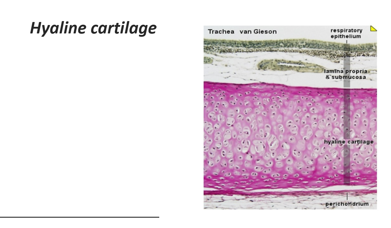
Characteristics: "Hyalos" means glass; it is the most common type, appearing homogeneous and semitransparent.
Major cells: Chondrocytes and Chondroblasts
Structure: Homogeneous matrix with Type II collagen and Aggrecan. Cells (chondrocytes/chondroblasts) are isolated or in small isogenous groups.
Presence of Perichondrium: Yes, except at the epiphyses and articular cartilage.
Main Locations: Respiratory tract (nose, larynx, trachea, bronchi), articular ends of long bones, ventral ends of ribs, epiphyseal plates, and the temporary fetal skeleton.
Main Functions: Provides smooth, low-friction surfaces in joints; provides structural support for the respiratory tract.
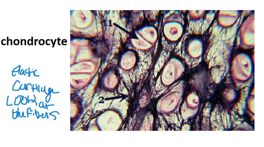
Organized: Isolated or in small isogenous groups
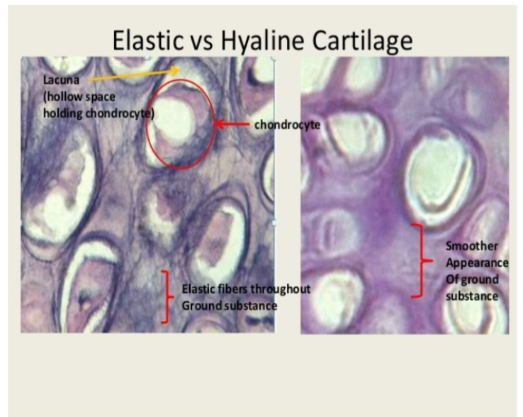
Elastic Cartilage
Characteristics: Similar to hyaline but contains an abundant
Major cells: Chondrocytes and Chondroblasts
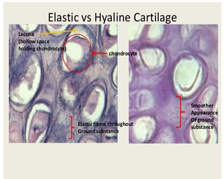
Structure: Type II collagen, aggrecan, and darker elastic fibers. Usually found in small isogenous groups.
Presence of Perichondrium: Always present.
Main Locations: External ear (auricle), external acoustic meatus, auditory (Eustachian) tube, epiglottis, and specific laryngeal cartilages.
Main Functions: Provides flexible shape and support to soft tissues.
Organized: unevenly distributed network of elastic fibers. It is yellow in its fresh state and highly flexible. Usually, in small isogenous groups
Fibrocartilage
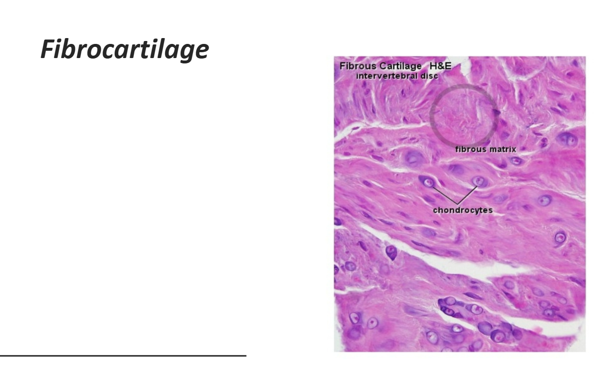
Characteristics: A combination of hyaline cartilage and dense regular connective tissue.
Structure: Contains Type II collagen and large areas of dense connective tissue with Type I collagen. Type I collagen is the most abundant and then type II.
Major cells: Chondrocytes and fibroblasts
Arrangement of the CHC: Chondrocytes and fibroblasts are isolated or in isogenous groups arranged axially (in rows).
Presence of Perichondrium: No.
Main Locations: Intervertebral discs, pubic symphysis, meniscus, certain other joints, and insertions of tendons.
Main Functions: Provides cushioning, tensile strength, and resistance to tearing and compression.
Organized: is organized in thick, parallel bundles of Type I collagen fibers, with chondrocytes arranged in rows between the collagen bundles.
Chondrocytes
✓Single
✓Aligned isogenous aggregates
Synthesize type I collagen and other usual components of ECM
• Regions with fibroblasts and type I collagen
✓Separate areas with chondrocytes and hyaline matrix
✓Provide extra tensile strength
• Matrix is acidophilic
✓Less proteoglycans
✓Type I collagen because of this abundance
Repair, Growth, Calcification
Cartilage Formation (Chondrogenesis)
Embryonic mesenchymal cells retract processes, round up, and multiply rapidly.
New chondroblasts become densely packed.
Synthesis and swelling of the ECM separate the cells into chondrocytes.
Mitosis of single chondrocytes leads to the formation of isogenous groups.
Growth Mechanisms
Appositional Growth:
Differentiation of chondroblasts from progenitor perichondrial cells.
Synthesis of matrix contributes to growth.
Primarily important during postnatal development.
Does not occur in articular cartilage.
Appositional growth requires a perichondrium. Therefore, it does NOT occur in articular cartilage because articular cartilage lacks a perichondrium.
Interstitial Growth:
Mitosis of pre-existing chondrocytes.
Synthesis of matrix contributes to growth.
Crucial for increasing the length of long bones.
Occurs in articular cartilage.
Isogenous Groups in Cartilage
Isogenous groups are clusters of 2 or more chondrocytes found within cartilage.
They are formed when a single chondrocyte divides by mitosis.
Since all cells originate from one parent chondrocyte, they remain grouped together.
Isogenous groups are evidence of interstitial growth (growth from within the cartilage).
Cartilage can grow in two ways:
Appositional growth → growth from the surface (via chondroblasts in the perichondrium).
Interstitial growth → growth from within the cartilage (via chondrocyte division forming isogenous groups).
Cartilage Repair
Repair is a slow and incomplete process (except in young children).
Limited new cartilage is formed from perichondrial cells.
Often results in a scar of dense connective tissue instead of new cartilage.
Difficult repair is due to avascularity and low metabolic rates.
Why Cartilage Heals Slowly
Avascular → no blood vessels, so nutrients and repair cells reach cartilage slowly.
Relies on diffusion for oxygen and nutrients.
Low metabolic activity of chondrocytes.
Anaerobic glycolysis is the main source of energy (less efficient than aerobic metabolism).
Limited ability of chondrocytes to divide and replace damaged tissue.
Thick extracellular matrix makes repair and cell migration difficult.
Cartilage Calcification
Hyaline cartilage is specifically prone to calcification under three conditions:
The portion of articular cartilage in contact with bone tissue.
During endochondral ossification (cartilage replaced by bone during growth).
As part of the natural aging process (calcification of adult hyaline cartilage).
Only at a certain point of growth is there no more growth and the cartilage is ultimately transformed into bone; there is no more length.
Consequences of Calcification:
Calcified matrix impedes the diffusion of nutrients.
Chondrocytes swell and eventually die.
The calcified matrix is eventually removed and replaced by bone tissue, often leading to marrow cavity formation.