9b: Cortical pathways
CORTICAL PATHWAYS
Definition: Cortical pathways refer to the neural pathways in the brain responsible for processing visual information.
Key Components:
V1 is the major cortical projection site for LGN output.
Beyond V1, many specialised visual cortical areas exist, approximately 30 linked to various aspects of visual processing.
Post-V1 Processing:
Two Main Pathways:
Ventral Pathway
Dorsal Pathway
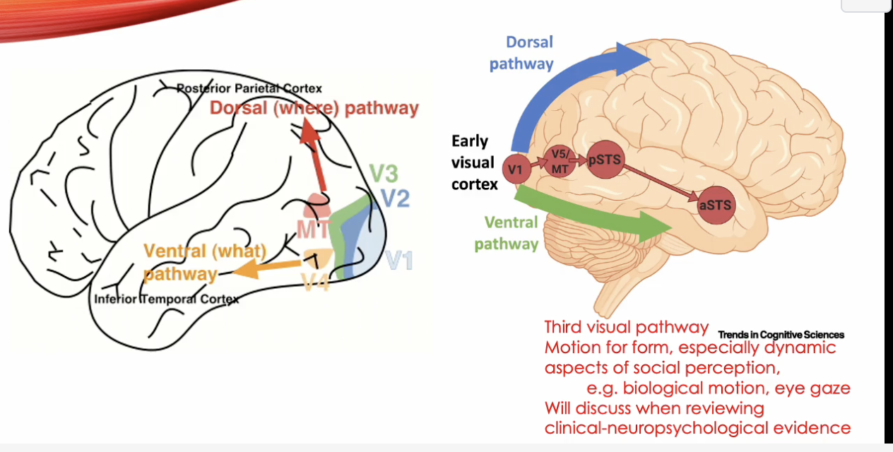
Evidence suggests a potential third cortical pathway related to visual processing.
VENTRAL AND DORSAL STREAMS
Ventral Stream:
Projects to the Inferotemporal Cortex.
Includes cortical area V4.
Dominated by parvocellular (P cell) inputs, responsible for color and fine detail.
Dorsal Stream:
Projects to the Posterior-Parietal Cortex.
Includes V5 (MT) area involved in motion perception.
Dominated by magnocellular (M cell) inputs, responsible for motion and spatial awareness.
V4
Originally Thought To Be: A color processing area due to the presence of numerous color-sensitive cells.
Cell Arrangement: Cells organized into clusters that are selective for color and orientation.
Performance Variation: Color and orientation tuning become more precise when animals engage in difficult discrimination tasks.
Subsystems: Possible existence of color and form subsystems within V4.
involved in processing colour and form
colour-sensitive cells
organised into clusters
some clusters respond to specific colours
others respond to specific orientations
v4 is adaptive, especially when doing a difficult visual task (telling apart simiilar colours or shapes)
INFEROTEMPORAL CORTEX
Contains multiple subdivisions with specialised functions.
Cell Sensitivities:
Respond to shape, color, or texture within their large receptive fields.
FFA (Fusiform Face Area): Selective for face recognition, heavily functioning under parvocellular input.
V5 (MT)
Functionality:
Most cells in this region are selective to motion.
Plays a significant role in the processing of motion information.
POSTERIOR-PARIETAL CORTEX
Function:
Involved in locating objects in space.
Directs attention to specific areas of the visual field.
Input to this area is mainly magnocellular dominated.
VENTRAL AND DORSAL PATHWAYS: DISTINCTION
What vs. Where Distinction:
(Ungerleider & Mishkin)
What: Object vision, linked to the ventral pathway (temporal cortex), P cell dominated.
Where: Spatial vision, motion processing, linked to the dorsal pathway (parietal cortex), M cell dominated.
Dorsal pathway also contributes to motion processing.
EVIDENCE FOR DISTINCTION
Research Types:
Studies examining functional distinctions in ventral and dorsal streams.
Focus on investigations in primates, including humans and non-human species.
Humans:
Utilization of PET & fMRI studies.
Comparison of spatial vs. face-matching tasks revealing activity in occipital lobe:
Spatial tasks activate the posterior parietal region (dorsal, where and how).
Face-matching engages temporal lobe (ventral, what ).
Additional insights through clinical studies of discrete lesions indicating functional losses.
Non-Human Primates:
Single Cell Recording Studies reveal:
Ventral pathway sensitivity to color and shape (FFA for faces).
Dorsal pathway sensitivity to motion (not to patterns).
Lesion Studies:
Dorsal lesions in (V5) affect motion perception without impacting other qualities.
Ventral lesions (V4) impair color and pattern perception while sparing motion.
BEHAVIORAL TASKS
Object Discrimination Task: Animals learn to identify one object from a pair then select the new object presented (ventral impairments will affect).
Landmark Task: Animals identify the food well closer to a landmark object (e.g., a tall cylinder) (dorsal impairments will affect).
Which pathway lesion (dorsal or ventral) would affect performance on which task?
MULTIPLE PROCESSING PATHWAYS AND STAGES
Key Aspects of Brain Processing:
Presence of parallel pathways: Multiple processing streams exist to extract various information types.
Multiple processing stages ensure information transformation at each pathway stage.
Clear implications for understanding functional localization within the brain.
TRANSFORMATION AT THE CORTICAL LEVEL
Transformation of information progresses through the visual processing hierarchy:
From light captured by cones to retinal ganglion cells, then to LGN and finally V1.
Transformations Include:
Luminance -> Dots of light (variation of light) -> Bars/Edges (spatial frequency components) -> Motion/Texture.
Consideration of tuning properties of V1 cells indicates further processing needs to represent typical objects of interest.
SMALL RECEPTIVE-FIELD SIZE OF V1 CELLS
V1 cells possess small receptive fields, approximately 2 degrees of visual angle.
This limitation necessitates pooling outputs from several V1 cells to process larger objects effectively.
each v1 cells can only see a tiny part of the visual field (2 degrees)
combine/pooling signals from many cells to build a full picture
NEED FOR POOLING
Implication of Small RFs: Objects are generally larger than the receptive fields of individual cells, thus requiring:
Pooling outputs from multiple V1 cells for full object processing.
POOLING IN FORM PROCESSING
Stages of Processing (built in layers):
Local edge/line extraction -> Combination to form contours/outlines -> Formation of shapes -> Development of high-level representations (e.g., face recognition).
STIMULI FOR BEHAVIOURAL STUDIES
Logic of Behavioral Studies:
Design stimuli targeting specific brain areas/functions and assess performance based on their interactions with those stimuli.
Tasks designed need to necessitate pooling of localized information from V1 cells.
DETECTING SHAPES
Task Example: Determine if the narrow side of an ellipse points left or right. This examines:
How the network that extracts overall shape aligns with the contour's smoothness and alignment.
Insights from studies on long-term ecstasy users indicate impact on network orientation tuning.
NEUTRAL MODELS FOR CONTOUR INTEGRATION
Cells with varying orientation alignment weight inputs differently:
Cells having similar orientation tuning exhibit stronger connections and hence heavier weighting.
SEPARATE MOTION PATHWAY?
Discussion on whether the motion processing fits as an extension of the dorsal pathway or constitutes a separate pathway.
TESTING FOR SEPARATION OF MOTION PATHWAY
Testing methodologies can include functional imaging or single-cell recordings.
Behavioral measures should be considered to demonstrate spatial localization vs. motion pathways.
Referencing motion aftereffect demonstrations for experimental design.
MOTION AFTEREFFECT (MAE)
Definition: Viewing motion in one direction leads to a static image appearing to move in the opposite direction post-viewing; also known as the waterfall illusion.
Understanding of MAE highlights a distinction between perceived location and motion, suggesting separate processing pathways.
MOTION CELL LOCATIONS
Inquiry into whether motion-sensitive cells are located in the retina or cortex of humans.
Experimental approaches can help identify these sites through differences in functioning between retinal and cortical cells.
INTEROCULAR TRANSFER OF MAE
Adaptation to motion with one eye followed by testing the MAE with the other showcases:
Monocular cells in the retina vs. binocular cells in the cortex; demonstrating motion units are cortical.
Interocular Transfer Explained
• Interocular transfer means:
◦ You adapt to motion using one eye.
◦ Then you test the MAE using the other eye.
• If the second eye still sees the aftereffect, it means the adaptation happened after the point where both eyes’ signals are combined.
🧠 What This Reveals
• Retinal cells are monocular — they only respond to input from one eye.
• Cortical cells (especially in areas like V1 and MT) are binocular — they integrate input from both eyes.
• So if MAE transfers between eyes, it must be happening in the cortex, not the retina.
🧩 Conclusion
• Interocular transfer of MAE shows that motion processing units are cortical, because only cortical neurons can compare and adapt across both eyes.