MCB 150 chapter 12.2-12.4
12.2 Describe how the nuclear and cytoplasmic components of a cell are divided during M phase
M phase typically consists of 2 distinct events
division of the nucleus
division of the cytoplasm
Mitosis divides replicated chromosomes to form 2 daughter nuclei with identical chromosomes and genes. Cytokinesis follows mitosis and divides the cytoplasm of the parent cell to form 2 daughter cells.
Chromosomes can vary among different species
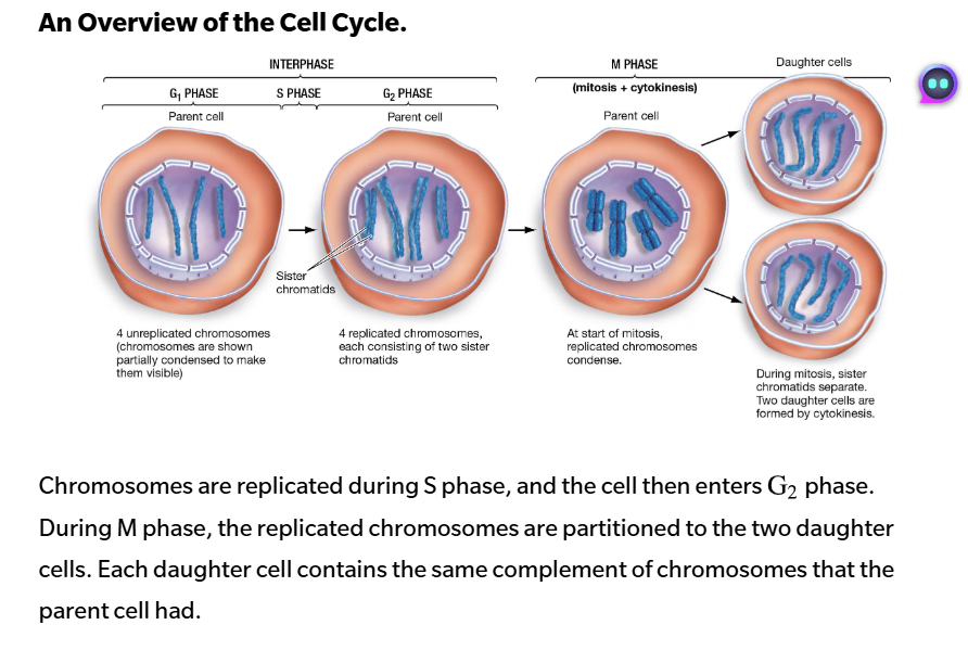
G2 phase is where the cell contains replicated chromosomes before mitosis. Each chromosome now consists of 2 sister chromatids. Each chromatid contains one long DNA double helix, and sister chromatids represent exact copies of the same genetic information.
Mitosis begins when chromatin condenses to form a much more compact structure.
During mitosis, the 2 sister chromatids separate to form independent daughter chromosomes. One copy of each chromosome goes to each of the 2 daughter cells. As a result, each cell receives the same complement of chromosomes as the parent cell had.
There are 5 subphases within mitosis based on distinctive events that occur
prophase
prometaphase
metaphase
anaphase
telophase
Before mitosis begins, chromosomes are replicated during S phase of interphase.
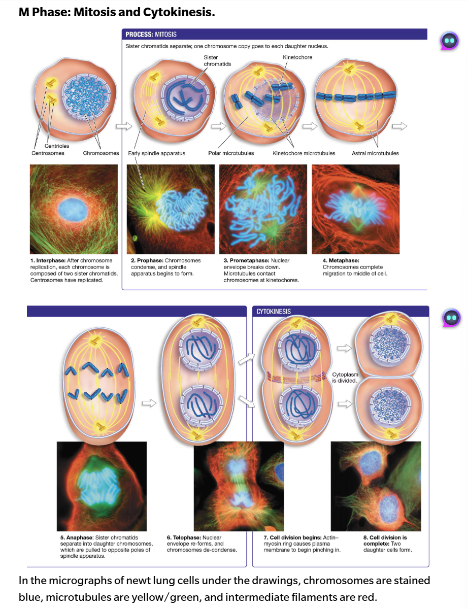
Prophase: mitosis begins with the events of prophase, when chromosomes condense into compact structures.
Prophase is also marked by the formation of the spindle apparatus. The spindle apparatus is a structure that produces mechanical forces that
move replicated chromosomes during early mitosis
pull chromatids apart in late mitosis
The spindle apparatus consists of microtubules—components of the cytoskeleton and have these characteristics:
are composed of a tubulin and b tubulin dimers
have a plus and end a minus ends—meaning they are asymmetric
the plus end is the site where microtubule rapid growth and disassemble normally occurs. the minus end rarely results in growth, but the disassembly is slower
Microtubules originate from microtubule-organizing centers (MTOCs). MTOCs define the 2 poles of the spindle apparatus and produce large numbers of microtubules, whole plus end grows outward through the cytoplasm. Although the nature of the MTOC varies among different organisms, the spindle apparatus has the same function.
During the S phase, the single centrosome replicates along with the DNA. At the start of prophase, the 2 centrosomes move to opposite sides of the nucleus to being forming the spindle apparatus. Some of these microtubules extend from each spindle pole and overlap with one another—these are called polar microtubules
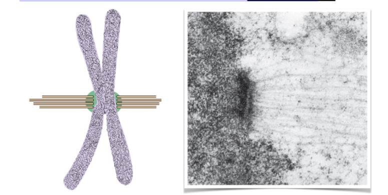
Prometaphase: once chromosomes have condensed, the nuclear envelope disintegrates. Removal of the envelope allows the cytoplasmic microtubules to attach to chromosomes at specialized structures called kinetochores. This defines the start of prometaphase.
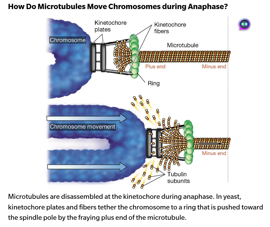
Each sister chromatid has its own kinetochore, which is assembled at the centromere.
In early prometaphase, kinesin and dynein motors attached to the kinetochores “walk” the chromosomes up and down the microtubules. When the chromosomes reach the plus ends of the microtubules, the kinetochore proteins secure their attachment.
Eventually, each chromosome will have its two kinetochores attached to microtubules that originate from opposite sides of the spindle apparatus. The chromosomes are then pushed and pulled by microtubules and motor proteins until they reach the middle of the spindle.
Metaphase: Once all the chromosomes have migrated to the middle of the spindle, the cell enters metaphase. The chromosomes are lined up on an imaginary plane between the 2-spindle poles called the metaphase plate.
Formation of the spindle apparatus is now complete. Because the sister chromatids of each chromosome are connected to opposite poles, a tug of war between the poles begins during metaphase.
Anaphase: at the start of anaphase, the cohesins that hold sister chromatids together at the centromeres are cleaved by an enzyme. Each replicated chromosome is pulled apart, creating 2 independent daughter chromosomes.
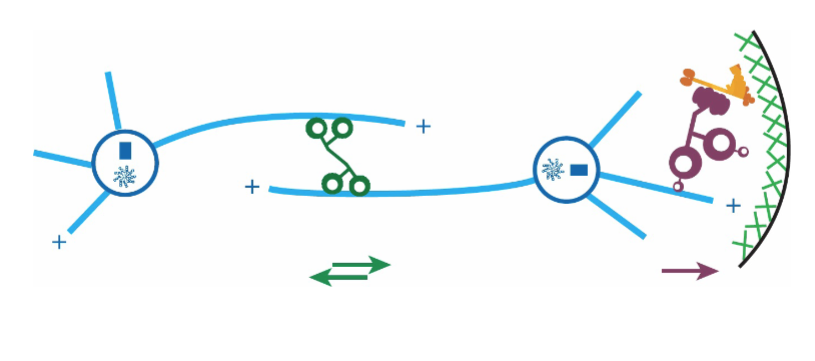
2 types of movement occur during anaphase. First, the daughter chromosomes move to opposite poled via the attachment of kinetochore proteins to the shrinking kinetochore microtubules. Second, the 2 poles of the spindle are pushed and pulled farther apart.
When anaphase is complete, 2 complete sets of chromosomes are fully separated, each set identical to that of the parent cell before chromosome replication.
Telophase: during telophase, the nuclear envelope re-forms around each set of chromosomes, and the chromosomes begin to de-condense. Once 2 independent nuclei have formed, mitosis is complete.
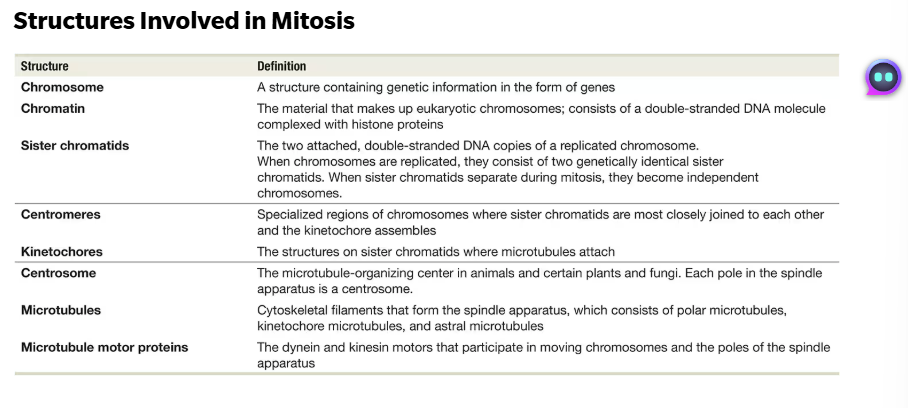
During mitosis, the microtubules originating from the spindle poles are highly dynamic. When cells transition from metaphase to anaphase, the plus ends of the kinetochore microtubules switch from adding tubulin dimers to removing them. As these plus ends shrink back to the spindle poled, the chromosomes are pulled along.
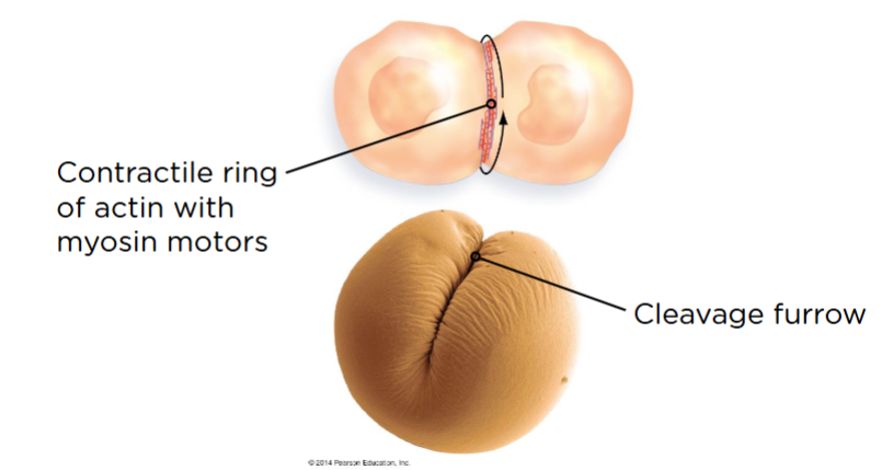
Whole the cell was in interphase, the cytoplasmic contents, including the organelles, increased in number or volume. During cytokinesis, the cytoplasm divides to form 2 daughter cells, each with its own nucleus and complete set of organelles. In most types of cells, cytokinesis directly follows mitosis.
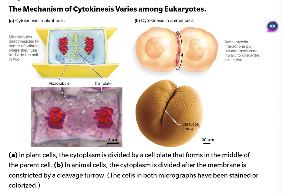
As myosin moves the actin filaments, the ring shrinking and tightens. Because the ring is attached to the inside of the plasma membrane, the contracting ring pulls the membrane with it.
Bacteria cells divide but do not undergo mitosis.
The eukaryotic cell cycle:
Our model system is cultured human cells
divide once every 24 hours (in human cells)
Consists of M-phase (~5%) and Interphase (~95%)
M-phase is mitosis (nuclear division) and cytokinesis (cytoplasmic division); Interphase is DNA replication and (mostly steady) growth in preparation for next M-phase
DNA is only synthesized in a specific portion of interphase, so the cell cycle is broken down into 4 distinct phases...
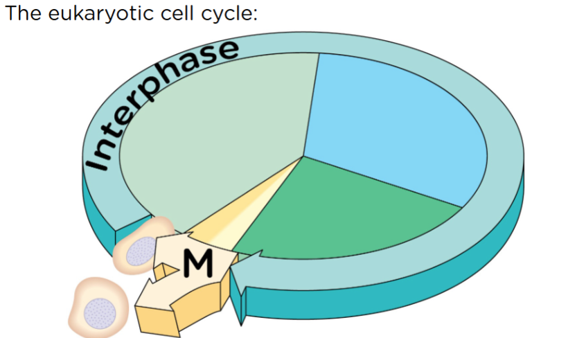
Within interphase: when you synthesize DNA is in S phase. G - gap phases (not missing anything). it is for cells to grow and getting ready for M phase.
Time spent in each phase (S, M, G1, G2) depends on the organism (and often on the cell type in a given organism):
DNA is not being worked on in the M phase - this is why it is quick
Cultured human cells:
11 hours in G1, 8 hours in S, 4 hours in G2, 1 hour in M
It takes a while in G1 because you need nutrients and stuff to prepare for M phase
Some budding yeast cells:
All 4 phases in 90 min
Time spent in each phase depends on the organism (and often on the cell type in a given organism):
only a cell would go into replication if they were going to divide
most cells are in G0
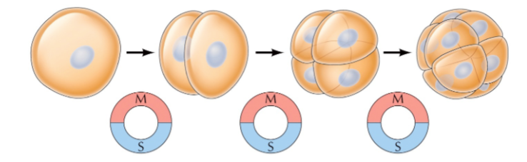
Early embryonic cells divide without growing:
Some adult cells cease dividing altogether, but are still metabolically active (not dead)
exit G1, enter what is called G0. Checkpoints are there to make sure cells do not go through the cell cycle so that there is not a surplus of cells.
the first 12 is just replication and dividing, but no gap phase
M-phase: mitosis and cytokinesis
Visually the most dramatic period of the cell cycle
During M-phase:
Chromosomes condense into x shaped structure
Cytoskeleton (mostly microtubules) reorganizes to form mitotic spindle
Nuclear envelope disappears
Chromosomes moved to opposite poles
Reformation of nuclear envelope
Cytokinesis
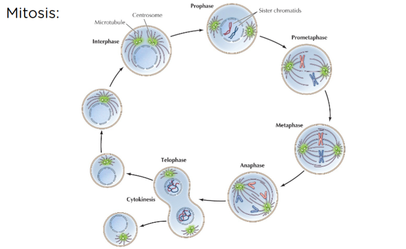
Mitosis is conventionally divided into 5 stages: (cytokinesis is a separate thing)
Prophase
Prometaphase
Metaphase
Anaphase
Telophase
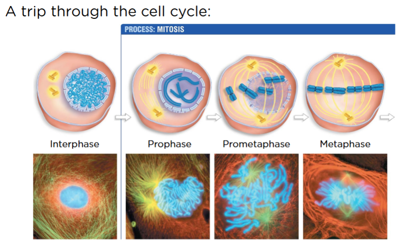
Also replicate your centrosome. Blue - DNA green - microtubule
Prophase - spindle poles separate from each other
Prometaphase - grab on to DNA and want to get into the middle
Metaphase - sister chromatids are in the middle of the cell
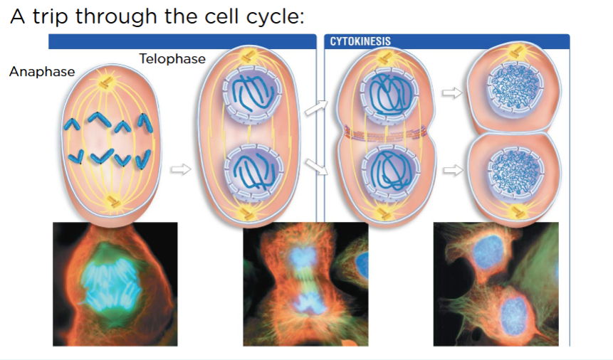
Anaphase - separating of sister chromatids
telophase - reform nuclei and nuclear envelope (cytokinesis happens synonymously with telophase)
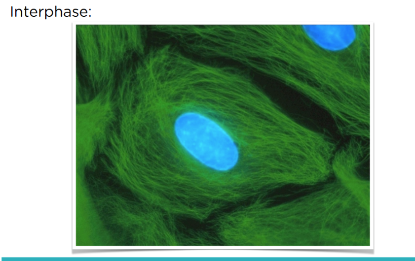
Clearly defined nucleus and can’t see individual DNA
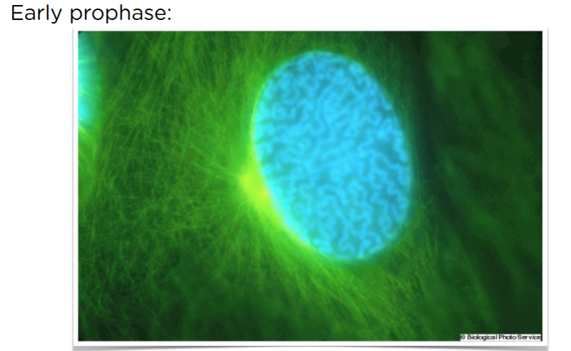
Still see the “bean”, but can start seeing individual DNA. Two very bright centrosomes
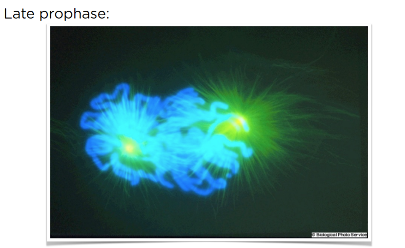
can’t see nucleus anymore and you have assessable DNA, and spindles are on opposite sides.
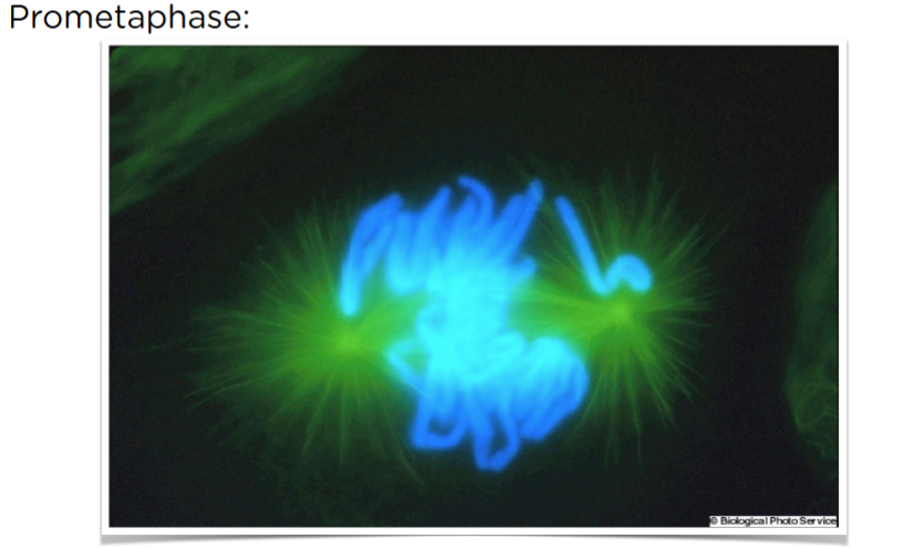
Middle of the “dance”
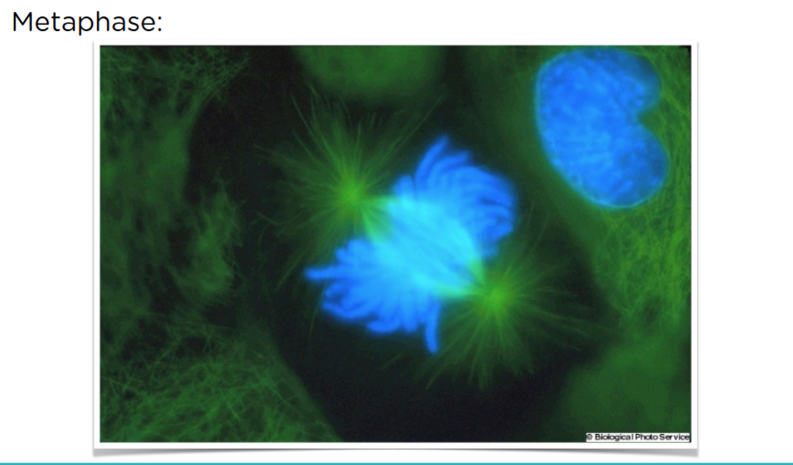
about ready to start pulling
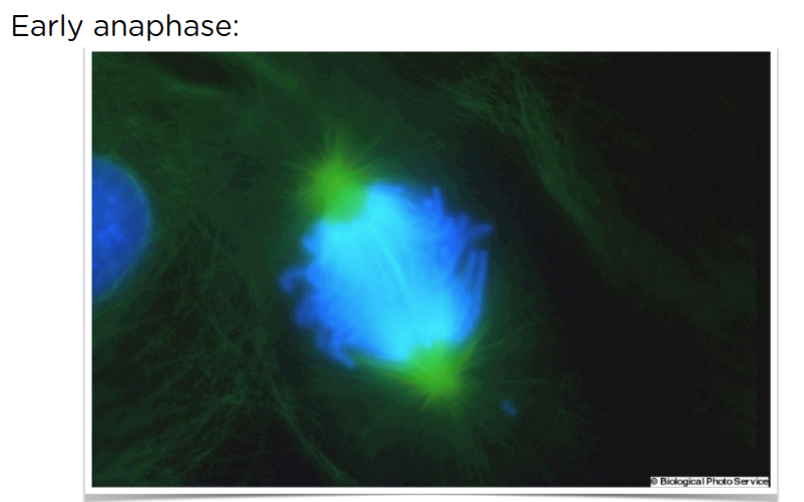
Can’t see pulling yet
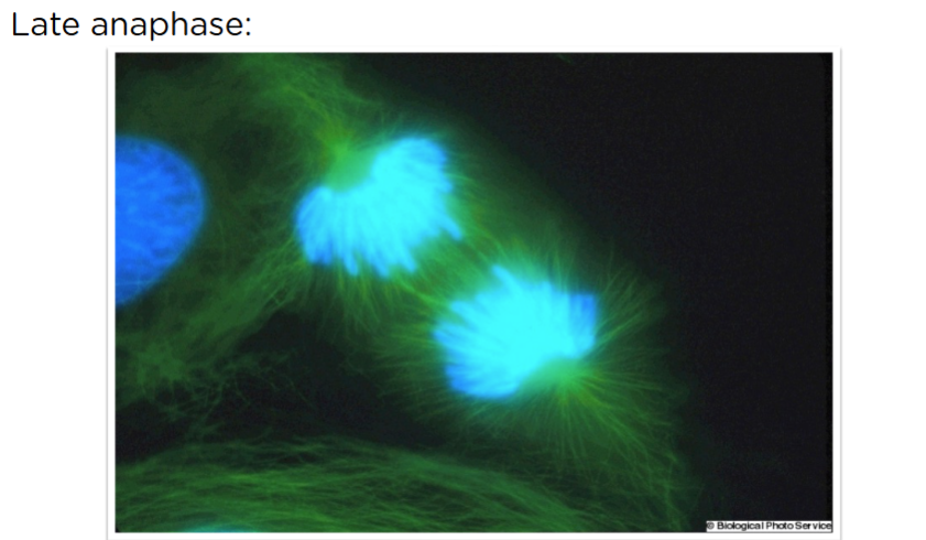
see separation, moving in different directions
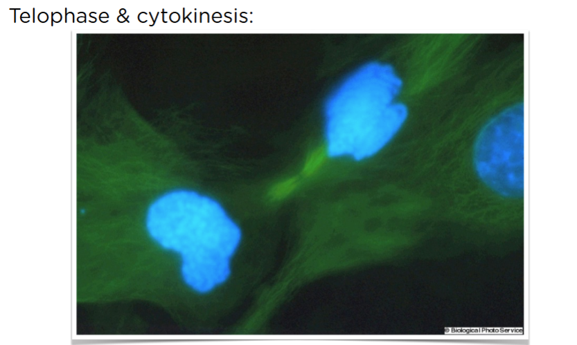
Depolymerization in microtubules because we don’t need them. Nucleus and nuclear envelope is formed again
In late prophase, the nucleus is disassembled:
Nucleus being disassembled - pretty big thing
Nuclear lamina - intermediate filaments, which is why the nucleus does not collapse. Gives you something to hang on your chromatids
Goal is to break down the nucleus - to do this, we make it temporarily lose its affinity by phosphorylating it, so they don’t latch on to each other anymore. This is reversible. made the sister chromatids available
In telophase, the nucleus is reassembled:
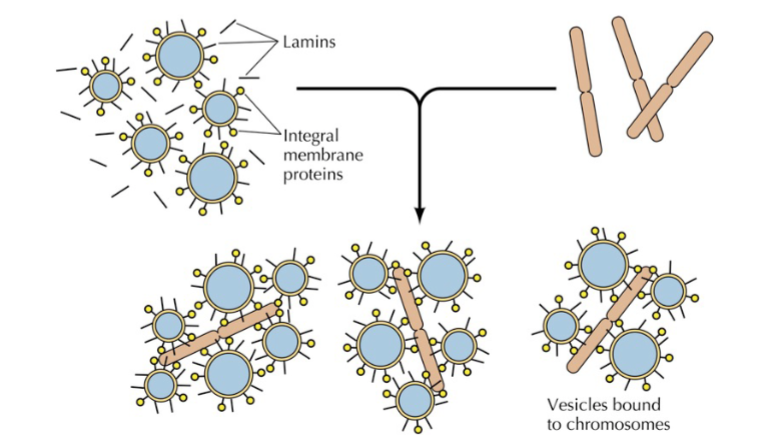
To regain their affinity, we just dephosphorylate it. when we do this, the chromatin regains their affinity, and they come back together. The chromosomes are coated in nuclea
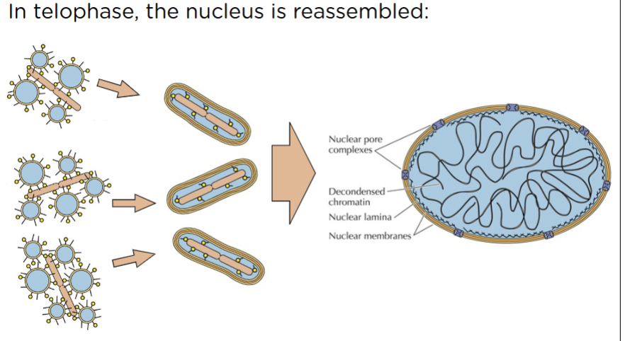
When the vesicles fuse and come together, they form the nucleus. Chromatin starts to decondense.
we fuse our vesicles and the assembly of nuclear lamina and nuclear pore complexes
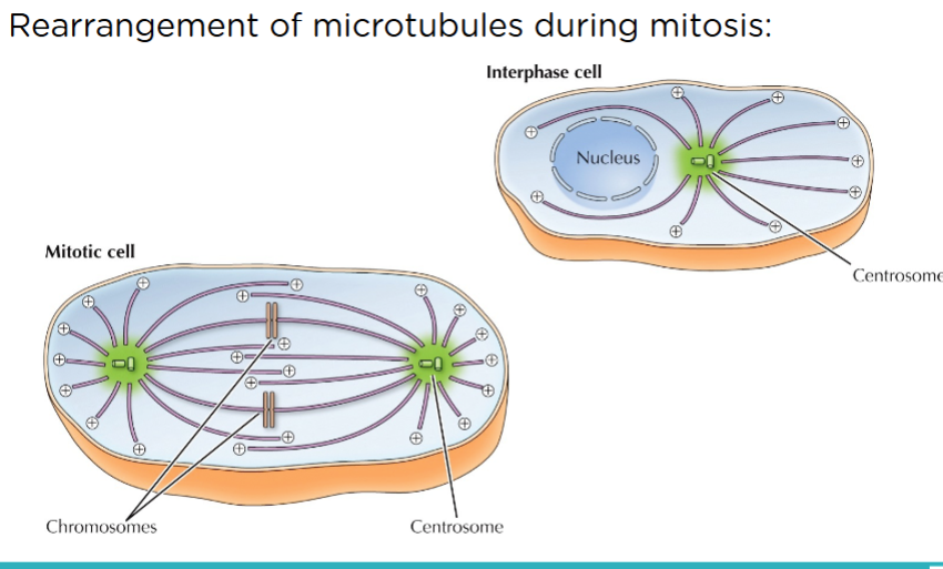
spider legs looking thing are microtubules. plus ends are away. Middle of chromosomes are the kinetochore.
Kinetochores:
Movement of sister chromatids to opposite poles via kinetochore microtubules is called Anaphase A. DO NOT CONFUSE ANAPHASE A AND ANAPHASE B. They happen simultaneously.
Aggregation of different types of proteins) motor, structural, regulatory) attached to the centromere
Microtubules gets stuck in the kinetochore. Tip of microtubule (plus) gets stuck. It gets pull either through kinesis or dynein (depending on which way). this is the “dance”
Spindles distance themselves from each other, further separating sister chromatids, in Anaphase B:
M phase ends with cytokinesis (cytoplasmic division):
Prophase
Disassemble interphase MT array, form mitotic spindle
Centrosomes move to opposite poles
Chromatin condenses
Nuclear envelope dissociates
Prometaphase
Kinetochore MTs move pairs of sister chromatids back and forth until they reach the metaphase plate
Metaphase
All pairs of sister chromatids are lined up on metaphase plate; connection between chromatids broken
Anaphase
Anaphase A: kinetochore MTs separate sister chromatids
Anaphase B: polar MTs "push" and astral MTs "pull" poles
Telophase
Undo what was done during Prophase
Cytokinesis
Cytoplasmic division using contractile ring of actin and myosin
12.3 Explain how cells monitor and regulate the progression of the cell cycle
Although the events of mitosis are similar in all eukaryotes, control of the cell cycle often varies—even among cells in the same organism.
Most of these differences are due to variation in the length of the G1 phase. In rapidly dividing cells, G1 is essentially eliminated. Most nondividing cells, in contrast, are permanently stuck in G1. Researchers refer to this arrested state as the G0 state.
A cell’s division rate can also vary in response to changing conditions.