Circulation Through the Heart
A) Circulation through vessels + heart

2 major loops through the body
Pulmonary loop: sends deoxygenated blood to the lungs, and returns oxygenated blood to the heart.
Systemic loop: sends oxygenated blood to tissues, and returns deoxygenated blood to the heart.
Blood cells always travel from the pulmonary to the systemic loop and back.
→ Pulmonary and systemic loops are interconnected by the heart.
Major parts of the heart
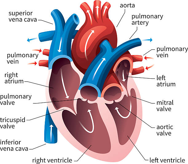
Septum: divides the right and left halves of the heart
Atrium (right & left): a thin-walled chamber at the top of the heart that receives blood from the veins and sends blood to the ventricles below.
Ventricle (right & left): a thick muscular chamber that receives blood from the atrium and sends blood to the arteries.
Atrioventricular valve (AV valve): ensures one-way blood flow from the atrium to the ventricle.
Semilunar valve: ensures one-way blood flow from the ventricle to the artery.
B) Blood flow through the heart
Blood enters the right atrium from the 2 systemic veins.
Superior (anterior) vena cava: collects blood from the head, arms, and upper torso.
Inferior (posterior) vena cava: collects blood from the lower body parts.
The atria contract, sending blood from the right atrium through the right AV valve, and then into the right ventricle.
The AV valve is supported by the chordae tendinae, an elastic connective tissue fibre that prevents the valve from inverting (flipping inside out) under pressure.
The right ventricle contracts, sending blood through the right semilunar valve to the pulmonary trunk (a major blood vessel that sends blood through the pulmonary loop).
The pulmonary trunk splits into the left and right pulmonary arteries.
→ Then towards the pulmonary arterioles, capillaries, venules, and then the pulmonary vein.Blood then enters the left atrium.
The atria contract, sending blood through the left AV valve, and then into the left ventricle.
The ventricles contract, sending blood from the left ventricle through the semilunar valve into the aortic arch.
→ Aortic arch splits into the carotid and subclavian arteries, and becomes the dorsal aorta as it continues down behind the heart.
→ Dorsal aorta divides into iliac arteries, and then into femoral arteries.
→ Systemic arteries branch into arterioles, capillaries, venules, and veins.
→ ALL VEINS FUSE INTO THE INFERIOR VENA CAVA, AND THE CYCLE BEGINS AGAIN!
The heart muscle requires O₂, and therefore, its own arteries, capillaries, and veins.
Coronary arteries can be blocked, preventing the O₂ delivery to the heart.
→ This is called a heart attack.If the coronary arteries are too blocked by plaque, this requires a coronary bypass operation.
→ Graft vein from the leg into position in the heart to bypass blockage.