Cytoskeleton
Key Terms
Cytoskeleton - is the internal skeleton of the cell.
Actin Filaments - Long rope-like cables and fibres that run around the outside of the cell, providing strength.
Centrosome - Organelle near the nucleus that has to centrioles arranged at right angles to eat other, surrounded by pericentriolar material (PCM)
Cell Migration - Process by which a cell moves from one place to another using actin filaments and motor proteins.
Cells
Key Facts
There are around 10 trillion cell in the human body (1013) cells
How where this amount of cells means there can be challenges with communication.
Key Functions:
Cells must connect strongly and coordinate functionality for proper operation of tissues and organs.
The supporting structure must be flexible, allowing for stress adaptation, environmental changes, and repair.
Cytoskeleton
Cytoskeleton - is the internal skeleton of the cell.
it is essential for joining cells together
it provides rigidity, however it can reorganise itself inside the cell.
Made up of:
Actin filaments in red
Microtubules in green
Nucleus in Blue
Intermediate filaments
The Dynamic Cytoskeleton
The cytoskeleton is highly dynamic and undergoes constant re-organisation:
Actin filaments extend and contract, pushing the plasma membrane as cells sense the environment.
The overall actin network can rapidly remodel during movement.
Microtubules adapt to facilitate the transport of organelles or during cell division (mitotic spindle formation).
Actin Filaments
Actin Filaments - Long rope-like cables and fibres that run around the outside of the cell in the plasma membrane
, providing strength.
Key Facts
they can also run through the cell for support.
Dictate cell shape
Myosin Motor units travel along then to transport substances around the cell.
critical for connecting cells in tissues
Can help cell movement.
it is needed as cellular contents put pressure on the plasma membrane, which can lead to lysis.
Structure
it is long thin fiber made up lots of monomers called actin (protein )
it is 5-8 nm thick ( very thin )
can be bundled together to make them thicker and increase strength
They have a + and - end.
THE + END IS WHERE THE ACTIN FILLAMENT GROWS FROM.
Actin Polymerisation
Free actin monomers bind ATP.
Profilin (green) helps load ATP onto actin and promotes delivery of ATP-actin monomers to the barbed end.
ATP-actin monomers are incorporated into the growing filament.
ATP is hydrolyzed to ADP.
Phosphate (Pi) is released.
Older regions of the filament become enriched in ADP-actin.
ADP-actin subunits are less stable in the filament.
They dissociate preferentially from the pointed (-) end.
ADF/cofilin (blue) binds ADP-rich regions of the filament.
Cofilin weakens actin-actin interactions and promotes filament severing and depolymerization.
ADP-actin monomers are released.
ADP is exchanged for ATP, often aided by profilin.
ATP-actin becomes ready for reincorporation at the barbed end.
Component | Function |
|---|---|
ATP-actin | Preferred form for polymerization |
Barbed (+) end | Rapid assembly of actin |
ATP hydrolysis | Converts newer ATP-actin into older ADP-actin |
Pointed (-) end | Preferential disassembly |
Profilin | Promotes ATP loading and addition to filament |
ADF/Cofilin | Binds ADP-actin and enhances depolymerization |
Treadmilling | Simultaneous growth at + end and shrinkage at − end |

Microtubules
Microtubules - Extend all through the cell.
Key Facts
they can provide strength
involved in transport with Dynein and Ki
involved in cell division during mitosis as they disappear and reappear as spindle fibers.
Structure
thicker than actin
diameter of 25nm
hollow cylinders with distinct ends (nothing goes down the tubules
the minus end is anchored near the nucleus
Has a Microtubule organising centre (MTOC) which is organised by the centrosome
The MTOC is a big complex of proteins that help organise our microtubules.
the microtubules then spiral around the cell so the - ends are bunched in the MTOC
MICROTUBULES GROW AND SHIRNK FROM THE + END.
Microtubule Polymerisation
to grow you add tubulin molecules onto plus end, once the cell is happy with there the microtubule ends they put on a cap.
to shrink again you remove the cap.
Monomers of the protein tubulin are added at the plus end,
Intermediate Filaments
Intermediate because their side is in between actin and microtubules.
Key Facts
more diverse than actin Filaments or microtubules.
types include Keratin or cytokeratin in hair
Functions
Provide mechanical strength to the cell.
Connect cells in tissues.
extend to the nucleus, so can bind to DNA and help anchor chromosomes.
Comprise rope-like fibers, typically stronger and more resilient than other cytoskeletal components.
Structure
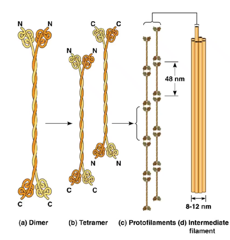
with the intermediate filaments you start with a dimer ( 2 molcules of your intermediate filliment)
then 2 dimers fuse to form a tetramer, these can then attach to another tetramer , repeatedly until it kind of forms a polymer
however this is import complex than a polymer
Cell Migration
Cell Migration - Process by which a cell moves from one place to another using actin filaments and motor proteins.
How it happens
The cell extends its membrane in the direction of movement.
Actin polymerisation pushes the membrane forward.
The cell attaches to the surface.
Myosin contracts the cell body and pulls it forward.
The rear of the cell detaches.
Examples
Neutrophils towards bacteria
chemotaxis
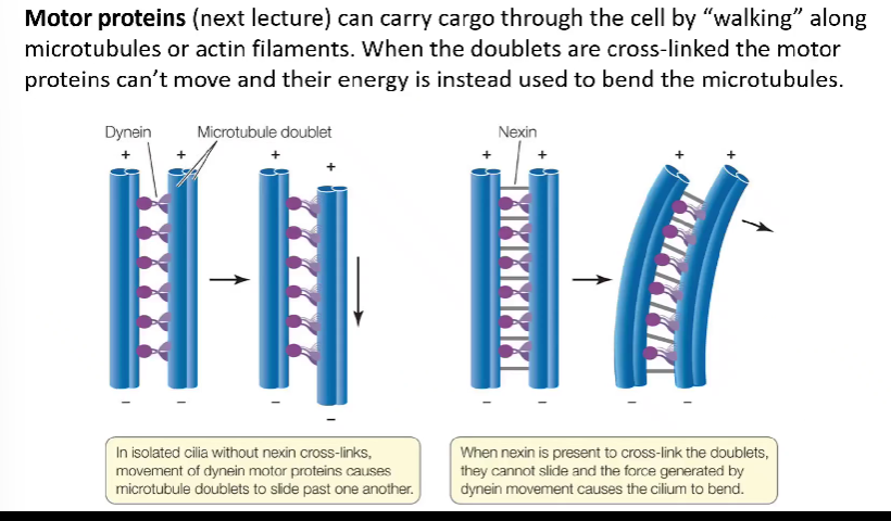
Flagella Movement - Motor proteins (next lecture) can carry cargo through the cell by "walking" along, microtubules or actin filaments. When the doublets are cross-linked the motor, proteins can't move and their energy is instead used to bend the microtubules.
Cilia - Shorter than flagella, beating in a coordinated manner, with cells often containing several cilia or flagella.
Principles of Studying Cells
Methods
Microscopy:
Light
Electron
Super resolution
Bright field
Phase contrast
Differential interference contract
Transmission Electron
Scanning Electron
Fluorescence
Confocal
Green Fluorescent Protein
Stains
H&E Stain
Azan trichrome stain


Microscopy Type | What it Does | How it Works | Advantages | Limitations |
|---|---|---|---|---|
Light Microscopy | Views cells and tissues using visible light. | Light passes through lenses to magnify specimens. | Cheap, simple, can view living cells. | Lower resolution (~200 nm) than electron microscopes. |
Electron Microscopy | Produces highly detailed images of cell ultrastructure. | Uses a beam of electrons instead of light. | Very high magnification and resolution. | Expensive, specimens must be dead and prepared. |
Super-Resolution Microscopy | Visualises structures smaller than the diffraction limit of light. | Uses specialised fluorescence techniques and image processing. | Higher resolution than conventional fluorescence microscopy. | Expensive, complex, often requires fluorescent labelling. |
Bright-Field Microscopy | Observes cells against a bright background. | Light passes through the specimen. | Simple and widely available. | Poor contrast in transparent cells, often requires staining. |
Phase-Contrast Microscopy | Allows viewing of unstained living cells. | Converts differences in light phase into brightness differences. | Good for live cells and intracellular structures. | Produces halo artefacts around structures. |
Differential Interference Contrast (DIC) Microscopy | Produces high-contrast, 3D-like images of cells. | Uses polarized light and prisms to detect refractive index differences. | Excellent detail in living, unstained cells. | Expensive and cannot provide true 3D images. |
Transmission Electron Microscopy (TEM) | Examines internal cell structures. | Electrons pass through an ultrathin specimen. | Extremely high resolution; shows organelles clearly. | Specimens must be thin, dead, and heavily prepared. |
Scanning Electron Microscopy (SEM) | Examines surface features of specimens. | Electrons scan the specimen surface. | Produces detailed 3D-like surface images. | Shows surface only; specimens must be dead. |
Fluorescence Microscopy | Detects specific molecules or structures within cells. | Fluorescent dyes or proteins emit light when excited. | Highly specific; can identify particular proteins. | Requires fluorescent labels; photobleaching can occur. |
Confocal Microscopy | Produces sharp optical sections and 3D images. | Uses lasers and a pinhole to eliminate out-of-focus light. | High contrast, excellent for thick specimens, 3D reconstruction possible. | Expensive and slower than conventional fluorescence microscopy. |
Green Fluorescent Protein (GFP)
Originally derived from the jellyfish Aequorea Victoria, GFP has been optimised for specific experimental conditions.
Florence means you absorb light of one colour and emit light from other.
this one absorbs blue and emits green
It looks like a short barrel
which has a chromophore in the center which has a set of peptides that absorbed blue and emits green.
They make this single protein
Meaning there is a gene that codes for this GFP.
The genetic code is universal you can put this gene in a human cell.
however this gene and protein have evolved to have a optimum temp at sea level.
GFP Details:
Commonly presents as a tetramer, may be weakly fluorescent and prone to photobleaching.
Mutations have improved stability and fluorescence.
Fusion Proteins with GFP:
Fusion can occur at the C-terminus (after the start codon) or the N-terminus (before the stop codon); C-terminal attachment recommended for proper protein processing.
Limitations of Fluorescent Proteins
Photobleaching: Destruction of fluorescent proteins by high-intensity laser light.
Phototoxicity: Toxicity caused by laser light generating free radicals.
Overexpression Effects: Tagged proteins expressed at excess levels may not behave similarly to naturally occurring proteins due to cellular processing differences.
Folding & Distribution Effects: Large fluorescent proteins may impact the folding and localization of tagged proteins, potentially altering their cellular function.