BIOL 200 Learning Objectives
Unit 1
1.1 Eukaryotic Cells & Organelles
Critically discuss classical definitions of cells and organelles
-eukaryotic cells
membrane bound unit capable of carrying out essential life processes
-plasma membrane
lipid bilayer that controls what enters and exits the cell
-cell wall
rigid structure around plasma membrane that provides shape and support
-nucleus
membrane bound organelle containing DNA and controls cell functions
-nuclear envelope
double membrane surrounding nucleus, has pores that regulate molecule transport in and out of nucleus
-nucleolus
region inside nucleus where ribosomes are made
-chromatin
DNA and protein complex within nucleus → condenses into chromosomes during cell division
-mitochondria
generates ATP
-chloroplast
performs photosynthesis to convert sunlight to energy
-endoplasmic reticulum (ER)
protein synthesis
-rough er
has ribosomes; synthesizes and processes proteins
-smooth er
lacks ribosomes; synthesizes lipids
-golgi apparatus
modifies, sorts and packages proteins and lipids for transport
-vesicle
small membrane bound sac that transports materials in cell
-lysosome
contains digestive enzymes to break down waste
-peroxisome
breaks down fatty acids and detoxifies harmful substances
-ribosome
synthesizes proteins using mRNA instructions
-cytoskeleton
network of protein filaments providing structural support and helps with movement
microtubules
hollow tubes that maintain cell shape and aid in cell division (spidle fibers)
microfilaments
filaments involved in movement and support
-vacuole
stores water, nutrients, waste; helps maintain cell structure (plant cells)
Compare and contrast major characteristics of bacteria and eukaryotes
Feature | Prokaryotes (no nucleus no membrane bound organelles) | Eukaryotes (nucleus with membrane bound organelles) |
Cell Structure | simple, small | complex, larger |
Nucleus | absent; DNA in nucleoid | present; DNA in membrane-bound nucleus |
DNA Organization | circular; single chromosome | linear chromosome within nucleus |
Organelles | no membrane-bound organelles | membrane-bound organelles present (mitochondria, golgi apparatus, endoplasmic reticulum, nucleus) |
Cell Wall | peptidoglycan | cellulose, chitin, absent |
Reproduction | asexual | sexual/asexual |
Ribosomes | 70S | 80S |
Cytoskeleton | minimal/absent | extensive (microtubules, microfilaments, intermediate filaments) |
Organelles that eukaryotes have but prokaryotes don’t | nucleus, nuclear envelope, endoplasmic reticulum (rough er, smooth er), golgi apparatus, mitochondria, chloroplasts, lysosomes, peroxisomes, vacuoles, cytoskeleton, centrioles |
1.2 Microscopy
Distinguish between 4 major classes of microscopy: brightfield light microscopy, fluorescence light microscopy, transmission electron microscopy (TEM), scanning electron microscopy (SEM)
Discuss the major advantages and limitations of each of the 4 major classes of microscopy
Understand the difference between magnification and resolution and how each can be used to identify the type of microscopy used
-magnification
enlarges image
-resolution
determines clarity
high resolution → clear more details
low resolution → blurry, less details, things mushed together
Recognize the major organelles in light and electron micrographs
Predict the type of microscopy that can be used to detect and study cellular components based on their size and functional aspect being studied
Microscopy Type | Features | Advantages | Disadvantages | Best for Observing |
Light Microscopy | light passes through specimen and viewed through lens | -living cells -whole tissues -track cells | -low resolution (~200 nm/0.2 um)
-only certain organelles visible -thick tissues must be sliced to thin sections | -whole cells -large organelles
|
Fluorescence Microscopy | -black background -fluorescent dyes to tag structures to be visible -fluorescent molecules absorb light at specific wavelength and emits light | -can view specific labelled structures -live and dead cells -track cells | -must tag structures -low resolution | -specific proteins & molecules -cytoskeleton -DNA -nucleus -plasma membrane -mitochondria -golgi apparatus |
Transmission Electron Microscopy (TEM) | -plane of section -detailed organelles -speckled background | -high resolution (~0.1-2 nm) -details of cytoplasm seen | -dead, fixed cells -extensive prep
-difficult to know 3D shape from 2D slices -heavy metal stains used to increase contrast | -ER -mitochondria -ribosomes -nucleus -nucleolus -plasma membrane -endoplasmic reticulum -golgi apparatus -chloroplasts |
Scanning Electron Microscopy (SEM) | -3D image of cell -surfaces only | -high resolution | -dead, fixed cells -extensive prep -can’t see internal structures | -cell surfaces |
Interpret the results in micrographs based on scale, magnification, resolution and plane of section
Unit 2
2.0 Macromolecules
-Describe the terms redundant and ambiguous in terms of genetic code
redundant
multiple codons can code for same amino acid
ensures that genetic code is robust against mutations
if mutations occur in 3rd position of codon → may still result in same amino acid being made into protein
ambiguous
genetic code isn’t ambiguous
single codon always codes for same amino acid
no one codon can specify more than one amino acid
-What machinery is involved in tRNA activation and why are tRNA molecules considered the translators of genetic code
tRNA activation is where amino acids are attached to corresponding tRNAs
aminoacyl-tRNA synthetase
ensure correct amino acid attached to right tRNA
tRNA, ATP used in tRNA activation
tRNA molecules interprets genetic information encoded in mRNA and translates it into corresponding sequence of amino acids during protein synthesis
-How is it possible that mRNA can be simultaneously producing several copies when there is only one start codon
multiple ribosomes simultaneously translate a single mRNA molecule
multiple ribosomes can bind to same mRNA and translate it simultaneously
Types of non-covalent bonds
-ionic bonds
atoms with opposite charges
-van der waals
dipoles formed when electrons move around atoms
Types of covalent bonds
-polar bonds
electrons unequally shared between atoms with different electronegativities
-non-polar bonds
electrons equally shared between atoms with similar electronegativities
What is the monomer of a protein, nucleic acid, and carbohydrate?
-protein
amino acids
-nucleic acid
nucleotides
-carbohydrates
monosaccharides
How are lipids different than other macromolecules when considering polymers?
-lipids don’t form true polymers like proteins, nucleic acids and carbohydrates do
-lipids aren’t made from repeating monomers
they are small nonpolar molecules that don’t form long chains through polymerization
aggregate into structures like membranes or fat deposits
What type of reaction brings monomers together?
-condensation reaction
2 monomers are joined together by covalent bond and a molecule of water is released as a byproduct
removal of H from one monomer & OH from other monomer → combines to form H2O
What is amino acid residue?
-individual amino acid incorporated into a protein after water is removed during formation of peptide bond
2.1 Biological Membranes
Discuss the structural and functional features of biological membranes and relate how the different macromolecular components (lipids, proteins, carbohydrates) interact to form biological membranes
-molecular components of membranes
lipids → phospholipids, sterols, glycolipids
proteins found on both side of membrane
carbohydrates
found outside (non-cytosolic) of membrane; not cytosol side
glycocalyx
sugar coat on plasma membrane
protects cell
acts in cell communication/identification
-lipids form the basic structural framework of the membrane (phospholipids)
in charge of the barrier, fluidity, ins and outs of the membrane
-proteins are embedded in or attached
in charge of transport, signaling, cell recognition
amphipathic nature of lipid bilayer allows a hydrophilic head to face extracellular environment and cytoplasm
allows protein transports to be spread around bilayer → selective transport of the efflux and influx of molecules
allows for the maintenance of ion concentrations to maintain necessary osmotic pressure of the cell
-carbohydrates attached to proteins or lipids on outer surface
in charge of cell recognition, communication, glycocalyx formation
-membrane features
bilayer
organized but fluid
different permeabilities for different molecules
asymmetric
-membrane functions
barrier to outside environment
protects cells from outer attacks
form cellular compartments
ionically distinct from extracellular space
allows for different environment compared to outside the cell
controls transport process
pumps & pores in membranes
controls what comes in & out of cell
localize reactions
digestion in lysosomes can’t occur if digestive enzymes loose in cytoplasm
regulate cell communication
receptors embedded in membranes
signals cross barriers
-membrane is selectively permeable
some molecules can pass through while others can’t
gases, hydrophobic, small nonpolar molecules cross lipid bilayer easily
oxygen, CO2, NO
large, uncharged polar molecules can’t cross bilayer due to size
amino acids, nucleotides, glucose
small, uncharged, polar molecules cross bilayers at slower rates
H2O
charged ions can’t diffuse across lipid bilayer
-thermodynamics of hydrophobic effect → forms bilayer in water
most stable form of phospholipids
minimizing exposure of hydrophobic groups to water
phospholipids spontaneously form bilayer because it’s most stable
most energetically favourable
Describe the chemical properties of phospholipids, cholesterol, and glycolipids and how these properties contribute to the lipid bilayer structure and function
-phospholipids
amphipathic
hydrophilic head (phosphate)
polar
interacts with water
hydrophobic tail (fatty acid chains)
nonpolar
avoids water
in aqueous solution → bilayer formation
hydrophilic heads face outward → interacts with aqueous environment
hydrophobic tails face inward → avoid water
allows fluidity → movement of molecules across membrane
gives membrane self-sealing & self-healing property
if hole in membrane → lipids heal and seal hole because its more favourable and stable
-cholesterol
amphipathic
polar hydroxyl group (-OH) at one end → slightly hydrophilic
hydrocarbon ring structure → mostly hydrophobic
in aqueous solution → -OH polar groups orients itself to be in water
-glycolipids
lipids with carbohydrate attached via glycosidic bond
amphipathic
hydrophilic head (carbohydrate)
interact with water
hydrophobic tails (fatty acid chains)
avoids water
located on outer leaflet of lipid bilayer → contributes to glycocalyx
-fatty acids in aqueous environment → micelle
-triacylglycerols in aqueous environment → oil droplets
Recognize the 20 amino acids according to their chemical properties and their 3-letter and 1-letter abbreviations
Amino Acid | Chemical Property | 3-Letter Abbreviation | 1-Letter Abbreviation |
Alanine | nonpolar | Ala | A |
Arginine | polar; positive | Arg | R |
Asparagine | uncharged polar | Asn | N |
Aspartic Acid | polar; negative | Asp | D |
Cysteine | polar | Cys | C |
Glutamine | uncharged polar | Gln | Q |
Glutamic Acid | polar; negative | Glu | E |
Glycine | nonpolar | Gly | G |
Histidine | polar; positive | His | H |
Isoleucine | nonpolar | Ile | I |
Leucine | nonpolar | Leu | L |
Lysine | polar; positive | Lys | K |
Methionine | nonpolar | Met | M |
Phenylalanine | nonpolar | Phe | F |
Proline | nonpolar | Pro | P |
Serine | uncharged polar | Ser | S |
Threonine | uncharged polar | Thr | T |
Tryptophan | nonpolar | Trp | W |
Tyrosine | uncharged polar | Tyr | Y |
Valine | nonpolar | Val | V |
Differentiate between the primary, secondary, tertiary, quaternary levels of protein structure
Protein Structure | Description | Bonds | Covalent bonds between backbone atoms | Covalent bonds between R-groups | Non-covalent bonds between backbone atoms | Non-covalent bonds between backbone and R-group | Non-covalent bonds between atoms and 2 R-Groups |
Primary (sequence) | sequence of amino acids in polypeptide chain | peptide bonds between amino acids (covalent bonds) | X | ||||
Secondary (alpha helix, beta sheets) | folding of amino acids into alpha helices and beta-pleated sheets -secondary structure elements fold into domains within tertiary structure -different domains associated with different functions | hydrogen bonds between backbone atoms | X helix-outwards beta-alternating | ||||
alpha helix -rotating structure -repetitive H bonds along backbone
-interior doesn’t form hydrophilic pore for passage of small molecules | |||||||
beta sheet -side chains up and down
-flattened arrows pointing towards C-terminus -can fold and form beta-barrel | |||||||
Tertiary (protein fold) | overall 3D shape of a single polypeptide chain -stabilized by
| H bonds, ionic bonds, hydrophobic interactions, disulfide bonds | X | X | X | H bonds, ionic bonds, van der Waals, disulfide bonds | |
Quaternary | assembly of multiple polypeptide subunits into a functional complex -stabilized by
| H bonds, ionic bonds, hydrophobic interactions, disulfide bonds | X | X | X | X |
-domain — secondary structure fold into domains within a tertiary structure
List and discuss the role of non-covalent bonds in protein folding and stabilization of protein structure
Illustrate how the primary sequence and environment of a protein influenceits final 3D structure
-primary sequence of a protein provides the initial blueprint for its structure
environmental factors fine tune the final 3D shape
pH
temperature
ionic strength & salts
-interactions between amino acid residues within protein and surrounding environment results in protein’s 3D conformation
2.2 The Lipid Bilayer
Describe factors affecting or regulating the fluidity of lipid bilayer
-better to have ↓ van der waals, ↑ fluid
-lipid composition
degree of unsaturation of lipids
↑ saturated lipids, ↓ fluid, ↑ van der waals
tightly packed
↑ unsaturated lipids, ↑ fluid, ↓ van der waals
more kinks in fatty acid tail
-sterol content (cholesterol — animal, phytosterol — plant)
low temperature, ↓ fluid, no sterol
add sterol → ↑ fluidity
prevents tight packing of fatty acids → ↓ van der waals
high temperature, ↑ fluid, no sterol
add sterol → ↓ fluidity
sterol ring structure stiffens cell membrane →↑ van der waals
-fatty acid tail length
↓ tail length, ↑ fluid, ↓ van der waals
less surface area
< 18 carbons
↑ tail length, ↓ fluid, ↑ van der waals
more surface area
14-24 carbons (~18-20)
-temperature
cold temperature
↓ fluid, ↑ van der waal
membrane bilayer freeze
crystalline-like structure → very fragile
phospholipids stuck together
trapped by van der waals
Predict the relative fluidity of membrane bilayers based on their lipid composition
-membrane fluidity
essential for cell movement, membrane fusion and activity of membrane proteins
how easily molecules within a cell membrane can move around
fluid membrane
lipids and proteins in membrane move around easily
rigid membrane
lipids and proteins packed tightly together → hard to move around
membranes can self-heal if they are punctured
self-seal when in close proximity to a second membrane
Explain how Fluorescence Recovery After Photobleaching (FRAP) works and interpret results of FRAP experiments
-green fluorescent proteins (GFP) bleached via laser
no more fluorescence
black patch of proteins as a result
-tagged proteins diffuse into the area
-untagged proteins diffuse out of area
-over time → area becomes fluorescent again
-proteins moving because it’s fluid → if tagged proteins don’t diffuse back into area → membrane not fluid
-fluorescence fully recovers to pre-bleached intensity → molecules are mobile and can diffuse quickly into bleached region → high diffusion & little immobility
-fluorescence partially recovers → molecules have some degree of mobility, constrained by various factors (molecules may be immobile due to being bound in clusters, complexes, macromolecular assemblies)
due to adhesion to proteins inside cell/ECM/neighbouring cells
-no fluorescence recovery → molecules in bleached region are immobile (trapped or tightly bound within structure)
due to adhesion to proteins inside cell/ECM/neighbouring cells
Describe membrane asymmetry and explain the origin of the asymmetric distribution of membrane lipids
-membrane is asymmetric
arrangement of components is not the same on 2 sides of bimolecular leaflet
always an inside (cytoplasmic) and outside (external)
non-cytosolic side faces extracellular side
cytosolic side faces intracellular space
carbohydrates are major contributors to membrane asymmetry
2 halves of membrane are different from each other
orientation of membrane remains constant — even throughout transport
asymmetry established by lipid flipping enzymes
flippases
enzymes in membrane that moves phospholipids from one leaflet to another
uni-directional movement
floppases
uni-directional movement
scramblases
bi-directional movement
-lipid rafts
microdomains in plasma membrane rich in specific types of lipids
sphingomyelin, cholesterol
more ordered than surrounding membrane
thicker than other regions of cell membrane
-attachment to structures inside/outside cell
adhesion to proteins inside cell
inside cell cortex
RBCs → membrane proteins attached to cell cortex
plasma membrane reinforced by association of membrane proteins with cell cortex
adhesion to extracellular matrix (ECM)
integrin proteins connected to ECM
adhesion to neighbouring cells
cell to cell adhesion molecules linking plasma membranes of neuronal cells
cells attached to each other so don’t flow away
barriers to diffusion
tight junctions
adhesions between neighbouring epithelial cells forming attachment points between 2 cells so nothing leaks between cels
segregates tops and sides/bottoms of cells to distinct membrane domains
*important for FRAP
2.3 Membrane Proteins
Distinguish between integral and peripheral proteins with regard to their solubility, structure and manner of attachment to membranes
-integral proteins
proteins directly attached to membrane
amphipathic
monomeric
one polypeptide
multimeric
multiply polypeptide
can’t be removed without destroying membrane completely
insoluble
hydrophobic regions; embedded in bilayer
amino acids found in transmembrane (hydrophobic) region
nonpolar amino acids (Leu, Iso, Val, Phe, Ala, Met, Try)
hydrophobic amino acids interacts with lipid tails of bilayer → stabilizes protein within membrane
amino acids found on extracellular/cytoplasmic surface (hydrophilic)
charged polar amino acids (Asp, Arg, Glu, Lys, His)
uncharged polar amino acids (Ser, Thr, Gln, Tyr)
transmembrane proteins
proteins that pass through lipid bilayer
uses secondary structures to pass through membranes (alpha helices, beta sheets)
alpha helices
1+ alpha helical transmembrane domains
domains - stretches of 20+ amino acids with hydrophobic side chains
can form pores & channels but several alpha helices required
beta-sheets
beta strand/flat beta sheet not stable enough to be used in transmembrane protein
edges of sheet has exposed backbone that doesn’t interact with hydrophobic core of bilayer
beta sheets transform in to beta-barrels
hydrogen bonds between beta sheets form a cylindrical structure
backbone of polypeptide chain always polar
transmembrane domain
part of membrane protein that passes through bilayer
mostly alpha-helices; some beta-barrels
need multiple alpha helices/beta-barrels to make a pore
monolayer-associated proteins
only attached to one side of membrane
-peripheral proteins
bound to membrane surfaces via non-covalent association with other membrane proteins
not attached to membranes → attached to something attached to membrane
can be removed using mild salt washes
soluble in water
hydrophilic regions; loosely associated with membrane
amino acids found in peripheral membrane proteins (hydrophilic)
hydrophilic amino acids (Glu, Asp, Lys, Arg, His, Ser, Thr, Glu, Asp)
charged polar amino acids help peripheral proteins bind to hydrophilic heads of membrane lipids
no amino acids found in membrane → proteins not embedded in membrane
lipid-linked proteins
proteins covalently bonded to lipid that is attached to membrane
-plasma membrane polysaccharides
glycocalyx
glycolipids
integral parts of membrane
fatty acids hold glycolipid firmly in membrane
synthesized on cytosolic side of ER (like other membrane lipids)
glycoproteins
proteins that carry attached polysaccharides
integral membrane proteins that extend through or into membrane
attached to exterior part of protein
Interpret results from SDS-PAGE, fluorescence and glycoprotein staining experiments to determine orientation and location of proteins and glycoproteins in a membrane
-SDS-PAGE
treatments
beta mercaptoethanol
breaks disulfide bonds within proteins
salt solution
breaks ionic interactions of peripheral proteins and structures they are bound to
brief detergent
pokes holes in plasma membrane by solubilizing phospholipids
digestive enzyme
digests parts of proteins accessible
if combined with detergent → digestion of proteins on cytosolic side of membrane
protein isolated from membrane (SDS)
proteins treated with protease enzyme
enzyme that break peptide bonds, can’t cross lipid bilayer
peripheral proteins treated with high salt concentrations to wean interactions
integral proteins treated with harsh treatments (detergents) to solubilize proteins
proteins are now separated from membrane
filter proteins through polyacrylamide gel matrix (PAGE)
-polyacrylamide gel matrix → gel electrophoresis separates proteins based on size
large proteins = top
small proteins = bottom
more proteins = thick bands
less proteins = thin bands
-fluorescence staining
fluorescent dyes used to tag membrane proteins
dye only tags protein on outer membrane (dye can’t cross lipid bilayer)
poke holes in membrane → dye can enter
comparing staining before and after permeabilization → can tell which proteins are outside vs inside cell
fluorescence microscope can be used to see where proteins are located in cells
fluorescence only on cell surface → protein is outer membrane protein
fluorescence inside cell (after permeabilization) → protein on inner leaflet or inside organelle
-glycoproteins staining
glycoproteins (proteins with sugars attached) usually found on outer leaflet of membrane
special stains that bind to sugars are applied to cells
if stain binds membrane without needing permeabilization → glycoproteins facing outwards
confirms which proteins are exposed on outer cell surface
Predict the hydropathy profile for a given protein given its amino acid sequence or for a given hydropathy plot, predict the amino acid composition of the protein domains within a membrane protein.
-bioinformatics (hydropathy plots)
only works on alpha-helices
doesn’t work on beta barrels
amino acid sequence of beta-barrels have alternating hydrophobic and hydrophilic R groups facing in and out of the pore → hydropathy plot ends up oscillating between hydrophobic & hydrophilic regions
shows no significant peaks → near zero hydropathy plot
determines whether a protein is a transmembrane protein, how many transmembrane regions (helices) it has, orientation of protein (which parts are inside vs outside membrane)
can predict if protein is embedded in cell membrane
based on properties of amino acids in peptide sequence
hydrophobic regions → positive peaks → membrane spanning alpha helices (proteins embedded within bilayer — interacts with tails) → line above 0
hydrophilic regions → negative peaks → extracellular or cytoplasmic alpha helices (proteins on outer side of head) → line below 0
if first region is hydrophilic → N terminus in cytoplasm or extracellular space
if last region hydrophilic → C terminus in cytoplasm or extracellular space
-Fibronectin is a large glycoprotein present in extracellular matrix (ECM), if a cell contains a mutation that leads to a loss of fibronectin, how would plasma membrane attach to cytoskeleton and is the ECM affected?
plasma membrane’s attachment to cytoskeleton and ECM affected
cell has trouble holding its shape, moving or responding to signals from environment
cells have a harder time sticking to ECM → weaker tissue structure → ECM disrupted
Unit 3
3.1 Nuclear Structure & Protein Import
Describe the structure of an interphase nucleus and recognize structural elements in different kinds of microscopy
-nuclear envelope
double membrane with perinuclear space in between
surrounded by 2 lipid bilayers
outer nuclear membrane
continuous with rough ER
ribosomes attached
inner nuclear membrane
cytoplasm and nucleoplasm connected through nuclear pores
-nucleolus
not membrane bound
site of rRNA synthesis
assembly site of ribosomal subunits made from rRNA and ribosomal proteins
1-10 nucleoli in nucleus depending on cell cycle
disassembled during mitosis so chromosomes can be condensed
ribosomes
made of protein and RNA
ribosomal proteins made in cytosol
then imported to nucleus and assembled with rRNA molecules in nucleolus
-nuclear lamina
provides structural support
important role in DNA replication, transcription, gene regulation
made of strong nuclear lamin proteins
proteins in inner nuclear membrane can be anchored to it
aids in disassembly and reassembly of nuclear envelope during cell division
disassemble → phosphorylation
reassemble → dephosphorylation
Describe nuclear pore complex (NPC) and explain the different types of nuclear transport mechanisms
-nuclear pore complex (NPC)
nuclear pores — very small
protein subunit complex that controls what enters and leaves nucleus
pore complex ~90-120 nm
import in (proteins required for)
DNA replication, transcription, gene regulation, mRNA processing, DNA structure, ribosomal subunit assembly
cytosolic fibrils
guide molecules in and out of nucleus
export out
assembled ribosomal subunits
mRNA
tRNA
Explain how proteins are transported into and out of the nucleus, including the roles of nuclear localization signal (NLS), nuclear transport receptors and NPC itself
-nuclear transport
bidirectional movement
free diffusion
ions & small molecules can pass (~9 nm) freely through NPC
non selective import via NPC
active transport
energy required → hydrolysis of GTP
transport molecules (>9 nm) in/out of nucleus
NLS binds to NIR
proteins without localization signal in primary sequence will end up localized in cytosol because no signal to enter nucleus
NLS of protein binds to NIR
complex binds to cytosolic fibril on annular ring → guides protein into pore
protein moved through pore
protein to target location (KKKRK)
signal sequence in transported protein → protein that wants to enter nucleus must have targeting signal in primary amino acid sequence
nuclear transport receptor → protein receptor that recognizes protein’s signal
-components for import/export
cargo proteins
signal sequence NLS = KKKRK
energy (GTP)
required for import/export of large molecules
signals
import → nuclear localization signal (NLS)
must be accessible on surface of protein to be recognized
stays on protein (doesn’t dissociate and return to cytosol)
NLS necessary & sufficient for protein import
export → nuclear export signal (NES)
receptors— recognizes signal sequence & binds cargo protein
import → nuclear import receptors (NIR)
once inside nucleus → NIR dissociates from protein and returns to cytosol
binds to nuclear signal sequence (NLS)
export → nuclear export receptors (NER)
NES in protein bound by protein receptor
-mature mRNA for export must be bound by proteins or else it can’t leave nucleus
finds a protein with a signal that is leaving the nucleus → hops onto protein and gets a ride out of nucleus
-protein targeting
targeting signals
encoded within proteins
direct protein to specific organelle
must be present for protein to leave cytosol
-loss of function
removing something from system to see if removed component is necessary
necessary — required, essential
system can’t function without it → necessary
eg. mutation of NLS on nuclear protein is no longer found in nucleus → NLS necessary for nuclear import of that protein
-gain of function
adding something not normally present to see if sufficient
sufficient — enough, adequate
system can function with or without it → sufficient
eg: addition of NLS to a cytosolic protein → protein found in nucleus → NLS sufficient for nuclear import of that protein
Analyze experimental evidence from fluorescence microscopy and explain how it provides evidence that the primary sequence of a protein contains all of the necessary information to determine whether a protein is imported into the nucleus
-How is the nuclear envelope’s double membrane different than a plasma membrane?
-How is the structural integrity of nucleus maintained? How does this facilitate the breakdown and reformation of nucleus during mitosis?
-What are the macromolecular components of the nucleus organizer regions (NORs)?
-What kinds of molecules are required for transport into/out of nucleus?
-What major factor determines whether a molecule requires active or passive transport through nuclear pore?
-Why do proteins require a targeting signal to enter nucleus? When do proteins acquire the targeting signal?
-What is the difference between something being necessary vs something being sufficient for a process?
-Why is it important for NLS to remain part of the protein after it has entered the nucleus
-is export from nucleus controlled in same way as nuclear import?
3.2 Chromatin & Chromosomes
Discuss how proteins and DNA interact to form chromosomes
-primary structure — DNA sequence
nucleotides
nitrogenous bases
-secondary structure — double helix, base pairs
-tertiary structure — DNA packs into chromatin
nucleosomes
DNA wraps around histone proteins (H2A, H2B, H3, H4) to form nucleosomes
beads on a string
10 or 11 nm fiber
chromatin
nucleosomes further organized into chromatin
H1 interacts with nucleosome by binding to outside of nucleosome and pulls adjacent nucleosomes together
can be relaxed or packed state
heterochromatin
euchromatin
30 nm fiber
nucleosomes form 30 nm fibers
compact chromatin
stabilized by H1 linker histones
-quaternary structure — higher order chromatin organization
chromatin loops
30 nm fiber further organized into loops
anchored to scaffold of non-histone proteins
essential for organizing genome in nucleus
allows for gene regulation
Explain how primary, secondary, tertiary and quaternary levels of histone structure contribute to nucleosome assembly
-histones
basic proteins that interact with DNA
important for regulation of chromosome structure
5 histones used to pack DNA and produce chromatin
core histones (H2A, H2B, H3, H4)
interacts with each other and DNA to form nucleosome
4 pairs of H2A, H2B, H3, H4
DNA & core histones = nucleosome core particle
in between each nucleosome core particle = linker DNA
nucleosome core particle + linker DNA = nucleosome
linker histones (H1)
binds to outside of nucleosome
packs nucleosome together
tightly packs DNA to 30 nm fiber
Interpret experimental results providing quantitative information about spacing of nucleosomes and amount of DNA associated with these structures
-Discuss the general structure of core histones (H2A, H2B, H3, H4) and their role they have in histone protein structure & chromatin assembly
-What is the purpose of core histone tails
-How does histone H1 fit into packaging of DNA
-How does 11 nm fiber form and how is it different from 30 nm fiber
-What is the level of packaging of DNA in interphase nucleus
-Difference between gel electrophoresis of DNA vs membrane proteins
-What is linker DNA
-What do nucleases do and what is its substrate and how are they useful in determining chromatin structure and nucleosome size
-How does micrococcal nuclease work and what does the time length of exposure of chromatin to micrococcal nuclease change results of the experiment?
Nuclease Digestion Experiment
-separating DNA and histone proteins
via ionic salt washes
multiple washes at increasing concentrations
-breaks down chromatin into its components
Summary of Nuclease Digestion Experiment
lightly digest chromatin with nuclease
remove all associated proteins
separate DNA molecules by size using gel electrophoresis
-lightly digest chromatin with enzyme that selectively digests exposed DNA only
-enzyme used for this is from bacteria → micrococcal nuclease
enzyme will cut exposed DNA but can’t reach DNA sequences covered by other molecules
-if chromatin is digested for a short time → a few cuts in DNA will form but not enough time for all exposed DNA to be degraded
some nucleosome would still be joined by uncut linker DNA and others would not be → partial nuclease digestion of chromatin
Micrococcal Nuclease
enzyme that can cut DNA that is not tightly bound to proteins
can’t properly bind and cut DNA that is tightly bound to surface of proteins
can be used to determine what DNA is tightly bound to proteins
protected from cutting by nuclease
all unprotected DNA eventually destroyed if nuclease is left to digest for long enough
3.3 Regulation of Gene Expression
Discuss the different types of DNA and histone modifications and their roles in the regulation of gene expression
-euchromatin
lighter regions on TEM
lightly packed chromosomes (less dense)
interphase chromatin can form higher-ordered loops of 30nm fiber
chromatin fibers can form chromosomes
attached to nuclear envelope via nuclear lamina
genes are transcriptionally active
-heterochromatin
darker regions on tEM
densely packed chromosomes
not many active genes
genes are transcriptionally inactive
~10% of interphase chromosome
loops of 30 nm chromatin
formed by non histone chromatin proteins that form a scaffold
-chromosome territories
interphase chromosomes spatially organized
specific regions of chromosomes attached to nuclear envelope or nuclear lamins
Gene Regulation
-histone tail modifications
interphase chromatin regulated by chemical modifications of histones
acetylation, methylation, phosphorylation
addition or removal of acetyl, methyl, phosphate
gene activation → histone acetylation relaxes chromatin structure → increases access to genes
gene repression → histone methylation compacts chromatin structure → blocks access to DNA
histone tails are short, flexible extensions of histone proteins that stick out from nucleosome
controls chromatin structure (affects gene expression)
loosen or tighten chromosomes
loosened chromatin (euchromatin) → DNA is accessible to RNA polymerase and transcription factors
genes active and transcription can occur
tightened chromatin (heterochromatin) → DNA is packed tightly and inaccessible
genes inactive
-chromatin remodeling complex
large protein complexes that alter nucleosome structures
makes chromatin more or less accessible for transcription
complexes binds to histones/DNA to slide nucleosomes along DNA to expose DNA for transcription
depends on ATP
can be recruited by transcription regulators
-transcription regulators
inactive genes → nucleosomes packed tightly
active genes → nucleosomes destabilized
enhance or inhibit transcription by destabilizing nucleosomes and opening DNA regions
regulatory transcription factors
bind to specific DNA sequences to activate or repress transcription
control gene expression in response to signals
activators
bind to enhancer regions to recruit RNA polymerase and increase transcription — boosts gene expression
bring co-activators that help RNA polymerase bind to DNA
repressors
bind to silencer sequences to block RNA polymerase and decrease transcription — reduces gene expression
recruit proteins that block RNA polymerase
tightens chromatins via histone modifications
competitive DNA binding
repressor that competes with activator for activation binding site
repressor binds to site where activator normally binds → activator can’t do its job
→ parking spot gets taken so you can’t park there
masking activation site
repressor binds to activator instead of allowing activator to bind to general transcription factors
repressor blocks activation sites so activator can’t bind to it
→ if repressor covers keyhole (activation site) the activator can’t fit its key in → no transcription
direct interaction with general transcription factors
repressor binds to general transcription factors so activators can’t bind to it
→ manager (repressors) tell workers to not do their job so nothing gets done
Transcription & Translation
-Where does the transcription initiation complex bind the DNA and where is this site relative to the transcription start site and what order is the complex assembled
transcription initiation complex binds the promoter region of gene
-what direction does the transcription complex move on the DNA and how does the transcription complex recognize which orientation to bind the DNA
template DNA
read in 3’ to 5’
strand that RNA polymerase uses to synthesize mRNA
complementary strand
coding strand/non-template strand
read in 5’ to 3’
same sequence as mRNA
RNA polymerase moves along template strand in 3’ to 5’ direction
adds RNA nucleotides to growing mRNA strand in 5’ to 3’ direction
orientation of promoter region (TATA box) and specific transcription factors ensure that transcription complex binds the DNA in correct direction to allow RNA polymerase to synthesize mRNA in 5’ to 3’ direction
key DNA sequences in transcription unit
5’ flanking sequence
DNA found upstream of transcribed region
regulatory sequences found here
transcription factors bind to regulatory sequences in this region
3’ flanking sequence
downstream from end of transcription
regulatory regions (DNA sequences)
provides binding sites for transcription factors (3.2)
enhancer region
located thousands of base pairs away either upstream/downstream of transcription unit
can bind with transcription factors and increase levels of transcription of related gene
promoter region (TATA box)
binding site for RNA polymerase
adjacent to transcription start site (5’ area)
-general transcription factors
binds to promoter region (TATA box)
help RNA polymerase bind to DNA at start site of transcription
essential for all genes
Types of RNA polymerases in nucleus
polymerase I
transcribes majority of rRNA genes
polymerase II
transcribes mRNA and genes that code for non-coding RNAs
polymerase III
transcribes small stable RNAs
Distinguish between the different types of transcription factors/regulators and explain how their interaction with specific regulatory regions of DNA regulate transcription
-transcription factors are proteins that turn genes on or off by binding DNA and recruiting or blocking RNA polymerase
controls when and how transcription happens
Discuss how mRNA processing events (cap, tail) can regulate overall gene expression
RNA Processing
takes place in nucleus before mRNA is transported to cytoplasm for translation
5’ capping — methylated guanine added to 5’ end
added after transcription starts
protects mRNA from degradation by nucleases (stabilizes mRNA)
helps with ribosome recognition & binding (translation initiation)
assists in nuclear export of mRNA to nucleus
splicing
introns (non-coding regions) always removed
exons (coding regions) joined together
destined for export to cytoplasm
ensure only correct and proper protein-coding sequence is translated
75-80% initial primary mRNA transcript lost as result of splicing
exon skipping
entire exon may be skipped → leaving out part of gene sequence
snRNPs
small nuclear ribonucleoproteins
bind to splice sites and forms spliceosome
recognition & binding
spliceosome recognizes splice sites by binding to conserved sequences
intron looping
intron folds and brings splice sites together
branch point A attacks 5’ splice site and cuts mRNA to form lariat loop
exon joining and intron removal
3’ splice site is cut and 2 exons are joined
intron released as lariat structure
alternate splicing
different combinations of exons can be spliced together/genes spliced in different ways → one gene can code for multiple proteins/produce variants of same protein
occurs in nucleus before mRNA transported to cytoplasm for translation and occurs at different time points
reduces number of genes needed to express different proteins
splice donor site (5’ end of intron) to splice acceptor site (3’ end of intron)
3’ polyadenylation — poly-A tail added to 3’ end
added shortly after transcription starts
protects mRNA from degradation by nucleases (stabilizes mRNA)
helps with mRNA export to cytoplasm
helps with translation termination
Unit 4 Mitochondria & Chloroplasts
4.1 Introduction & Protein Import
Explain the unique characteristics of mitochondria and chloroplasts that are the result of their evolutionary origin as bacterial endosymbionts
-endosymbiont theory based on structural & genetic similarities between bacteria and mitochondria & chloroplasts
evolved from theory that mitochondria and chloroplasts are able to divide themselves via binary fission
circular genomes
ribosomes that produce proteins
mitochondria: reduction in size of organelle genome by
gene loss of unrequired genes
transfer of genes to nucleus (genes became nuclear coded)
-evidence suggests that mitochondria & chloroplasts originate from other prokaryotic cells via phagocytosis
-bacteria → mitochondria → chloroplasts
-new mitochondria & chloroplasts can only be produced via binary fission
if cell’s mitochondria or chloroplasts are removed or destroyed → cell can’t create new ones
-mitochondria & chloroplasts generate energy like bacteria
mitochondria uses ETC to produce ATP
chloroplasts use ETC in thylakoid membrane to produce energy via photosynthesis
-structural similarities
chemical components found in mitochondria and/or chloroplasts that are only found in bacteria but not other eukaryotes
cardiolipin (membrane lipid) found in inner mitochondrial membrane & bacterial cell membranes
proteins translated by mitochondria & chloroplasts use N-frmylmethionine as initiating amino acid (eukaryotes use unmodified form of methionine)
double membrane
inner membrane from engulfed bacterial ancestor
outer membrane from engulfing eukaryotic cell
porins (transport proteins) found in outer membranes of mitochondria and chloroplasts also found in bacterial cell membrane
ribosomes (70S) similar to bacterial ribosomes (eukaryotic ribosomes 80S)
-genetic similarities
carry circular DNA molecules similar to bacterial DNA
genome and protein synthesizing systems
synthesize components of ETC & photosynthetic systems
mitochondrial genome
small (16.5 kb - 2500 kb)
genes encode for parts of
transcription/translation machinery (tRNAs, ribosomal proteins, RNA)
mitochondrial ETC
ATP synthase complex
proteins encoded in genome
hydrophobic subunits of membrane proteins
proteins translated by mitochondrial ribosomes
remain in mitochondria
chloroplast genome
circular(~120 kb - 160 kb)
gene encode for parts of
transcription/translation machinery (tRNAs, ribosomal proteins, rRNA)
ATP synthase complex
large subunit of RuBisCo (enzyme in carbon fixation)
some of the 20 components of chloroplast ETC
-almost all proteins in mitochondria/chloroplasts are synthesized on free ribosomes in cytosol then targeted to move to mitochondria/chloroplasts
Describe the post-transitional targeting of proteins to mitochondrial and chloroplast depending on specific domains in its primary sequence and its final destination within these organelles
-specific targeting signal located at N-terminal end of proteins destined for mitochondria/chloroplasts
signal sequence always found at N-terminus of proteins
-chloroplast & mitochondria have different sequences
-mitochondrial matrix targeting
one way movement → sequence is cleaved after it enters mitochondria
protein with signal sequence synthesized in cytoplasm
signal sequence binds to receptor on outer mitochondrial membrane
chaperones help maintain protein in unfolded state
pulls protein through channel then refolds protein on inside
receptor-protein complex diffuses within membrane to a contact site
2 membranes close in proximity
translocation channel present
protein moves across membrane
energy required
ATP hydrolysis and harnessing energy stored in proton gradient across inner mitochondrial membrane drives protein import
carried out by translocation channel (protein complex)
channel has 2 major components
outer membrane (TOM - transporter outer mitochondrial membrane)
inner membrane (TIM - transporter inner mitochondrial membrane)
signal sequence is first part of protein to enter matrix
signal sequence cleaved off by specific peptidase once inside
proteins refold with help of chaperones
-chloroplast matrix targeting
plants must send different proteins to mitochondria & chloroplasts → localization sequences needs to be different
precise targeting sequence that fits into a pocket in membrane receptor
one way movement → sequence is cleaved once it enters chloroplast
stromal proteins fed across a translocation channel and passes through inner & outer membrane at the same time
channel has 2 major components
outer membrane (TOC - transporter outer chloroplast membrane)
inner membrane (TIC - transporter inner chloroplast membrane)
proteins destined for thylakoid lumen ends up in stroma first
after second sequence revealed when initial chloroplast targeting sequence is cleaved → directs protein to thylakoid lumen
energy required
ATP (not proton gradient)
protein meant for stroma → TIC complex
protein meant for thylakoid membrane → second targeting pathway within stroma
sequence cleaved and protein folds into proper shape once it reaches final location
Compare and contrast protein targeting to mitochondria and chloroplasts with targeting to other organelles/destinations already described
-similarities
use targeting signals
special sequence of amino acids that tells cells where to send protein
start in cytoplasm
proteins made by ribosomes in cytoplasm before sent to destination
use protein transport complex
mitochondria: TOM/TIM complex
chloroplast: TOC/TIC complex
nucleus: nuclear pore complex
-differences
mitochondria/chloroplast: proteins unfolded until inside
nucleus: proteins can be fully folded when entering
mitochondria/chloroplast sequence: only at N-terminal
nucleus sequence: anywhere on protein
mitochondria destination: OM, IM, intermembrane space, matrix
chloroplast destination: OM, IM, intermembrane space, stroma, thylakoid
nucleus destination: inside nucleus
mitochondria/chloroplast reversibility: one way (can’t leave)
nucleus reversibility: two way (can exit if have export signal)
4.2 Mitochondria
Explain and interpret micrographs of mitochondria in cells
Relate mitochondrial structure to the chemiosmotic coupling of proton pumping and ATP formation in mitochondria during the process of oxidative phosphorylation
-outer membrane
controls what enters and exits mitochondrion
-inner membrane
contains ETC & ATP synthase
folded into cristae → increases SA for more ATP production
-intermembrane space
area between inner and outer membrane
fills with protons (H+) during energy production
stored with protons to create proton gradient
-matrix
citric acid cycle occurs here
molecules that feed the ETC
-substrate level phosphorylation
produces ATP y combining substrate with phosphate group
doesn’t produce enough ATP to meet energy needs of complex organisms
all ATP produced by glycolysis
-chemiosmotic coupling (oxidative phosphorylation)
produces enough energy to meet energy needs of complex organisms
proton motive force (pmf)
usage of H+ protons
actively being transported against their gradient so that they can flow back down their gradient to do work
ATP synthase
aids with production of ATP from ADP
large protein complex
same enzyme used by bacteria to produce ATP on plasma membrane
portion is embedded in membrane
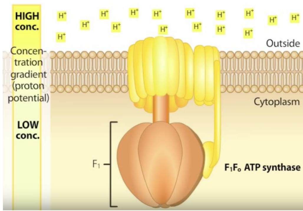
electron transport & proton pumping (electron donors drive ETC)
occurs in inner membrane
electrons energized with food enters ETC
electrons move through ETC → energy is released and used to pump protons into intermembrane space
proton gradient formed (more H+/protons on outside than inside)
proton flow drives ATP formation (proton gradient drives ATP production)
energy in electrochemical gradient drives photophosphorylation of ADP to produce ATP
proton gradient stores potential energy
protons pass through ATP synthase complex and turns ADP → ATP
oxygen is the TEA
O2 combines with electrons and H+ to form H2O
electrons from food → ETC (inner membrane)
energy from electron pumps H+ into intermembrane space (proton gradient)
O2 as TEA forms water
H+ flows back to matrix via ATP synthase where ADP → ATP
4.3 Chloroplasts
Recognize chloroplasts and its various compartments on electron micrographs
Discuss the chloroplast structure and its relevance to the process of photophosphorylation and photosynthesis
-inner and outer membrane
-thylakoid membrane
third internal membrane system not present in mitochondria
flattened sac-like structures where photosynthesis occurs
has proteins that help in electron transport → H+ protons pumped into lumen to create high concentration of protons → protons rush back via ATP synthase → ATP used in Calvin cycle
-plastids
family of organelles in plants that chloroplast is part of
-lumen
space inside of thylakoid
-production of
purines, pyrimidines for nucleic acids
complex lipids (all fatty acids)
most amino acids for plant cells
except cysteine, methionine, sulfur containing AA
stress response chemical involved in triggering plant immune response
-chemiosmotic coupling (photophosphorylation)
occurs in thylakoids
sunlight required and energy captured by chlorophyll
electrons with energy moves through ETC → powers ATP synthesis → water split to O2, H+ and electrons → NADPH + ATP made
ATP made in chloroplast stays in chloroplast
used in Calvin cycle
-Calvin cycle
light not required
ATP & NADPH from photophosphorylation converts CO2 into glucose → for plant growth & energy storage
-inputs H2O & CO2 → outputs O2 & glucose
electrons energized from sun enters ETC (thylakoid membrane)
energy allows H+ pumped into thylakoid lumen
H2O split to release more H+ and O2
H+ flows back into stroma via ATP synthase where ADP → ATP
ATP powers Calvin cycle to make glucose
Compare and contrast chemiosmotic coupling of proton pumping and ATP formation in mitochondria and chloroplasts
-similarities
use ETC
electrons move through proteins in
mitochondria: inner mitochondrial membrane
chloroplasts: thylakoid membrane
powers proton pumping to create H+ gradient
create a proton gradient
higher proton concentration on one side
ATP synthase complex used
protons flow back to low concentration via ATP synthase
converts ADP + phosphate → ATP
membrane to separate protons
mitochondria: inner membrane
chloroplast: thylakoid membrane
-differences
mitochondria energy: food
chloroplast energy: light
mitochondria electron source: NADH + FADH2
chloroplast electron source: H2O
mitochondria TEA: O2 to form H2O
chloroplast TEA: NADP+ to form NADPH
mitochondria proton pump: matrix to intermembrane space
chloroplast proton pump: stroma to thylakoid lumen
mitochondria ATP production: matrix
chloroplast ATP production: stroma
mitochondria ATP usage: cellular work (muscle movement, metabolism, etc)
chloroplast ATP usage: making glucose for Calvin cycle
Explain the relationship between mitochondria and chloroplasts in plant cells
-mitochondria and chloroplasts work together
-plants remove CO2 in atmosphere and put it back into biosphere
helps combat climate changes, CO2 accumulation
humans rely on plants
-sugars produced by chloroplast in plants are used by mitochondria to produce ATP required for cellular function
-mitochondria → oxidative phosphorylation
-chloroplast → photophosphorylation
Unit 5 Endomembrane System
5.1 Introduction & Protein Transport
Explain the structural & functional relationships between the different compartments of the endomembrane system and identify them on micrographs
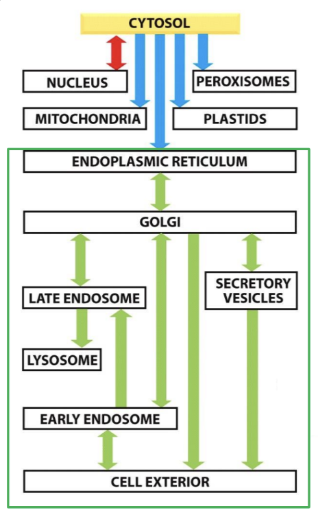
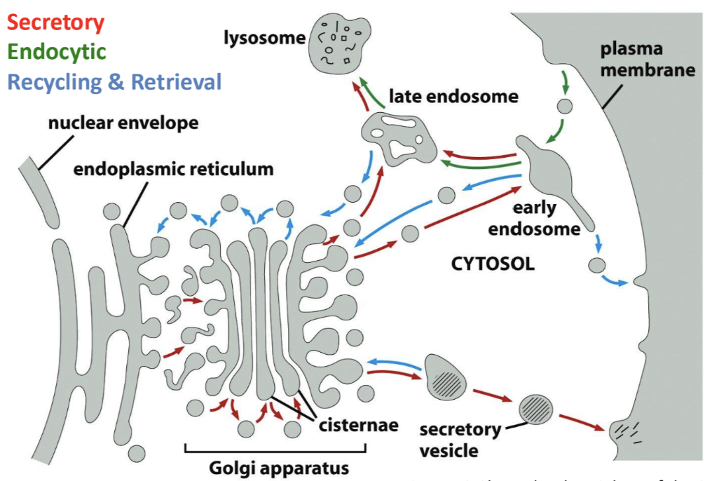
-endomembrane system
membrane limited compartments involved in processing & movement of proteins and membrane
consists of ER, Golgi apparatus, lysosomes, endosomes, secretory vesicles
involved in
processing of proteins for export from cell
proteins destined for lysosomes
proteins entering cell from surface
3 major subdivisions
secretory pathway
lysosomal pathway
endocytic pathway
3 types of proteins enter endomembrane system after translation
proteins destined for secretion
plasma membrane proteins
proteins destined to be endomembrane resident proteins
lives in Golgi apparatus or ER
only points of entry into endomembrane system are ER and plasma membrane
proteins destined for secretory/lysosomal pathways must enter at ER
proteins for endocytic pathway must enter via plasma membrane
proteins that enter endomembrane never leave pathway planned for them until final destination has been reached
ONLY return to cytosol if they are misfolded (refolded or destroyed)
-endoplasmic reticulum (ER)
structure: network of membrane-bound tubules & sacs
rough ER: covered with ribosomes
smooth ER: no ribosomes
function
rough ER: synthesizes proteins and sends them to Golgi in vesicles
smooth ER: makes lipids & steroids; doesn’t participate in protein synthesis
relationship to endomembrane system: ER packages proteins and lipids into vesicles → Golgi
-golgi apparatus
structure: flattened membrane sacs
function: modifies, sorts, packages proteins & lipids before sending them to their destination
proteins enter from cis to trans
relationship to endomembrane system: receives vesicles from ER, modifies and packs protein into new vesicles → plasma membrane or lysosome or other parts of cell
-lysosomes
structure: small, membrane-bound organelles filled with digestive enzymes
function: break down worn-out cell parts, food particles, foreign invaders
relationship to endomembrane system: made by Golgi and contain enzymes that help break down waste materials inside vesicles
-endosomes
structure: vesicle-like membrane bound compartments; have acidic interior
function: sort and transport materials inside the cell (particularly those taken in from outside cell via endocytosis); decides whether materials should be sent to lysosomes, recycled, sent to other areas of cell
relationship to endomembrane system: connects plasma membrane, Golgi, lysosomes and vesicles
-vesicles
structure: small, membrane-enclosed sac
function: transports proteins, lipids, waste materials between organelles
relationship to endomembrane system: vesicles move between ER, Golgi, lysosomes, plasma membrane
-plasma membrane
structure: lipid bilayer that surrounds the cell
function: controls what enters and exits the cell
relationship to endomembrane system: vesicles from Golgi fuse with plasma membrane to release proteins outside the cell (exocytosis) or bring materials inside (endocytosis)
-protein processing
all proteins are processed after translation
folding
supported by disulfide bridges between cysteine residues
bonds formed as protein assumes its folded structure
disulfide isomerase in ER facilitates formation of disulfide bridges
disulfide bridges not common in cytosolic proteins
removal of first methionine (carried by first tRNA)
methylation
phosphorylation
acetylation
formation of disulfide bridges
glycosylation
new polypeptides from ribosomes fold immediately based on primary sequence
spontaneous
lowest possible energy conformation
proteins fold in different ways → different path each time
Describe how proteins are targeted and imported into the ER and compare these mechanisms to protein targeting and import into the nucleus
-endoplasmic reticulum
flattened membrane cisternae and tubules
continuous with nuclear envelope
controls calcium levels in cytoplasm (calcium store)
cell signaling, muscle contraction
-rough ER
cytosolic surface of membranes have attached ribosomes synthesizing proteins for import into ER
site of synthesis for proteins destined for secretion, lysosomes, membranes
quality control → checks for defective proteins
-smooth ER
continuous with rough ER
site of lipid and steroid synthesis
doesn’t participate in protein synthesis
-transitional ER
site where both ERs meet
vesicles form and newly synthesized proteins exit ER to be able to move to next destination
-protein targeting
proteins going to a specific destination containing a sorting/targeting signal in their sequence
-entry/exit from ER
cells control what is allowed to enter/exit er
only proteins with proper signals are allowed to enter
proteins are either
ER residents
proteins that live in ER
carries ER retention signal (KDEL) at carboxyl ends
if no KDEL → protein will travel through endomembrane system and secreted out of cell
passing along through to other destinations
once proteins enter ER → never return to cytosol (unless sent to proteasome)
proteins that get exported from ER must be properly folded by chaperone proteins, packaged into vesicles and moved into next organelle (usually Golgi)
-proteins enter ER co-translationally
ribosome that is translating proteins destined for ER becomes attached to surface of ER during translation
-soluble proteins entering ER
translocated into lumen of ER
need only a single transfer sequence (N-terminus)
peptides move through translocation channel into ER lumen
signal sequence remains embedded in membrane
cleaved by signal peptidase → protein free in lumen of ER
-membrane proteins entering ER
partially located in ER
stuck with part of it embedded inside membrane
proteins can be destined for
ER
resident proteins in Golgi
plasma membrane
once inserted into membrane → can’t be removed
Predict the signal sequence required to insert a protein into the ER membrane in any orientation and predict protein topology from a corresponding domain map
-2 things required for protein to be targeted to ER
signal encoded within protein
receptor that recognizes & binds signal
Signal sequences are necessary & sufficient to direct proteins to ER
necessary (lof): remove signal sequence from ER protein to see if it stays in cytoplasm — ER protein doesn’t enter ER
sufficient (gof): add signal sequence to cytosolic protein to see if it ends up in ER — supposed to be in cytoplasm but has sequence so it enters ER
-ER signal sequence
always at least 8-10 nonpolar amino acids in a row
N terminal start transfer sequence
internal start transfer sequence
stop sequence
-signal recognition particle (SRP)
binds to exposed N-terminal signal sequence and ribosome
-start transfer sequences
N-terminal start transfer sequence
~18 hydrophobic amino acids at N-terminal
always cleaved off by signal peptidase
initiates transfer of protein across ER membrane
if present → N-terminal end of protein in ER lumen
internal start transfer sequence
located internally
binds to SRP
initiates transfer of protein across ER membrane
membrane crossing domain
not cleaved after transfer of protein
if starts with internal start sequence and not N-terminal start sequence → N-terminal end of protein in cytosol
*can have N-terminal start and internal start transfer sequence BUT is separated by a stop sequence
-stop transfer signal
~18 hydrophobic amino acid residues
found internally
only occurs after N-terminal start sequence or internal start transfer sequence
stops transfer of protein across ER membrane
membrane crossing domain
remains in membrane
not cleaved
ER signal sequence translated as part of primary sequence
recognized by ribonucleic protein
SRP binds to ER signal sequence → inhibits translation
ribosome + mRNA + partially translated protein complex → ER → binds to SRP receptor protein in ER membrane
ribosome attaches to translocation channel for newly synthesized polypeptide
attachment facilitated by SRP receptor; energy required (GTP)
SRP removed and translation resumes
new protein pushed through translocation channel into ER lumen
C - cytosol; L = lumen
A: protein inserted in ER lumen
B: vesicle with protein buds off from ER
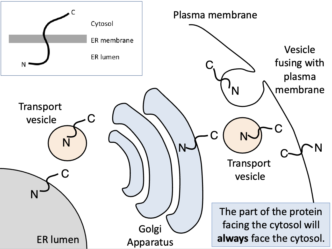
cytosolic portion of protein still in cytosol
C: vesicle travels to plasma membrane and starts fusing with membrane
vesicle successfully fuses with membrane → protein now part of membrane
cytosolic portion still in cytosol
-domain maps
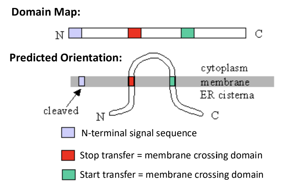
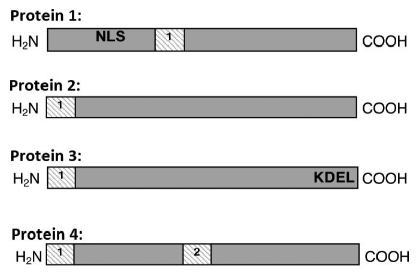
protein 1: ends up in plasma membrane via ER to be a integral membrane protein
protein has NLS (but signal is only recognized after protein has been completely translated & folded in cytoplasm)
internal start transfer signal sequence is recognized first
protein has
N-terminus in cytoplasm
transmembrane domain in ER membrane
C-terminus in ER lumen
protein 2: forms a soluble protein in extracellular space
has 1 hydrophobic sequence → N-terminal signal sequence → protein is co-translated into ER lumen → N-terminal signal sequence is cleaved → soluble protein in ER lumen BUT because no KDEL → moves to membrane
protein processed → vesicles → vesicle fusion with membrane → protein excreted as extracellular soluble protein
protein 3: ends up as ER resident protein (soluble)
has 1 hydrophobic sequence → N-terminal signal sequence → protein is co-translated into ER lumen → N-terminal signal sequence is cleaved → soluble protein in ER BUT because KDEL retention sequence → kept in ER lumen
protein 4: becomes integral protein in plasma membrane
has 2 hydrophobic sequences
N-terminal signal sequence
internal stop transfer signal sequence
protein has
N-terminus in ER lumen
transmembrane domain in ER membrane
C-terminus in cytosol
becomes an integral protein in plasma membrane after processing
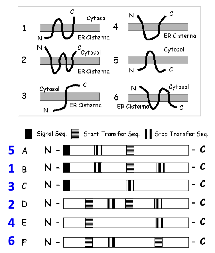
-hydropathy plots
shows protein sequence before N-terminal signal sequence is cleaved off
produced based on original protein sequence
not what they look like after protein processing
Discuss the role of chaperones in protein folding and role of proteasomes in quality control of misfolded proteins in the secretory pathway
-if cells make mistake in protein assembly → ER ensures properly folded proteins move on
misfolded proteins → refolded with chaperone proteins
-chaperones
facilitate and aid with proper polypeptide folding
binds specific regions of polypeptide
provides a protected space away from rest of cytosol so protein can fold in peace (in ER)
BiP (binding protein)
prevents hydrophobic domains of proteins from aggregating and promotes promotes proper folding
-proteasome (only time protein is sent back to cytosol after entering ER)
found in cytosol and nucleus
misfolded proteins destroyed
amino acids recycled and incorporated into new proteins
degrades proteins tagged for destruction with small polypeptide ubiquitin
Describe and interpret the results of the different experimental tools studying the secretory pathway
-microscopy
live cell imaging of GFP-tagged protein
5.2 Vesicle Transport
List the 4 stages of vesicle transport and list the protein machinery involved at each step
-vesicles
moves proteins (cargo) around the cell
from membrane-bound organelles to another compartment or plasma membrane of endomembrane system
produced by all organelles of endomembrane system
formation
regulated by GTPase
bound to GTP: active
allows adaptors and coat proteins to bind
assembles membrane coat
GTP hydrolyzes to GTP: inactive
GTPases, coat proteins & adaptors dissociate from vesicle
flippase — generated asymmetry
vesicle formation & fusion relies on same membrane thermodynamics
coat proteins
coats have to be shed off → naked vesicle for transportation
2 primary functions
force membrane to curve and form bud
capture cargo molecules for transport
COP → coatamer proteins
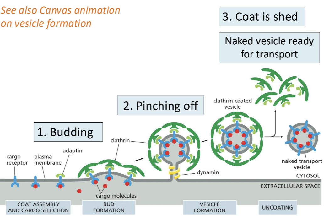
clathrin-coated vesicle
used for
endocytosis
traffic to lysosome
receptor recycling
between trans Golgi network (TGN)
endosomes/lysosomes
plasma membrane
requires
cargo receptor
adaptin
coat protein
dynamin
uptake of extracellular
COPI-Coated Vesicles
involved in moving cargo from Golgi → ER traffic (retrograde)
dynamin protein not required
COPII-Coated Vesicles
involved in moving cargo from ER → Golgi traffic (anterograde)
dynamin protein not required
cargo can things that were secreted
transport
short distance vesicles move via diffusion
long distance vesicles move along
cytoskeletal tracts
microtubules
motor proteins
kinesins/dynein for microtubules
myosin for actin
docking
vesicles MUST dock before fusing (vesicles directed to correct place for fusion)
dock via Rabs & Tethers
proteins that direct vesicles to the right place for docking
active Rab-GTPases are present on vesicular membranes
if right tethering (Rab-binding) → protein is present on target membrane → vesicle will tether
brings vesicle in close enough proximity to begin membrane fusion
Rabs
small GTPases that sit on vesicle surface when activated
identifies vesicle targeting to particular membrane
Tethers
binds to vesicle Rab and brings in vesicle from cytosol
fusion
fuse via SNAREs
proteins that mediate vesicle fusion with membrane
alpha-helix structures
vesicle-SNAREs (v-SNAREs)
on vesicle membrane
target-SNAREs (t-SNAREs)
on target membrane
must have at least
1 SNARE on each membrane
4 distinct SNARE coils to mediate fusion
orientation of membrane constant
cytosolic face of vesicles remains cytosol facing throughout vesicle transport
what happens in cytosol stays in cytosol
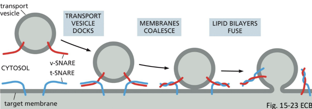
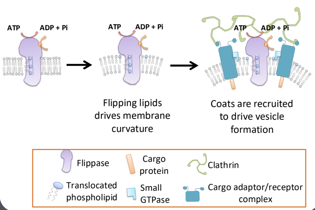
Explain how vesicle coats facilitate cargo loading and vesicle budding
Discuss the importance of maintaining specificity in both docking and fusion of vesicles at their target compartments, and how Rabs, tethers and SNAREs facilitate this process
Discuss and illustrate how the orientation of membrane proteins in the lipid bilayer is maintained during transport and after fusion of vesicles with target membranes
5.3 Golgi & Protein Processing
Discuss the functional compartmentalization of the Golgi structure and recognize the Golgi apparatus using different types of microscopy
-Golgi apparatus
center of endomembrane system
receives cargo from
ER, lysosome, plasma membrane, endosomes
receives cargo → modifies → repacks into new vesicles → sent to new destination
site of all major cellular work involving polysaccharides
site of all known glycosyltransferases
glycosyltransferase — enzymes capable of attaching sugars to each other
important for production of
glycoproteins
cell walls of plants and fungi
structure
varies between different kingdoms/species
polar
cargo enters from one end
cis face for newly synthesized proteins
cargo exists from other end
trans face
cisterna
each pancake of Golgi
series of flattened stacks
fluid containing sac
lumen
interior space of each cisterna
-Golgi resident proteins
distributed in Golgi
cis to trans
full time jobs in Golgi
different resident proteins present in different cisternae
resident proteins have retention signal so they know to stay in Golgi
-transient/cargo proteins
moves through golgi (cis → trans) to get to where they need to (final destination)
just passing through golgi
moving on to other destinations of endomembrane system
cis cisterna → medial cisterna → trans cisterna → into TGN to be packed into vesicles → sent to next destination
-trans golgi network (TGN)
protein sorting center
vesicles leave TGN for different destinations
maturing lysosome
condensing secretory granules released through regulated secretory pathway
plasma membrane
vesicles containing membrane and proteins are released to surface via constitutive secretory pathway
-golgi aids in production of cell wall
rosettes (proteins) too large for vesicle but has to be delivered to plant plasma membrane
rosettes must follow same path as a regular newly synthesized protein destined for plasma membrane
rosettes synthesized one sugar at a time as molecules move from cis to trans in Golgi
packaged into vesicles in TGN → plasma membrane → secreted and incorporated into plant cell wall
Describe the sequence of events occurring during protein glycosylation in the ER and Golgi
-glycosylation
post-translationational modifications of proteins
addition of polysaccharides
adding sugar molecules (glycans) to proteins
helps proteins fold properly, become more stable
enzymes that carry out reactions are located in lumen of ER and Golgi
2 types of glycosylation
N-linked glycosylation
occurs in ER & golgi
attaches sugars to N of Asn amino acid
starts in rER → Golgi
large sugar group built on lipid molecule (dolichol) in ER
sugar transferred to specific Asn-X-Ser/Thr) site on a protein
protein checked for proper folding
Golgi trims and modifies sugar to make mature glycoprotein
O-linked glycosylation
occurs in golgi AFTER protein has folded
attaches sugars to O of Ser/Thr amino acids
single sugar (N-acetylgalactosamine) added first
more sugars added in step-by-step manner
final glycoprotein sent to destination
-oligosaccharides assembled one sugar at a time
specific enzymes found in each compartment of Golgi stack that does its job
-secretion of glycoprotein
ribosomes binds to mRNA in cytoplasm
co-translational insertion of protein into ER
enzyme transfers oligosaccharide core from dolichol to protein
oligosaccharide has sugars trimmed and added by glycosyl transferases
protein packaged in trans-Golgi into secretory vesicle
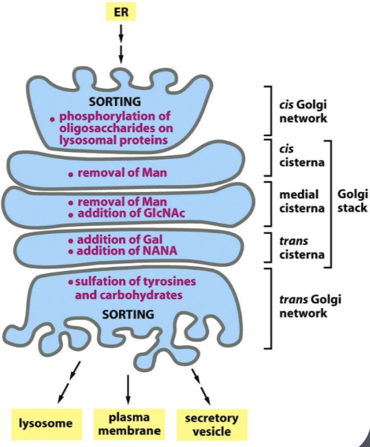
Use experimental evidence to determine how membrane and cargo are transported through the Golgi
-vesicle transport model (lecture 25)
cisternae stationary
resident proteins living in cisternae
cargo proteins moved from one cisterna to next via transport vesicles
vesicles need to be formed every time from cis to medial to trans
vesicles fuse every time they enter a cisterna
transient proteins in vesicles (vesicle formation via COPII coat proteins) → bud from periphery of Golgi cisterna → fuses with target cisterna
new vesicles formed every time
vesicle fuses with cis membrane → proteins enter cis cisterna→ protein forms new vesicle to exit cis → vesicle fuses with medial membrane → proteins enters medial cisternae → protein forms new vesicle to exit medial → vesicle fuses with trans membrane→ proteins enter trans → proteins sorted into new vesicles (in TGN) for final destination
*sugars added at every cisternae
*transient proteins moving forwards via vesicles; cisterna stationary
*resident proteins stay in place
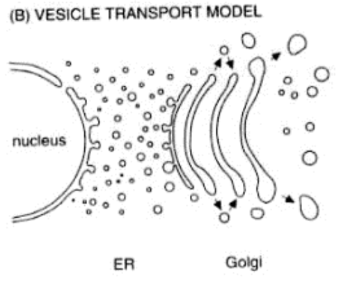
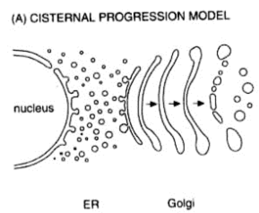
-cisternal maturation model
cisternae moves
resident proteins move backwards to the original cisterna location
new cis-cisterna forms → old cis-cisterna becomes medial-cisterna (and so on)
eventually trans-cisterna broken down to form vesicles
transient protein with vesicle fuses with cis membrane → transient proteins enter cis cisterna → cisternae shifts (cis → medial, medial → trans, trans → broken down, new cis comes in) → resident proteins move backwards to the original location of cisterna via vesicles (fuse) & transient proteins don’t move via vesicles, but they move with the cisternae → when transient proteins reach cisternae that has matured into trans cisternae → packaged into new vesicles (in TGN) for final destination
*resident proteins moving backwards via vesicles; cisternae not stationary
*transient proteins stay in place
5.4 Post-Golgi Traffic
Describe the secretory, lysosomal, endocytic pathways and list all components of these pathways
-secretory pathway (exocytosis)
proteins carry information needed to determine which pathway they belong within their primary amino acid sequence
constitutive secretion
occurring continuously
vesicles are continuously forming
vesicles continuously carrying proteins from Golgi to cell surface
leads to plasma membrane
supplies continuous stream of vesicles to plasma membrane for release outside of cell
vesicles containing lipids, proteins, polysaccharides
supplies plasma membrane with these
refreshes old lipids
contributes to extracellular matrix
no signal required for vesicle release
vesicle formation doesn’t require coat proteins
regulated secretion
vesicles only released when specific signal is received
binding of hormone
vesicles may or may not require coat proteins
pH may play a role in formation of vesicles when no coat used
proteins aggregates at low pH
aggregation allows vesicles to be highly condensed
proteins consolidated into vesicles that are stored in cell until secreted
leads to plasma membrane
release of vesicles with cargo requires signal from cell before final fusion with membrane
vesicle formation aided by proteins destined for this pathway
proteins clump in acidic conditions (aggregation)
helpful since Golgi is acidic as it moves from cis to trans)
TGN → large protein aggregates can push up against membrane and help to promote curvature
-lysosomal pathway
directs newly synthesized lysosomal proteins to lysosomes via endosomes
sends lysosomal proteins to late endosome
acidic enough to allow separation of enzyme from receptor that carried it there
late endosome can become a hybrid compartment before maturation to lysosome
-endocytic pathway
Compare and contrast constitutive and regulated secretion and explain the important of each
-secretory mutants
proteins needed for secretion
temperature-sensitive mutants are observed only at higher temperatures
proteins function normally at lower temperatures
secretory proteins with GFP can be tracked in live cells
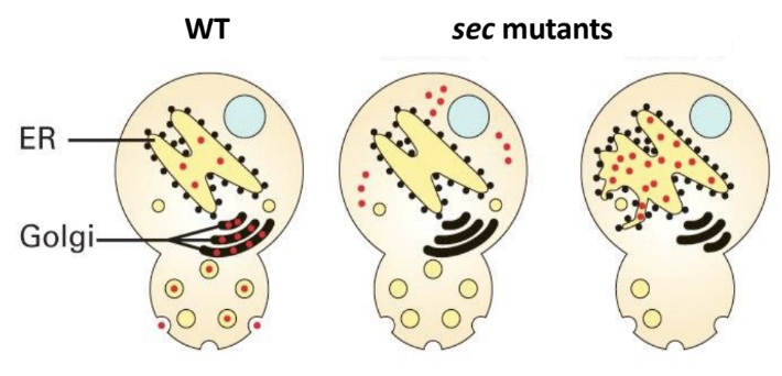
wild type: secretory proteins in vesicles, ER, Golgi
sec mutant left: secretory proteins in cytoplasm
transport of proteins into ER disrupted
mutation → components for ER targeting/co-translation
ER signal sequence & SRP amy be specific mutated components
sec mutant right: secretory proteins in ER
transport of proteins from ER → Golgi disrupted
mutation → vesicle formation error
COPII-vesicle formation (COPII coat, adaptor protein, cargo receptors)
*mutation due to ER retention signal is unlikely
Discuss the structure and function of lysosomes and explain how they mature from late endosomes
-lysosome
secretory vesicles that contain digestive enzymes
site of intracellular digestion
contains mixture of ~40 different types of digestive enzymes
degrades nucleic acids, proteins, lipids
membrane bound
very acidic (pH ~4-5)
maintained by proton pumps embedded in membrane
membrane of lysosome resistant to action of its own digestive enzymes
due to extensive glycosylation of proteins on inside of membrane
facilitate digestion of intracellular structure that cell needs to dispose
large polymers brought in from exterior via endocytic pathway
breakdown of bacteria engulfed by cell during phagocytosis
Trace the path of a newly synthesized lysosomal protein to its final destination and compare this to the path of proteins to all other destinations within the endomembrane path
-for protein to be directed to lysosome → specific sorting signal required
signal within chemical structure of that protein cell can recognize
targeting sequence (M6P - mannose-6-phohsphate)
M6P is built from oligosaccharide tree added to asparagine in protein in ER based on its amino sequence
in cis Golgi → phosphate added to 6th carbon of specific mannose in tree
2 fold result
M6P tag formed used later by receptor
added as result of stopping other glycosyltransferases in Golgi from being able to further modify sugar tree
signal recognized by M6P receptors in TGN
packaged into clathrin-coated at TGN → vesicles sent to endosome
lysosomal proteins separated from their receptors due to increased acidity in endosome
lysosomal proteins travel with endosome to lysosome and receptors are returned to TGN
5.5 Endocytosis
Discuss the structure and function of endosomes and differentiate between early and late endosomes
-endosomes
sorting center
junction between lysosomal pathway and endocytic pathway
differentiates between cargo going to lysosome and receptors that has to be returned to TGN/pM
cargo fate compared to receptor is dependent on acidic environment of endosome
cargo is one that gets released by acidic endosomal environment → cargo in lysosome & receptor free to be recycled
receives cargo from plasma membrane destined for different places
receptors returned to PM domain from where they came from while their cargo is passed onto lysosome
some receptors not recycled but sent to lysosome to be degraded
done with or without cargo
some receptors along with their cargo transferred to another plasma membrane domain
allows specific cargo to be passed from one side of cell to another (transcytosis)
pH
endomembrane more acidic than cytosol
increasingly acidic moving from point of origin to endpoint
culminates in lysosome (as acidic as stomac acid)
specific pH causes change in certain proteins
gives cell control over how and when certain events happen
proper functioning of TGN requires it to maintain appropriate pH
different from cytosol pH and nearby surroundings
-early endosome
first compartments that form when material from outside cell is engulfed into cell via endocytosis
material brought to cell in vesicle and fuses with early endosome
sorts internalized materials
decide whether substances should be sent back to cell surface, go to lysosomes (degradation), stored for future use
slightly acidic (6.0-6.5)
-late endosome
later stage of endocytic pathway
early endosomes mature into late endosomes after they undergo acidification and internal sorting
involved in preparing cargo for degradation in lysosomes
more acidic (5.0-5.5)
activates enzymes needed to break down internalized cargo before it transfers to lysosome for complete digestion
Compare and contrast between constitutive and receptor-mediated endocytosis
-constitutive endocytosis = pinocytosis
continuous process where cells remove excess membrane added by exocytosis
allowing recycling of plasma membrane
non-selective process
fluid & macromolecules from extracellular region taken up without regard to type of concentration of molecules present
no specific receptors for uptake of macromolecules
may or may not use coated vesicles
advantages
doesn’t require receptor proteins
vesicles may not be required → coat proteins, adaptors not required → less energy required
lower energy cost
disadvantages
accidentally bring in unwanted items into cells
cell membrane gets bigger
-receptor mediated endocytosis
receptors used to collect specific extracellular compounds
clathrin used for vesicle formation
endosomes act as sorting centers for endocytic pathways (from TGN & plasma membrane)
helps sort
proteins/materials enter cell that are destined for
lysosome
cell surface
other endomembrane compartments
proteins that leave TGN destined for lysosomes
early endosomes mature into late endosomes → form lysosomes or fuse with existing lysosomes
Trace a molecule from the exterior of the cell to the lysosome along the endocytic pathway
uptake via endocytosis
molecule from outside of cell binds to receptor in cell membrane
vesicle created that engulfs molecule (early endocytic vesicle) via clathrin protein
early endosome
vesicle formed from membrane fuses with early endosome inside cell
slightly acidic pH dissociates cargo from receptors
early endosome sorts material and decides where it should go
maturation into late endosome
undergoes acidification
internal environment is more acidic → activates enzymes to break down internalized material
lysosome fusion
late endosome fuses with lysosome
lysosome contains hydrolytic enzymes that degrade wastes
fusion creates mature endolysosome
material is broken down into small components by enzyme in lysosome
degradation and recycling
simpler molecules recycled within cell
pathogen/damaged cellular material is ingested by lysosome → resulting molecule used/expelled by cell
Explain with examples how receptor proteins facilitate endocytosis
-low-density lipoprotein (LDL) receptor
complex that makes lipids/cholesterol water soluble so they can be carried in bloodstreop
receptors used to bring LDL into cell
receptor: LDL receptor on cell membrane recognizes and binds LDL (carries cholesterol in bloodstream)
binding: LDL molecule binds to LDL receptor on cell surface
clathrin coat formation
binding triggers clathrin formation
internalization
clathrin-coated vesicle buds off from plasma membrane
carrying LDL and its receptor inside the cell
uncoating
clathrin coat removed once inside cell
vesicle → early endosome
sorting
acidic environment of early endosome sorts molecule
causes LDL to dissociate from receptor
receptor recycled back to plasma membrane
LDL directed to late endosomes → lysosome for processing
Compare and contrast receptor recycling in the lysosomal and endocytic pathways
Unit 6 Cytoskeleton
6.1 Overview of Cytoskeleton: Intermediate Filaments
Compare and contrast the structure and function of three types of cytoskeletal elements: actin filaments, intermediate filaments and microtubules
-cytoskeleton
gives cell its shape
resists mechanical stresses
cells aren’t static → change shapes all the time
cytoskeleton coordinates the change while maintaining tissue integrity
acts as an anchor for many proteins and organelles
provides tracks for transport of vesicles, organelles, other cargo
helps with division of both cytoplasm and DNA during mitosis
3 major types of filaments in most cells
actin filaments
intermediate filaments
microtubules
-dynamic instability
actin filaments, microtubules are dynamically unstable
grow, pause, shrink in the span of a few seconds
unstable, constant state of influx
-actin filaments (AF)
smallest of the three (~ 7 nm in diameter)
polymer (f-actin/filamentous actin/microfilaments) of repeating subunits (g-actin/globular actin)
undergoes dynamic instability
attaches to plasma membrane proteins
shapes cell
respond to cellular environment
animal cells
involved in cellular locomotion, cellular adhesion & shape, cytokinesis, cargo trafficking, muscle contraction
close to membrane
cortical arrangement
plant cells
cellular arrangement is different
found throughout cytoplasm
not just underneath plasma membrane
involved in cargo trafficking and cell shape
actin binding proteins (APBs) (MARCH 26 LECTURE FOR FULL DEPTH OF WHAT APBS DO)
uses proteins to control when and where actin filaments form
allow cell to control how actin filament grows
nucleating protein allows cell to control where actin grows
severing proteins cut actins
capping proteins (plus end) allows actin to stay the same length
side-binding proteins stabilize actins
bundle proteins form actin bundles
work with actin to form different actin networks in cell
parallel bundles
filaments closely spaced and have some polarity
ABPs (formin & profilin) build array
contractile bundles
in which filaments are arranged anti-parallel and cross-linked by stabilizing proteins
molecular motors drive contraction of these arrays
cross-linked gel
random orientation of fibers, linked at crossing by filamin
actin gel major component of cell cortex of most cell types
plays role in locomotion
ARP 2/3 used to build filaments in these arrays
additional proteins used to crosslink and stabilize
can be controlled by
controlling where an actin filament forms
provide nucleation sites
Arp 2/3 binds to side of a pre-existing actin filament and makes a branching network
formin & profilin work together to make parallel networks
stabilizing actin filaments using protein caps
structures formed by actin are required long term
-intermediate filaments
middle size of the three (~10 nm in diameter)
only found in animal cells
very strong
family of proteins classified into 5 major types
keratins, vimentin, neurofilaments, nuclear lamins
only 1 or 2 expressed in each cell types
nuclear lamins important in maintenance and organization of cell envelope
single filament always made from same type of subunit
different cell types express different intermediate filament proteins
doesn’t undergo dynamic instability (actin & microtubules do)
can be assembled and disassembled as needed
helps with anchoring non-motile cells
resists mechanical strain in tissues
vital to maintenance of cell shape
long, twisted strands that are fibrous
structure depends on twisting of coiled-coils via hydrophobic amino acids
twisting due to van der waal interactions
intertwined alpha-helices form coiled-coil dimers
whatever is structurally stable/favoured
-microtubules
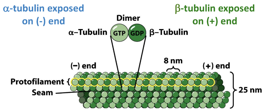
largest of the three (~25 nm in diameter)
hollow tubes made of repeating subunits (tubulin)
tubulin made of 2 smaller subunits (monomers) that carries a GTP molecule
alpha-tubulin
carries GTP
exposed at minus end
doesn’t hydrolyze GTP → GDP
beta-tubulin
hydrolyzes GTP → GDP
exposed at plus end
have polarity → ends are different from each other
undergoes dynamic instability
uses GTP as its energy source
constantly growing/shrinking
growing via addition of new monomers at the end of existing microtubule
addition of tubulin dimers is faster at plus (beta) end than the minus (alpha)
more growth on plus end vs the minus end
if microtubule required for a long period of time in same place → must be stabilized
dynamic instability reduced or eliminated
gamma-tubulin acts as a cap that stops microtubule from being able to depolymerize from minus end
protein cap can be added to microtubule at plus end so that it can’t deploymerize
microtubules that are capped at both ends are stable
remain in place for as long as cells need them
involved in mitosis in all cells, cargo trafficking, maintenance of cell shape
pathways where vesicles and cargo travel on
controls and maintains specific cellular shapes and location
provides sites to anchor complexes and proteins in specific regions of a membrane
cell controls conditions in order to promote growth, maintenance or shrinkage as required
Identify the different types of cytoskeletal elements on different type of micrographs
Discuss different types of intermediate filaments and correlate their structure with the strength of the assembled, functional cytoskeletal elements
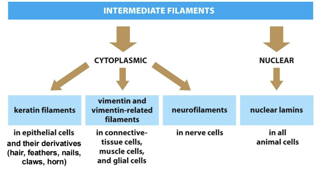
-intermediate filaments
only found in animal cells (vertebrates)
within one filament → single kind of subunit
keratin
protein found in epithelial cells and their derivatives
hold skin cells to each other and to underlying membrane
provides resistance against mechanical tension
vimentin
found in intracellular fibers in connective tissue cells
including muscles
helps to hold tissues together
provides strength
neurofilaments
found in neurons (expresses neurofilaments)
neurons have a distinct shape
helps to form and support characteristic shape of nerve cell
long axon projecting out one side of cell
longest axon in human body (sciatic nerve — base of spinal cord → big toe)
if neurofilaments or microtubules become disrupted → axon loses its shape, retracts and dies
dementia as a result
lamins
components of nuclear lamina
in interphase, assembled nuclear lamins forms a lattice work under nuclear envelope
provides structure to nuclear envelope
aids with chromatin organization
nuclear lamina
maintains attachments to cytoskeleton on exterior of nucleus
during mitosis
destabilization of nuclear lamina (via phosphorylation of lamins) drives disassembly of nuclear envelope
after mitosis
lamins dephosphorylated and lamins reforms
since its attached to membrane and chromosomes → everything ends up back where it’s supposed to be
form a meshwork beneath nuclear envelope
maintain integrity of nuclear envelope
-all intermediate filament subunits undergo self-assembly to spontaneously form final filament
energy not required
-amino acid sequence itself promotes formation of alpha-helix that makes up bulk of structure
formation of first dimer facilitated by this process
-chemical analysis of alpha-helix shows that they have a line of non-polar amino acids on one side of alpha-helix
-central helical domain is highly conserved among the different intermediate filaments
N & C terminal domains on either side of central domain can differ in size, sequence & function
differences in N & C terminal allows for diversity of functionality amongst the 3 different intermediate filaments
-in order for skin to resist everything it gets put through
cells need to be stronger than plasma membrane
keratin (proteins) provides protection in skin
keratin ropes pass from one side of cell to other and bind to desmosomes in plasma membrane
keratin + desmosome complex transmits mechanical stress along ropes instead of through membrane
-desmosomes & hemidesmosomes
cell junctions that connect cells to each other or to the extracellular matrix
anchored to intermediate filaments
strengthen tissues
desmosomes
connect adjacent cells together
cell-to-cell connection
provides mechanical strength to tissues under stress
binds to membrane layer under skin (hemidesmosome)
cadherins involved
adhesion molecules
hemidesmosomes
attaches cells to extracellular matrix
cell-to-matrix connection
anchors epithelial layers
anchors cells to basal lamina
integrins involved
adhesion molecules
diseases → mutations in intermediate filaments affect cell’s ability to resist tensile strength
epidermolysis bullosa (EB)
skin blisters and tears at the smallest touch
skin extremely fragile
6.2 Microtubules
Compare in vivo (in cells) and in vitro (in test tube) microtubule polymerization
-in vitro (test tubes)
tubulin removed from cells to see how it works
tubulin dimers have affinity for each other → over time → polymerize on their own into polymer
polymers form via non-covalent interactions
initiating cytoskeleton polymerization (nucleation) to build microtubule or actin polymers is a slow process in vitro
active tubulin dimers
GTP bound β-tubulin
higher affinity for microtubules
inactive tubulin dimers
GDP bound β-tubulin
can bind to microtubules, but less efficiency than active tubulin dimers
once bound to microtubule, over time GTP β-tubulin hydrolyzed into GDP β-tubulin
kinetics of spontaneous microtubule assembly in vitro
no enzymes required
rate of polymerization dependent on location
slow growing end — (-) end — higher critical concentration (Cc)
doesn’t easily bind β-tubulin in an incoming dimer → not the right conformation
needs more tubulin for it to grow
fast growing end — (+) end — lower critical concentration (Cc)
adding new subunits causes a conformational change in β-tubulin that increases binding for more subunits
doesn’t need as much tubulin for it to grow
lag phase is slowest
initial nucleation dependent on molecules running into each other via random motion in aqueous solution
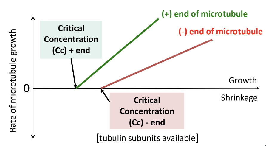
if concentration of tubulin is low → hard for tubulin subunits to find each other in solution to start the process
when concentration is so low that process doesn’t start → lag phase only → no polymerization
critical concentration (Cc) → concentration of tubulin required for polymerization to occur
minimum concentration of monomers required for polymer formation to begin
concentration of monomers that is maintained when the reaction is at an equilibrium
concentration of monomers where the microtubule switches from shrinkage to growth
growth rate = disassembly rate
rate of assembly increases with increasing concentration of tubulin subunits in solution
difference in both critical concentration AND rate of assembly at plus and minus end
plus end starts growing more easily than minus end (grows at a faster rate)
when bound as part of a microtubule
beta-subunit of tubulin dimer hydrolyzes its GTP
hydrolysis of GTP → GDP reduces affinity of binding of tubulin dimers to each other within microtubule
over time, affinity of tubulin dimers for each other decreases
formation of GTP cap
encourages microtubule growth
microtubule assembly happening faster than rate of GTP hydrolysis
occurs when high enough [free tubulin] is available
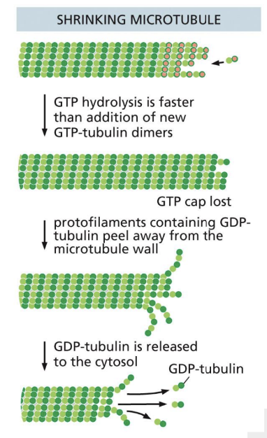
loss of GTP cap → rapid shrinkage of molecule
lower affinity
if polymerization slows down → GTP hydrolysis catches up and is now converted to GDP → GTP cap disappears
if there is high enough local concentration of activated tubulin → microtubule can be rescued and start growing again
GTP hydrolysis changes subunit confirmation forcing the protofilament into a curved shape
destabilization of microtubules through GTP hydrolysis results in instability in microtubule structure
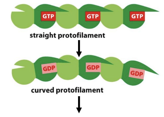
destabilization of microtubules via GTP hydrolysis results in unstable microtubule structure
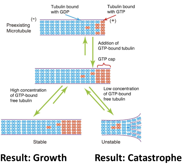
catastrophe
when GDP tubulin units accumulates too much → microtubule undergoes catastrophe
-in vivo (cells)
polymerization of microtubules influenced by environment
local tubulin concentrations may go up or down as nearby microtubules grow and shrink
other kinds of proteins bind to both monomers and polymers to influence their behaviour
microtubule organizing centers (MTOCs) in cells provide right conditions for rapid nucleation of microtubules
grow from centrosome
sites where microtubules growth is promoted via gamma-tubulin
gamma-tubulin
promotes microtubule growth
provides pre-made template for tubulin subunits to bind to
reduces lag phase of growth curve
controls exactly where microtubule is grown
side effect: bound to minus end of microtubule
minus end no longer free for growth or shrinkage
only plus end (beta) undergoing dynamic instability
microtubules can be stabilized via binding to proteins (microtubule associated protein — MAPs)
some MAPs bind microtubules to neighbouring microtubules but can still grow
establishes a network of microtubules going in the same direction
eventually microtubules can be capped (capping proteins) at the growing end by different types of MAPs
microtubules not growing or shrinking
specialized MTOC
spindle poles/fibers found in all cells during mitosis
cilia & flagella found in specialized cell types from different kingdoms
basal bodies are the MTOC
centralized MTOC
during mitosis → all eukaryotic cells have 2 centralized MTOCs (spindle poles)
coordinate proper placement of microtubules for separation of chromosomes
interphase → only some cell types have centralized MTOC
mammalian cells — centrosome (pair of centrioles and associated proteins
eukaryotic cells with cilia and/or flagella have additional MTOC (basal bodies)
located at base of each flagellum or cilium
decentralized MTOC
in plants and fungi → don’t have centralized MTOC in interphase
gamma-tubulin rings carried along pre-existing microtubules is motor proteins
during mitosis → cells collect all gamma-tubulin into 2 MTOCs
acts as spindle poles for mitotic spindle
GTP-bound tubulin dimers are in activated form
assembles at a faster rate compared to GDP-bound tubulin
GTP is in excess in cytosol
GDP-bound tubulin dimers that are bound in microtubules are reactivated by replacing GDP in beta-subunit with GTP
concentration of free-floating inactive tubulin is low in cell
→ only consider GTP-bound tubulin during microtubule assembly
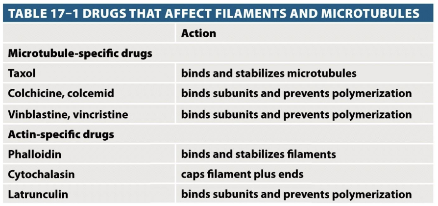
Interpret in vitro microtubule polymerization curves in terms of microtubule dynamics
Explain the process of dynamic instability, focusing on how tubulin dimers (alpha/beta) interact non-covalently to make microtubules with a ‘plus’ and ‘minus’ end
-dynamic instability
process where cells are constantly growing and shrinking
crucial for cell shape, movement and division
moving cargo around
helps cells divide by pulling chromosomes apart
allows cells to change shape and move
-microtubules made of tubulin dimers
alpha-tubulin
beta-tubulin
-microtubules have 2 ends
plus (+) end
grows and shrinks quickly
minus (-) end
more stable
usually anchored inside cell at MTOC
-growing microtubules (polymerization)
tubulin dimers add onto the plus end in a head-to-tail fashion
each beta-tubulin carries GTP
when GTP-bound tubulin dimer attaches → stabilizes the growing microtubule
Interpret live-cell images of microtubules undergoing dynamic instability and explain the current model of how GTP binding to tubulin can bring about this dynamic behaviour
Explain how motor proteins work and how their movement relates to the polarity of the microtubule
-motor proteins
able to bind to microtubule and move along it in a specific direction
energy required; ATP
microtubule has directionality (plus and minus end)
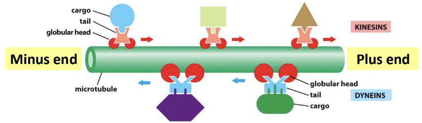
bind to microtubule in an orientation so that it moves cargo toward
minus → plus end (kinesins)
transports vesicles, organelles, proteins toward edges of cell
closer to cell
maintains structure of ER
plus → minus end (dyneins)
bringing cargo/materials back toward cell center/nucleus
moves structures (cilia, flagella)
farther from cell
maintains structure of Golgi
→ kinesis & dyneins are protein complexes with multiple chains
globular heads that attach to microtubule
tail region attached to cargo
two light chains part of molecule
located away from region of molecule that interacts with microtubule
powered by ATP
stepping cycline linked to ATP hydrolysis
may transport
vesicles
proteins and/or protein complexes
protein-mRNA complexes
RNP particles
membrane vesicles and organelles
other microtubules
flagella, cilia, other motile microtubule arrays
Provide examples of how proteins can interact with microtubules or tubulin to influence their structure, which in turn, will influence function
6.3 Actin Filaments
-polymerization of g-actin requires energy
g-actin subunit carries a single molecule of ATP in its core → hydrolyzed slowly once activated monomer gets added to polymer
ATP-cap found when polymer is adding g-actin at high rate
-dynamic instability
in vitro (test tube)
plus end has higher affinity for monomers than minus end
lower critical concentration
increased rate of assembly once polymerization starts
in vivo (cell)
cell has high concentration of ATP in cytosol
easy to keep unpolymerized g-actin in cell in activated states
Describe in vitro (in test tube) actin filament structure and polymerization and the role of monomer concentration and ATP in dynamic instability
-in vitro (test tube)
actin monomers (G-actin) assemble into actin filaments (F-actin)
kinetics of spontaneous actin filament assembly in vitro
same polymer assembly kinetics as microtubules
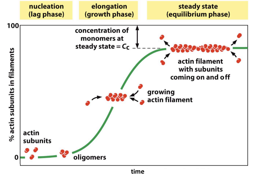
monomer accumulation → increases then plateaus → monomers being used to form filaments
filament starts low but as monomers start forming filaments → filament mass starts increasing
filament mass not present until actin concentration reaches point where monomer mass stops increasing and stays constant
filament mass increases linearly with actin concentration
critical concentration found at ______ LECTURE 32 MARCH 26th
Correlate in vivo (in cells) actin filament polymerization and organization at the cell cortex with its function of deforming the plasma membrane and regulating cell shape
-in vivo (cell)
free actin is 50-200 um → cC is < 1 because proteins bind to free actin subunits
f-actin uses nucleating sites in cell
no MTOC equivalent to organize actin like microtubules have
actin has to be nucleated at multiple sites
various APBs control behaviour of F-actin polymers in cells
refer to top part about what APBs do
ARP 2/3 complex nucleates branched actin arrays
attaches at minus end
nucleases at 70° and branches (branching network/effect)
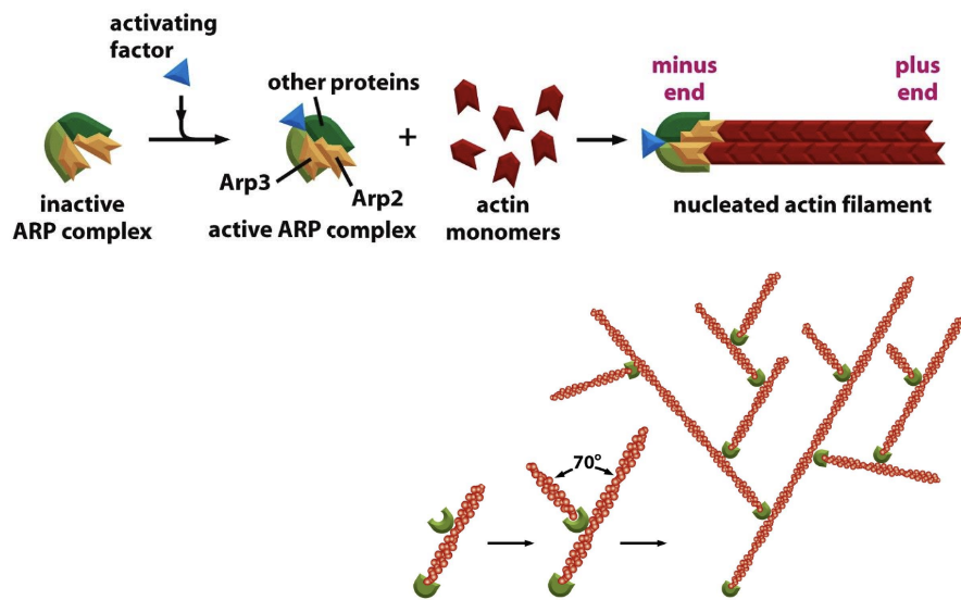
animal cells: actin filaments have different arrangements in different parts of the cell that performs different functions
formation of actin network at plasma membrane pushes membrane forward & allows cells to move
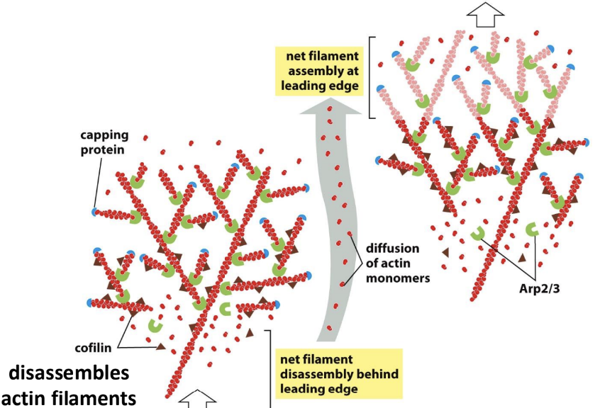
formation of actin network at plasma membrane pushes membrane forward allows cells to move
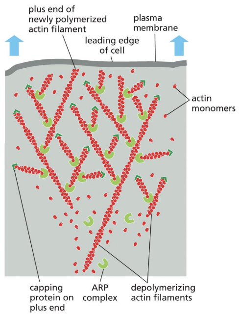
Relate how actin binding proteins influence actin filament polymerization and organization to regulate microfilament function (eg. cell motility)
-cellular locomotion
ABPs required
forming protrusion via formation of lamellipodium requires actin polymerization directly underneath plasma membrane
membrane at leading edge can be pushed forward
actin must be depolymerized as membrane moves forward near rear of network
ensures cell continues to have access to free actin for polymerization
actin polymerization in cell cortex results in cell movement
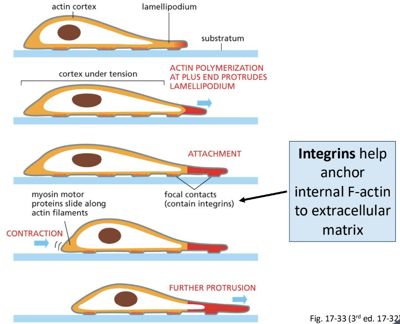
Interpret results from experiments using drugs that disrupt actin filaments and compare with equivalent experiments that disrupt microtubules
Explain the function of myosin motors and their role in contractile bundles as well as in other cellular contexts such as cytoplasmic streaming
Unit 7 Cell Cycle
7.1 Cell Cycle & Checkpoints
Review the overall organization of the cell cycle and its relation to cell division (mitosis and cytokinesis)
-cell cycle
divided into 4 stages
interphase consists of G1, S, G2
discrete process
events that only occur at certain points in the cycle
continuous process
occurs throughout the cell cycle
eg. nutrient assimilation, cell growth
-gap 1 (G1) phase
gap between end of cytokinesis and start of DNA synthesis
major period of cell growth → longest phase
duplication of organelles, volume of cytoplasm increases
cell gets bigger
ends with G1/S checkpoint
-synthesis (S) phase
initiation and termination of DNA synthesis
DNA replication → DNA doubled → sister chromatids formed
S-cyclin/CDKs active
ends at the end of replication
-gap 2 (G2) phase
second gap phase lasts from end of DNA synthesis to onset of mitosis
cell doubles in size in preparation for mitosis
S-cyclin/CDKs active
ends with G2/M checkpoint → commitment to divide
once cell enters mitosis → can’t go back
cell must be sure it can undergo mitosis properly
managed by M-CDKs
-mitosis (M) phase
division occurs
cell divides in 2
division of parental cell in 2 genetically identical cells
nuclear division which chromosomes condense & separate
cytokinesis
cytoplasm division during which cell actively divides in 2
M-CDK/M-cyclins active in first half of mitosis
to allow cells to exit mitosis
M-cyclin targeted for destruction by anaphase promoting complex (APC)
-cell moves past G1 into the next stage of cell cycle if and only if division is the eventual goal → cell must divide if they move to next phase
-growth & division requires a lot of resources → cell won’t unnecessarily enter into these cycles unless required
-there must be provision for cells to get out of cell cycle and into another developmental pathway (mating, meiosis, differentiation) or to arrest
necessary for these cells to get from these alternate fates back into cell cycle
-controls placed on the progression of cell cycle where the cell is required to meet certain criteria before it’s allowed to proceed
each checkpoint is controlled by gatekeeper protein that respond to cellular conditions → only allows cell to move forward into next phase of cell cycle if certain conditions are met
checkpoints help cell to ensure certain conditions are met before cell cycle is allowed to continue
Use experimental evidence to distinguish what phase of the cell cycle that a cell is in (ie. G1, S, G2, M phase)
-chemically synchronizing cells in a population
block stalls the cell cycle at a known location
cells continue to process through cell cycle until they hit a block where they can go no further
-fluorescence activated cell sorting (FACs)
used to measure DNA content of a cell
cells stained with fluorescent dye upon binding DNA
amount of DNA is proportional to amount of fluorescent emitted
using fluorescently labeled cell
flow cytometer measures the fluorescence using a laser and separates a mixed population of cells
separates cells based on amount of fluorescence cell is emitting
result is two-fold
synchronized population of cells without chemical inhibitors or temperature-sensitive mutations → used for further experiments
G1 phase = 1 full set of DNA
S phase = more than 1 set but less than 2 sets of DNA
replication started but not complete
in between peaks
G2/M phase = 2 full sets of DNA
cells in both peaks have the same number of chromosomes → chromatids is different
flow cytometer produces a graphical readout that summarizes how many cells were found with each amount of fluorescent material (DNA)
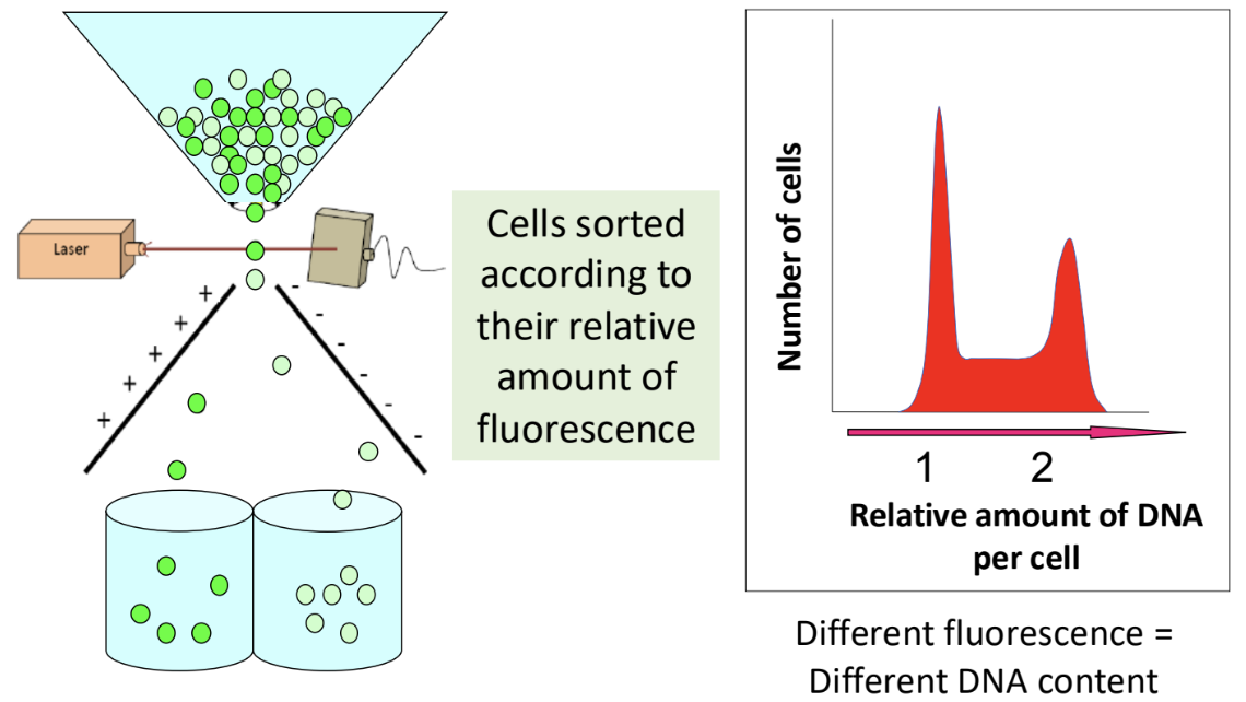
[x-axis]: peaks represent separate groups of cells
peak 1: 1 full set of DNA (G1 phase — after cytokinesis & before S phase)
peak 2: 2 full sets of DNA (after S phase & before cytokinesis → before cytokinesis since 2 full sets of DNA → if it’s after cytokinesis it would only be 1 full set of DNA)
[y-axis]: size of peaks represent amount of cells in each grou
peak 1: more cells
peak 2: less cells
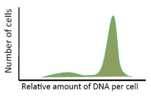
ex (above): FACs readout if chemically stopped cells at the G2 phase
interpreting FACs readouts (frequency histogram)
X-axis measures amount of DNA
Y-axis represents number of cells counted at each point on the X-axis
because amount of DNA correlates to phase of cell cycle → determine how many cells are in each stage of cell cycle at the moment at which they were measured
second peak = replicated DNA
2 phases that both have replicated DNA (G2 & M)
FACs can’t differentiate these → lumped together in graph
dip/section in between the peaks is important
Explain the concept at checkpoint control of cell cycle and give examples of what types of conditions should be met before S and M phases proceed
-cell cycle checkpoints
2 checkpoints
G1/S checkpoint
G2/M checkpoint
coordinates continuous processes (ie growth) with discrete processes (ie DNA replication) so that cell size is maintained
cells don’t move through stages of cell cycle on their own → signal always required
transition from one stage to next is controlled by proteins in cytoplasm
when no proteins present → cells stays in G1
proteins required to turn on and promote cells to next stage
-G1/S checkpoint
transition from G1 to S phase
restricts cells from beginning DNA synthesis before cell is ready
conditions that need to be met
nutrients (carbon source, energy source, Pi, S, N, vitamins) must be present at specific concentrations
chromatid separation (from previous mitosis) must be complete
no detectable DNA damage
cells must have reached a critical threshold size
external factors must be appropriate
-G0 phase
when cells aren’t ready to be divided → stalled in G0 phase for extended periods of time
cells remove themselves from cell cycle → arrested
senescent (permanent arrest)
cell to stay the same size and don’t divide
quiescent (temporary arrest)
long term deprivation of nutrients or other resources required for cell division → G0 phase allows cell to stay alive
-G2/M checkpoint
stops cell from entering mitosis before it’s ready
commitment to division (cell is 100% ready to divide) → cell can’t stop the process of mitosis once it has started
checkpoint controls transition from G2 to M phase
conditions that need to be met
DNA replication must be complete
DNA must be undamaged
cell must have reached a certain minimum size
cells can only move beyond checkpoint and into next stage of cell cycle when they have met required conditions
-xenopus cells demonstrated that something in cytoplasm controls entry into M phase
MPF (positive regulator that makes mitosis happen) → found to contain a kinase (enzyme that phosphorylates proteins using ATP bound)
MPF later became M-CDK (target protein involved in mitosis)
(cyclin-dependent) kinases activates/inactives target proteins by phosphorylating them and causing a conformational change → regulates progression into mitosis phase
7.2 CDK-Cyclin Regulation
Describe how specific protein modifications (eg. phosphorylation and ubiquitination) result in activation/reactivation of cyclin-CDK complexes to regulate cell cycle checkpoints
-cyclin-CDK complex work together to control the cell cycle
made of
cyclin (protein)
interacts with CDK
acts as a regulatory unit
cyclin-dependent kinase - CDK (protein)
regulates activity of a large number of other proteins required for cell cycle progression by phosphorylating them
only performs its function as a kinase when cyclin is bound
if cyclin removed from CDK → CDK inactivated
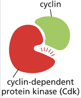
phosphorylation controls cyclin-CDK enzymatic activity
cyclin binding to CDK not sufficient on its own
cyclin-CDK complex gets phosphorylated by other kinases
ex: addition of phosphate group to M-cyclin by Wee1 (kinase/protein) → inhibition of complex
Wee1 responds to intracellular cues to ensure cell doesn’t go to G2/M phase checkpoint until big enough to divide
after cell is big enough → Wee1 degraded → can’t phosphorylate M-CDK → CDC25 (protein) removes original phosphate from M-cyclin-CDK complex → activates complex
ex: in yeast → Wee1 & CDC25 are key regulators of M-CDK
increasing gene dosage (gof) → increases enzyme concentration
decreasing gene dosage (lof) → decreases enzyme concentration
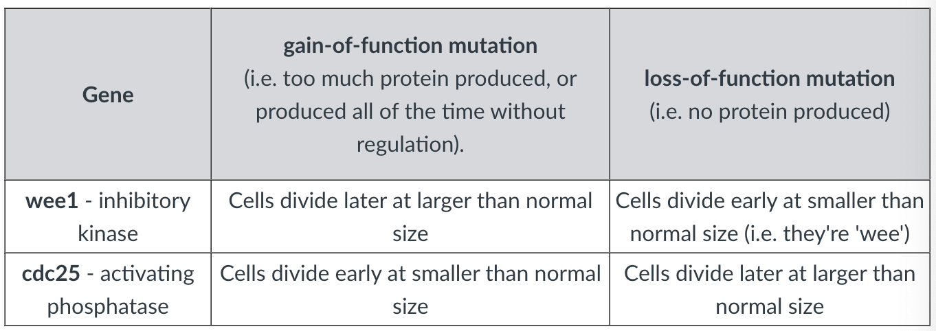
-anaphase promoting complex
signal that pushes cell from metaphase → anaphase during mitosis
large protein complex
functions as E3 ubiquitin ligase
adds ubiquitin tags to proteins to tag them for destruction by proteasome
triggers separation of sister chromatids
degrades cohesin proteins that binds sister chromatids together → chromatids pulled apart → anaphase begins
helps exit mitosis
activates ubiquitin that tags M-cyclin for degradation → deactivates M-CDK
causes M-CDK activity to drop → cell exits cytosis → cell returns to low-CDK state in G1
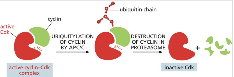
blocking APC function blocks its ability to activate proteins used to degrade cohesins
checkpoint in mitosis
protein inactive (securin)
active enzyme (separase) → not functional until activated
cuts cohesins so sister chromatids can separate from metaphase → anaphase
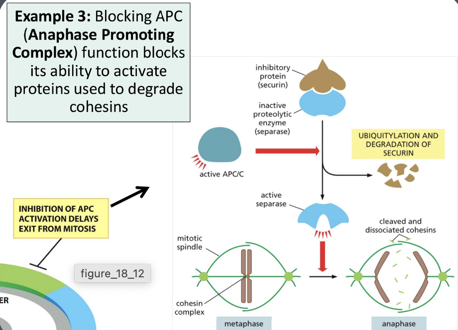
Explain how the activation of the cyclin-CDK complexes results in the start of the next phase of the cell cycle. Use examples of M-CDK complex and S-CDK complex to explain this
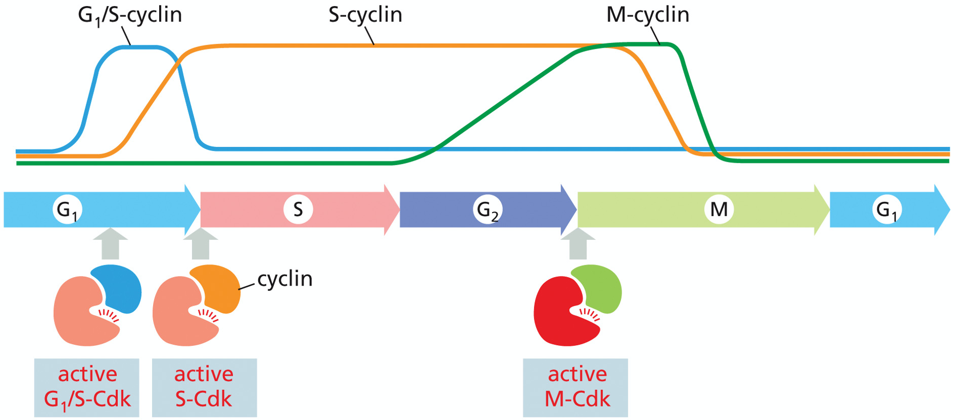
-cycling of cyclin concentration and CDK activity allows the cell to progress through checkpoints and the cell cycle
-cyclin-CDK activity must hit threshold before checkpoint is passed
-S cyclin
activated at start of S-phase
directly induces DNA replication
concentration of cyclins remains high through to M phase
activity of CDK activated at one checkpoint and stays high throughout cycle
-M cyclins
controls the G2/M checkpoint
active in first half of mitosis until destruction signalled by anaphase promoting complex (APC)
activity of CDK decreases rapidly not long after checkpoint has been passed
-G1 phase → S phase (G1/S checkpoint)
starts DNA replication
controlled by S-cyclin-CDK2 complex
forms toward the end of G1
remains active right until start of M phase despite S-phase being long over before that
once active → phosphorylates proteins involved in DNA replication so cells can enter S phase and begin DNA replication
S-CDK phosphorylates proteins at origins of replication
in order for cells to enter S phase (to replicate DNA) → must pass through G1/S checkpoint
no DNA damage
p53 (transcription regulator) protein that checks for damage
when DNA damage detected → p53 becomes phosphorylated → stops degradation → binds to promoter sequence for CDK inhibitor (p21) → activates transcription and subsequent translation (blocks S-phase entry)
p21 blocks cyclin-CDK complex activity & S phase entry
once checkpoint passes → G1/S cyclin initiates S phase via phosphorylation of replication machinery
G1/S cyclin degraded and S cyclin takes over
S-cyclin-CDK complex activates DNA helicase → promotes assembly of replication machinery
prevents replication from being able to happen more than once a cell cycle
via phosphorylating key enzymes in replication (Cdc6)
phosphorylation of Cdc6 → tagged for degradation → without Cdc6 → no replication
-chromosomes before replication
single (double stranded) DNA molecule (chromatid)
-chromosomes after replication
consist of 2 DNA molecules (sister chromatids)
connected at centromere (bound by cohesins - protein)
cohesins must be degraded prior to chromatid separation during anaphase
complete with full complement of histones and any other proteins required for chromosome packing
-G2 phase → M phase (G2/M checkpoint)
starts mitosis
shuts down transcription & translation
preparing organelles for separation → shut down
completely rearranges cytoskeleton
controlled by Mitotic-cyclin-CDK1 complex
cyclin accumulates during interphase
reaches maximum in mitosis then rapidly declines at the end of mitosis
forms in G2 but stays inactive until its needed
inactive at first because of inhibitory phosphate added by Wee1 (kinase)
Cdc25 (phosphatase - enzyme) removes inhibitory phosphate to activate M-cyclin-CDK complex
controls when M-CDK becomes active via dephosphorylation
removing inhibitory phosphate → activates M-CDK complex
positive feedback loop
phosphorylates and activates more Cdc25
inhibits Wee1
mitosis becomes fast and irreversible
ex: Wee1 puts lock on; Cdc25 unlocks → once running → engine powers itself by activating more keys (Cdc25) and disabling more locks (Wee1)
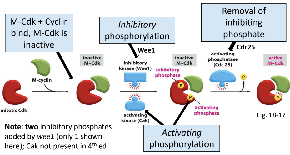
inhibitory phosphates added by Wee1 (inhibitory kinase)
Cdc25 removes inhibiting phosphate to activate M-CDK
complex activation of CDKs allow cells to ensure activation occurs under ideal cellular conditions
once cyclin bound to CDK → phosphated added/removed based on current cellular conditions
when all checkpoint conditions met → correct arrangement of phosphate groups added/removed from CDK so that it becomes active
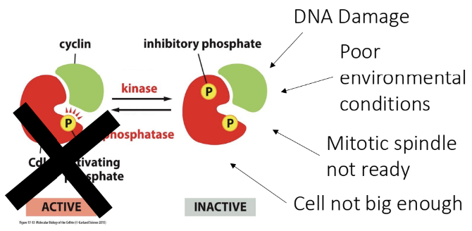
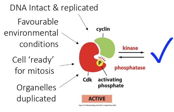
once active → triggers chromosome condensation (chromosomes become visible), breaks down nuclear envelope, activates spindle apparatus to divide the chromosomes
targets of activated M-cyclin-CDK complex (proteins phosphorylated by M-CDKs)
histone H1
changes in chromatin configuration → chromatin condensation
condensins
DNA binding proteins that binds to chromatins
aids with chromosome condensation
nuclear lamins
fall apart because phosphorylated lamins have lower affinity for each other
nuclear lamina disassembly → breaks up nuclear envelope into ER
nucleolus structural proteins (nucleolin)
dispersion of nucleolar proteins
nucleolus disintegrates
vital for chromosomal condensation
protein kinases that regulate the cytoskeleton
arrangement of cytoskeleton as the cell disassembles the interphase microtubule network and forms the mitotic spindle
Cdc5 - activating phosphatase
CDKs activate phosphatases by phosphorylating them
positive feedback loop - further activation of CDK
required for rapid rise in CDK activity after cell division commitment
anaphase promoting complex (APC)
degrades cohesin proteins that are keeping sister chromatids together → progeny cell chromosomes released
tags and deactivates M-CDK-cyclin
-cyclin-CDK complex deactivation
transcription of cyclin genes shut down
degrade cyclin proteins that already exist
negative feedback loops built so CDKs can be shut down
APC triggers ubiquitin to tag M-CDK for degradation by proteasome
cyclin concentration drops rapidly
cyclin degraded → CDK deactivated → next step of cell cycle begins
APC gets deactivated in G1
Interpret experimental results of experiments on cyclin-CDK regulation
-fission yeast studies
2 lof mutants with different sized cells after division
lof mutants
lof Cdc25 (knockout Cdc25)
results in big cells → Cdc25 mutants divide later than wild-type
wild-type allele of this gene advances cell into cell division
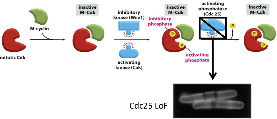
not as many activating phosphates added
when its added → cells are bigger → grows bigger than wild-type
lof Wee1 (knockout Wee1)
results in small cells → Wee1 mutants divide sooner than wild-type
wild-type allele of this gene slows entry into cell division
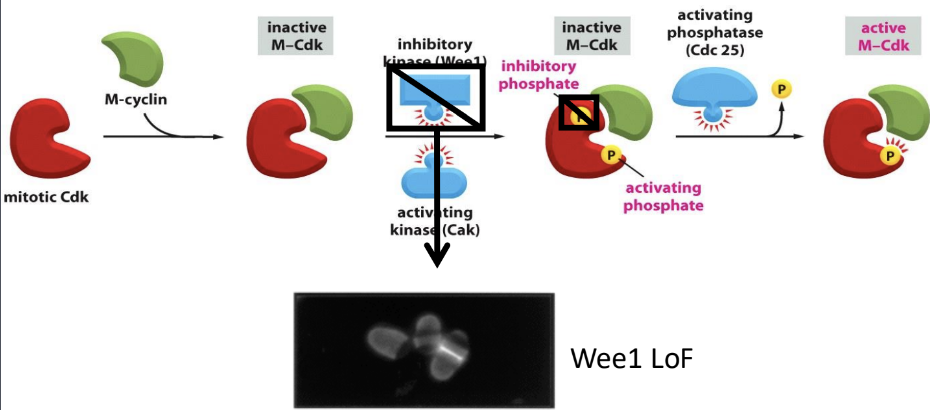
no inhibitory phosphates → G2 shortened → cells smaller and doesn’t grow as big as wild-type
7.3 Mitosis and Cell Division
Review the stages of mitosis and relate how the process of mitosis is driven by the activation of proteins by the mitotic CDK-cyclin complex
-M phase consists of
mitosis phase (nucleus division and its contents)
interphase
prophase
prometaphase
metaphase
anaphase
telophase
cytokinesis phase (rest of the cytoplasm gets divided)
-prophase
first stage of mitosis
prophase entry triggered by M-CDK activation by Cdc25
cell has passed G2/M checkpoint (M-CDK fully activated)
M-CDK phosphorylates
histone H1
serine residues at the ends of H1 histones are phosphorylated by M-CDK
adds negative groups to positive groups
decreases strength of H1 ends’ interaction with DNA
results in strong interactions between H1 molecules → pulls them together
chromatin fiber condenses → becomes shorter and fatter
condensin
helps with further packaging of interphase chromatin fiber
holds sister chromatids together (center point)
MT-associated proteins
nucleolin
for ^ those events to happen → 2 centralized MTOCs in cell must be produced
become the opposite poles of the spindle
mitotic spindle formation
mitotic spindle formed by selective stabilization of interacting microtubules
dynamic instability at the free plus-ends
interpolar microtubules that are interacting with other microtubules
motor proteins and other microtubule-associated proteins (MAPs) help stabilize the microtubules
relies on dynamic instability of microtubules
microtubules grow outwards from spindle pole → search throughout cytosol for things to grab onto
if it doesn’t find what its looking for in the cytosol → will depolymerize
nucleoli disappears
nucleolin
structural protein of the nucleous
M-CDK target
phosphorylation results in dispersion of the nucleolar proteins and disintegration of the nucleolus
-prometaphase
nuclear envelope breaks down
broken down by phosphorylation of nuclear lamins via activated M-CDK
nucleolus has disappeared
chromosomes now fully condensed and kinetochores formed at centromeres
spindle poles free to associate with chromosomes
by the end of
mitotic spindle formation during prometaphase
made up of
astral microtubules (green)
microtubules anchored at the plasma membrane
kinetochore microtubules (blue)
form when they attach to a kinetochore during random growth and shrinkage in the cytoplasm
kinetochore
specialized region of chromosome associated with centromere
plaque or button shaped structure composed of proteins
microtubules become inserted into kinetochore on each side of chromosome
one kinetochore associated with each chromatid
faces in opposite directions on the 2 sides of chromosome
interpolar microtubules (red)
forms when 2 of the microtubules from opposite poles interact with each other
stabilizes each other via motor proteins
forms a bridge between 2 MTOCs
XKCM1 is necessary for proper microtubule depolymerization since microtubule depolymerization is required for proper spindle assembly
formed via selective stabilization of interacting microtubules — motors play important role
kinetochore microtubules from both poles attach to sister chromatids
mitotic chromosomes can be pushed and pulled into place
kinetochore attachment via random growth/shrinkage of microtubules

-metaphase
chromosomal spindle fibers form after nuclear envelope breaks down
chromosomes aligned at equator of spindle/metaphase plate
after being simultaneously pulled toward both spindle poles by kinetochore microtubules
tension on microtubules and kinetochores provides even tension from both sides
small amounts of GFP-tubulin added to cell undergoing mitosis → GDP-tubulin becomes incorporated into spindle
fluorescence moves toward spindle pole
even though chromosomal fiber length remains the same
plus end has polymerization occuring at the same rate as tubulin addition as the other end so they remain the same length
mitotic spindle constantly assembling and disassembling
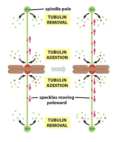
-anaphase
APC initiates anaphase
APC tags securin (regulatory protein) → releases separase (enzyme) → cleaves/degrades cohesins that allow sister chromatids to stay together → sister chromatids separate
sister chromatids synchronously separate
separates at the same time with same tension
each sister chromatid pulled toward opposite spindle pole → chromosome segregation
movement of kinetochores takes place via motor proteins (same motor proteins that aid with spindle assembly is now separating sister chromatids)
microtubules depolymerizing rapidly from plus ends
2 forces work together to separate sister chromatids and move them to opposite sides
kinetochore microtubules shortening (Anaphase A)
interpolar microtubules growing, pushing poles apart (Anaphase B)
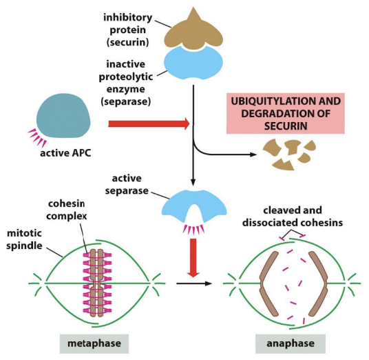
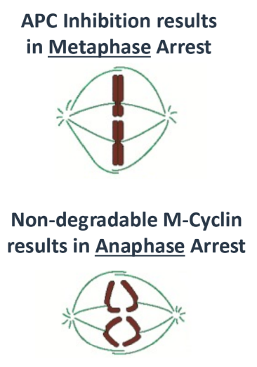
anaphase A
initial part of anaphase
chromosomes pulled poleward (centromere/kinetochore first)
kinetochore microtubules shorten & move chromosomes toward opposite poles
depolymerization of kinetochore microtubules required
motor proteins at kinetochore allow chromosomes to stay connected to shrinking microtubule
Taxol effect
microtubules still growing; stabilizes microtubules
prevents depolymerization of kinetochores
anaphase B
poles pushed and pulled apart
interpolar microtubules lengthen
astral microtubules shorten
kinetochore microtubule length remains mostly constant
3 separate forces
tubulin subunits added to plus ends of interpolar microtubules (longer tubulin subunits)
motor proteins move overlapping microtubules from 2 poles (longer spindle)
other motor proteins move astral microtubules (animal cells) against the cell cortex (peripheral cytoskeleton
shorter distance between boles of spindle and plasma membrane
Taxol effect
microtubules still growing; stabilizes microtubules
interpolar microtubules can still lengthen (but wouldn’t be able to disassemble once this phase passes); astral microtubules can’t depolymerize
-depolymerizing tubulin pulls things apart
kinetochore-attached fibrils bind to chromosome & to protofilament (PF)
protofilament undergoing depolymerization at plus end
gives ability to be pulled
protofilament bending will pull on fibrils & associated chromosome (attached via kinetochores)
affinity for fibril to soluble tubulin is low and allows for tubulin dimer from bending protofilament to be released
fibrils re-engage with protofilaments that are less bent or just about to bend, providing for a continuous pull
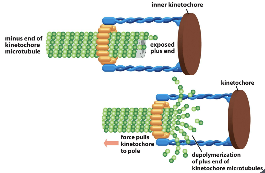
-telophase
division of nuclear content is complete
nuclear envelope reassembles around each set of chromosomes - 2 nuclei
spindle fibers disappear
division of cytoplasm
controlled by destruction of M-cyclin
shut down mitosis
end of M phase → M-CDK needs to be shut down
APC inactivates M-CDK via activating ubiquitin to tag M-cyclin for destruction by proteasome
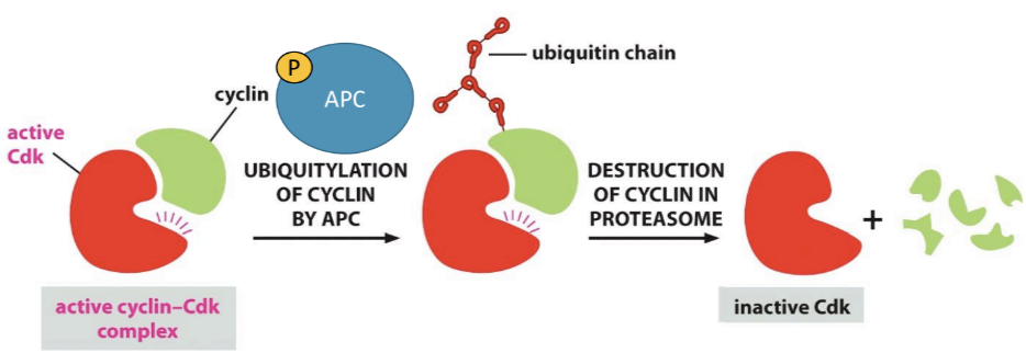
prepare for entry into G1
chromatin decondensation
reformation of nuclear lamina (and nuclear envelope) around chromosomes
centromeres and telomeres retain connection to nuclear lamina throughout cell cycle
reformation of nucleolus
disintegration of spindle and reformation of peripheral cytoskeleton
new nuclear envelope re-assembles around each chromosome set
lamin proteins dephosphorylate
vesicles of nuclear membrane first bind to chromosome
nuclear pores associate
membrane fuse together to form nuclear envelope (inner and outer)
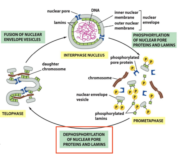
-cytokinesis
cytoplasm divides in 2
cell divides into 2 progeny cells
cytokinesis in animal cells
division via myosin/actin contractile ring that pulls plasma membrane inwards → splits cells in 2
contractile ring always forms around mid-zone of mitotic spindle
actin & myosin filaments pinch cell to divide cell into 2 daughter cells
dividing cytoplasm also divides organelles
vesicles fuse rapidly with plasma membrane → ensure there is enough plasma membrane to fit each progeny cell
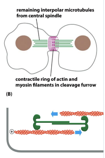
cytokinesis in plant cells (watch recording from april 4th)
new cell wall must be produced between 2 daughter cells
Golgi must remain functional during cell division
secretes cell wall compounds (via vesicles) rapidly to center
actin plays a role ensuring vesicles are correctly targeted to cell plate
cell plate grows from center of cell towards outer walls → fuses with walls of dividing cell
new cell formation uses interpolar microtubules to help guide this process via formation of a phragmoplast in the center of the cell
phragmoplast complex
comprise of
microtubules
actin filaments
membrane components
other proteins
new progeny cells fully formed with full complement of organelles and DNA
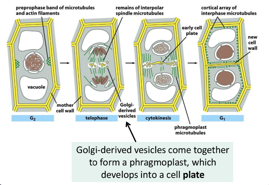
Illustrate how the process of mitosis is driven by the activation of proteins by the mitotic CDK-cyclin complex
-M-CDK-cyclin complex
condensin proteins
helps chromosomes tighten
nuclear envelope proteins
breaks down nucleolus
microtubule associated proteins
builds the mitotic spindle that pulls chromosome apart
cohesin regulators
activates APC to regulate cohesin to drive M phase
Explain the role of the cytoskeleton (and associated motor proteins) during mitosis including how dynamic instability of microtubules contributes to the formation and function of the mitotic spindle, and the role of the actin cytoskeleton during cytokinesis
-cytoskeleton (microtubules — tubulin, actin filaments — actin)
microtubules reorganize to form the mitotic spindle → pulls chromosomes apart
guided by centrosomes (MTOCs)
dynamic instability aids in formation
kinesins & dyneins (motor proteins) walk along microtubules, carrying cargo or sliding microtubules against each other
kinesins
moves toward plus end
push centrosomes apart
dyneins
moves toward minus end
pull spindle poles toward edges of cell
actin filaments form a contractile ring around middle of cell
pinches cell in 2 during cytokinesis
Explain how proteins and DNA interact to further condense the DNA to a mitotic chromosome
Describe how the regulation of cyclin via ubiquitination results in the end of M-phase and a transition to the next phase of the cell cycle
shut down mitosis
end of M phase → M-CDK needs to be shut down
APC inactivates M-CDK via activating ubiquitin to tag M-cyclin for destruction by proteasome