Detailed Study Guide on Molecular Cloning, Recombinant DNA, and Hybridization Techniques
Introduction to Recombinant DNA and Molecular Cloning
Recombinant DNA Technology: This technology allows for the direct manipulation of specific sections of DNA, such as individual genes.
Methodology: It is made possible by techniques designed to isolate specific sections of DNA and subsequently create multiple identical copies of them.
Gene Cloning: This is defined as the process of making multiple identical copies, known as clones, of each unique section of DNA.
Core Requirements for DNA Cloning:
Cutting Mechanism: A method to cut DNA into smaller, manageable pieces, which is achieved through the use of restriction enzymes.
Copying Mechanism: A method to replicate the DNA by inserting it into rapidly replicating organisms, such as bacteria or yeast, which act as biological factories.
Purpose and Applications of DNA Manipulation
Fundamental Principle: DNA is the universal genetic material. The ability to isolate and manipulate it allows scientists to understand and utilize biological functions.
Functional Understanding: Isolating and modifying genes is essential to understanding their specific functions within an organism.
Go from mutant phenotype to gene sequence: Researchers can move from observing a mutant phenotype to identifying the underlying gene sequence.
Protein Manufacturing: Recombinant DNA is used to manufacture vital therapeutic proteins, including: * Insulin (for diabetes treatment). * Factor VIII (for blood clotting).
Vaccine Generation: Developing modern vaccines depends on these cloning techniques.
Create Transgenic plants or Organisms: Facilitates the creation of transgenic plants or other modified organisms.
Diagnose genetic diseases: Used to diagnose genetic diseases accurately.
Develop Gene Therapy: Essential for the development of therapies that involve the insertion or alteration of genes within an individual's cells to treat disease.
Molecular Cloning Theory and the Cloning Vector
The Problem of Scale: After a genome has been reduced to smaller pieces of DNA, these fragments must be reproduced in large quantities (amplified) to allow for detailed study.
Recombinant DNA Molecule Formation: DNA fragments, such as the coding sequence of a specific gene, are inserted into a cloning vector.
Defining a Cloning Vector: A cloning vector is a DNA molecule capable of being introduced into a simple, single-cell organism (like bacteria) and possesses the ability to self-replicate within that host. * Example: A bacterial plasmid.
The Biological System: The recombinant DNA molecule is introduced into a biological system that replicates the DNA. Because all resulting copies are identical to the original fragment, they are termed DNA clones or molecular clones.
Restriction Enzymes (Molecular Scissors)
Definition: Restriction enzymes (RE) function as "molecular scissors" used to cut DNA.
Mechanism of Action: They recognize short, highly specific nucleotide sequences in DNA and cut both strands of the sugar-phosphate backbone.
Biological Origin: These enzymes were first identified in bacteria. In nature, their role is to protect the bacteria against viral infection by cutting (restricting) the invading viral DNA.
Specificity: Different restriction enzymes recognize different specific nucleotide sequences. Recognition sites are typically 4, 6, or 8 nucleotides (base pairs) long.
Palindromic Sequences: For many restriction enzymes, the recognition sequence is a palindrome, meaning the sequence reads the same on both strands when viewed in the 5’ → 3’ direction.
Cohesive (Sticky) Ends: Many enzymes create a staggered or asymmetrical cut. This leaves single-stranded "sticky ends" that are extremely useful because they can easily ligate (stick) to other single-stranded DNA ends that have been cut with the same enzyme.
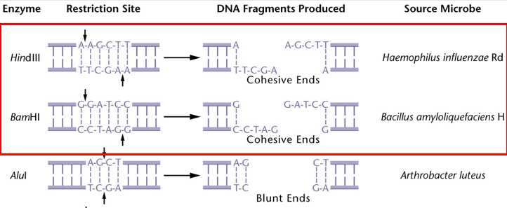
Creating Recombinant DNA with Restriction Enzymes and Ligase
Complementary Annealing: DNA molecules cut by the same restriction enzyme will possess the same cohesive (sticky) ends.
These ends have complementary base pairs that allow them to anneal to each other via hydrogen bonding.
Base Pairing Rules: A = T and C = G.
DNA Ligase: Once the strands have annealed, the free hydroxyl (-OH) and phosphate (-PO4) groups must be joined. This is accomplished by the enzyme DNA ligase, which covalently joins the DNA fragments.
number and size of fragments depend on which RE, the size of the genome, and its composition (GC content)
Enzymatic Requirements for Recombinant DNA:
Digestion: Performed by restriction enzymes to cut the DNA.
Ligation: Performed by DNA ligase to seal the fragments together.
Restriction Enzyme Mapping and Genomic Digests
EcoRI Example: The enzyme EcoRI recognizes a 6-bp sequence. Statistically, it cuts the human genome on average every 4,000 base pairs (4 Kbp).
Fragment Frequency: In the human genome, EcoRI generates approximately 780,000 different fragments.
Recognition Site Length vs. Frequency: Enzymes with 4-bp recognition sites (e.g., AluI) cut DNA more frequently than 6-bp cutters.
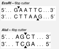
Non-random Distribution: The DNA sequence is not random. Restriction sites may be clustered closer together or spread further apart depending on the region.
Composition Factors: Bacteria with higher GC content will affect the frequency at which specific enzymes cut.
Genomic Digest Appearance: When genomic DNA (such as from Drosophila) is digested with EcoRI and separated on an agarose gel, it appears as a "smear." This is because the thousands of fragments range in size from 10 to thousands of base pairs in length.
Restriction Fragment: Each individual piece of DNA resulting from a digest is called a restriction fragment.
Mapping Procedure:
Cut DNA with specific restriction enzymes.
Separate fragments by size using agarose gel electrophoresis.
Estimate sizes by comparing the migration distance against a size marker (fragments of known sizes).
Determine specific site positions and distances between sites, often using pairs of enzymes to compare locations.
Bacterial Plasmids as Cloning Vectors
Plasmid Definition: A small, circular, double-stranded DNA molecule found naturally in bacteria. They are distinct from the main bacterial chromosome and can replicate independently.
Natural Function: They often carry genes beneficial to the bacteria, such as antibiotic resistance.
Plasmids are used as cloning vectors to carry DNA fragments into a host cell.
Engineered Key Features of Plasmid Vectors:
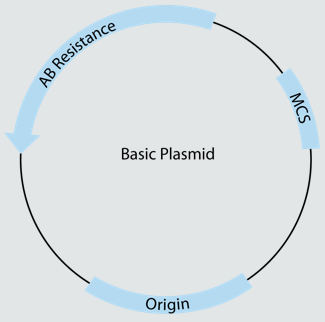
Origin of Replication (ori): A specific DNA sequence where replication begins, allowing independent copying of the bacterial chromosome within the host cell.
Antibiotic (AB) Resistance Genes: allows onlt the bacteria containing the plasmid to survive and grow in the presence of that antibiotic (selective growth).
Multiple Cloning Site (MCS): Plasmids used for DNA cloning also include a multiple cloning site for insertion of DNA fragments.
Plasmids for cloning DNA molecules
A DNA fragment can be cloned by inserting it into the MCS of a ‘cut’ plasmid vector to create a recombinant DNA molecule
achieved through
digestion (RE)
ligation (DNA ligase)
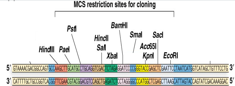
Plasmids are engineered to have individual RE recognition sites clustered together at the MCS.
DNA fragments can be easily inserted if cut with the corresponding RE
Amplification and Identification of Recombinant DNA
Creating the recombinant plasmid
The DNA fragment and cloning vector are cut with the same restriction enzyme
The vector and fragment will have the same complementary cohesive ends
Once the cut DNA fragments and the vector are mixed
DNA fragment will anneal to the cut vector via cohesive ends in
DNA ligase ligates the DNA fragment and the cloning vector together to form a recombinant DNA molecule
The he
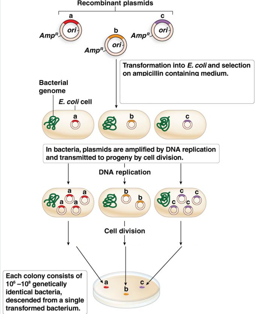
recombinant plasmid is ‘amplified’ in E.coli
Transformation: The process by which individual recombinant plasmids are taken up by E. coli bacteria.
Amplification: Inside the bacteria, the ori ensures that 100-200 copies of the recombinant plasmid are generated per bacterium. As the bacteria divide, each daughter cell inherits these copies.
Colony Formation: Bacteria grow as colonies; each colony represents millions of copies of the original DNA fragment.
Blue-White Selection:
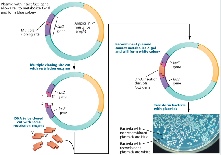
The MCS is often placed within the lacZ gene, which encodes the beta-galactosidase enzyme, which reacts with a chemical called X-gal to turn bacteria blue.
Disruption: If a DNA fragment is successfully inserted into the MCS, the lacZ gene is disrupted (insertional inactivation).
Outcome: Recombinant bacteria (with the insert) cannot produce beta-galactosidase and appear as white colonies, whereas non-recombinant bacteria appear blue.
Genomic Libraries and the Human Genome Project
Genomic Library: A collection of overlapping DNA fragments that represent an entire genome.
achieved by fragmenting and cloning all genomic DNA
Utility: These libraries enabled the mapping, identification, and sequencing of the entire human genome through the fragmenting and cloning of all genomic DNA.
Gene Expression and Protein Production
In Vitro Gene Expression: Cloning genes to express them synthetically in a laboratory to generate protein products.
Reasons for Synthetic Expression:
Producing therapeutic proteins (e.g., Growth Hormone, Clotting Factors).
Studying protein function.
Generating vast amounts of protein for manufacturing.
Testing mutation effects (normal vs. mutant versions).
Adding tags or reporters (e.g., GFP, His-tag) to track localization or simplify purification.
Expression plasmids:
Specialised cloning vectors are used to produce a protein from a cloned gene
differ from basic cloning vectors because they contain extra sequences to allow the expression of genes
1. bacterial expression plasmids - drive gene expression in bacteria to produce large amounts of protein
2. Eukaryotic expression plasmids - enable gene expression in eukaryotic cells and contain eukaryotic regulatory sequences required for transcription and translation
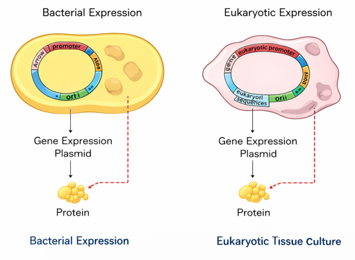
Cloning Vector vs. Expression Vector:
Cloning Vector: A
small piece of DNA maintained in the bacteria
ued to introduce cloned DNA into bacteria
can be plasmids, cosmids, phages, or BACS
includes ori, unique RE cut sites, and antibiotic resistance gene
Expression Vector:
used to introduce cloned DNA into bacteria to allow expression of the cloned gene
used to obtain the gene product of the cloned DNA (protein or RNA)
can only be plasmids
includes all features of cloning plasmid + regulatory elements - promoters, transcription initiation site, and translation initiation site
cDNA Synthesis for Eukaryotic Proteins
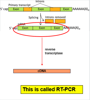
The Intron Problem: Eukaryotic genes contain large non-coding regions called introns, making genes very large (10s - 100s of kb). Bacteria (prokaryotes) cannot process or remove these introns.
Solution: Use mRNA, which is intron-free, as the starting material.
Reverse Transcriptase: An enzyme (reverse transcriptase) used in the lab to convert mature mRNA into complementary DNA (cDNA) for cloning and expression. This process is known as RT-PCR.
We can study which genes are expressed in which tissues by collecting mRNA from different cell types
cDNA Library: A collection of plasmids containing individual cDNA inserts representing the genes actively expressed in a specific tissue at a specific time.
1. Isolate mRNA from the tissue sample and convert it to cDNA
ligate cDNA into the RE-cut vector
transform bacteria with a recombinant plasmid
Amplify the library by growing bacteria
Each clone contains a cDNA that represents one of the mRNAs being expressed in that tissue.
Composition: Highly expressed genes are over-represented; non-expressed genes are absent. Each clone lacks introns and regulatory sequences.
Genomic vs. cDNA Libraries Summary:
Genomic: Represents the entire genome; needs computer assembly of fragments.
cDNA: Represents only expressed genes; varies between different tissue types.
Therapeutic Products Made via DNA Cloning
Human Insulin: Produced in E. coli or S. cerevisiae (yeast) to treat diabetes. Specific types include Insulin aspart, glargine, lispro, and glulisine.
Hormones:
Human choriogonadotropin (CHO cells) for superovulation.
Follicle-stimulating hormone and Luteinizing hormone (CHO cells) for infertility.
Somatotropin (E. coli) for growth failure.
Haemopoietic Growth Factors: Erythropoietin alpha, beta, omega (CHO/BHK cells) for anaemia; Filgrastim (E. coli) for neutropenia.
Blood Coagulation Factors: Factor VIII (CHO) and Factor IX (CHO).
Thrombolytics/Anticoagulants: Alteplase (CHO) and Lepirudin (S. cerevisiae).
Interferons/Interleukins: Interferon alpha-$2$b, beta-$1$b, gamma; Interleukin-$2$ and Interleukin-$11$.
Therapeutic Enzymes: Dornase alpha (cystic fibrosis) and Glucocerebrosidase (Gaucher disease).
Nucleic Acid Hybridization
Principle: Allows identification of nucleic acid fragments or clones containing specific sequences. Hybridization relies on the complementarity of nucleic acid strands and the specificity of base pairing.
Denaturation: Separation of the double-stranded DNA into single strands via heating or denaturation (High pH/alkaline conditions).
Hybridization: Cooling or neutralization allows complementary single strands to reanneal.
Single strands of DNA can also hybridise to complementary RNA molecules
we can use ssDNA to identify complementary fragments of both RNA or DNA
Probes: A probe is a single-stranded nucleic acid molecule (ssDNA) used to identify specific sequences within a complex mixture, such as a cDNA library.
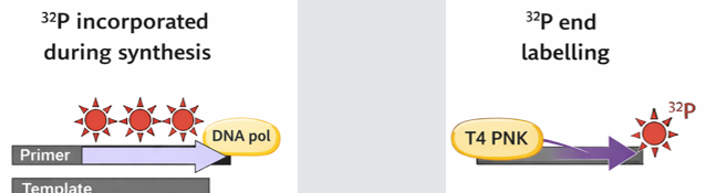
Labelling Probes: Probes must be labeled for detection using:
Radioactive isotopes (32P).
Fluorescent moieties.
Enzyme-conjugated labels.
Affinity-based labels (e.g., Biotin or Digoxigenin).
Labelling Methods:
During Synthesis: Incorporating labeled nucleotides (dNTPs) using DNA polymerase.
End Labelling: Adding labels to the ends of strands using T4 polynucleotide kinase.
Southern Blotting Technique
Origins: Developed by Ed Southern, it is used to identify specific restriction fragments within genomic DNA using complementary probes.
applies this principle to genomic DNA fragments separated by size on a gel
Step-by-Step Method:
Digest: Genomic DNA is cut with a restriction enzyme into a complex mixture of fragments.
Electrophoresis: Fragments are separated by size on an agarose gel.
Denature: The DNA in the gel is treated with alkali to make it single-stranded.
Blot/Transfer: The ssDNA is transferred (blotted) from the gel onto a nylon or nitrocellulose membrane.
Hybridize: The membrane is incubated with a labeled ssDNA probe complementary to the target sequence.
Detect: The membrane is washed, and the label is detected to reveal which band contains the target DNA fragment.