EMT Midterm Full Notes
Vital Signs + Intro
Body Substance Isolation (BSI)
Separates People From Bacteria
Hand Washing
Eye Protection (Goggles)
Gloves
Gowns
Masks
Best Prevention
Gloves
Hand Sanitizer
Hand Washing
Vital Signs
Provides Internal Picture Of Patient
Baseline Vitals = First Set
Everything Else Is Compared To The First Set
Respirations
Rate
Number Of Breaths In 30 Seconds*2
Quality
Normal
Labored (EX. After Running)
Noisy (Wheezing/Sick)
Depth
Shallow (Rapid And Slow)
Deep (Normal??)
AVG Breathing Rates
Adult (15+) = 12-20 Breaths/Min
Child (2-14) = 15-30 Breaths/Min
Infant (0-2) = 40-60 Breaths/Min
Pulse
Pumping Of Blood
Rate
Number Of Beats 30 Seconds*2
Strength
Strong (Easy To Find)
Weak (Hard To Find)
Regularity
Regular
Irregular
AVG Pulse Rates
A = 60-100 BPM
C = 80-100 BPM
I = 100-120 BPM
Blood Pressure
Pressure Of Blood Against Arteries
Systolic
Heart Contracts
Diastolic
Heart Relaxes
Sphygmomanometer (BP Cuff) & Stethoscope
Used To Find BP
Units
mm/Hg (Millimeters Of Mercury)
Written Format
SYSTOLIC / DIASTOLIC
EX.
Location
Slightly Bent Elbow (LVL With Heart)
Normal BP Rates
A =
C =
I =
All Vary By Genetics, Weight, Time, Age, ETC
EXTRA
Pulse In The Wrist = Radial Pulse
AVG Newborn Is 18 IN
SKIN
Color
Pale, Red, Blue, Or Yellow (Jaundice)
Blue Is Common In Infants Due To A Lack Of O2
Yellow Usually Caused By Liver Issues
Temperature
Cold, Warm, Cool, Or Hot
Moisture
Dry, Clammy, Moist, Or Wet
PULSE OXIMETRY
Monitoring Of The Oxygenation Of A Patient’s Hemoglobin
Normal Level
95-99% SpO2
Hypoxic Drive Problem
88-94% SpO2 ( Saturation Of Oxygen )
Carbon Monoxide Causes This To Drop
BLOOD GLUCOSE
Amount Of Glucose (Sugar) Present In The Blood
AVG Levels
Normal - 70-120 mg/dL
Elderly (50+) - Slightly Elevated
Doesn’t Work On Dark Nail Polish
PUPILS
P.E.A.R.L
Pupils Equal And Round/Reactive To Light
Pupils Can Tell If Patient Has Head Injury
Abnormal Pupil Reactions
Fixed (Not Moving) /Dilated/Sluggish/Unequal In Size
TEMPERATURE
Not Important For EMTS
AVG
98.6 F
Glasgow Coma Scale (GCS)
Objective Way Of Recording The Conscious State Of A Person
Eye Opening, Verbal, Motor
Highest = 15
Lowest = 3
CUPS
Used As A Tool To Prioritize The Patient For Transport
Critical, Unstable, Potentially Unstable, Stable
Critical
ABT To Die
Receiving CPR, ETC
Unstable
Unresponsive But Still Alive
Potentially Unstable
Difficulty Breathing
Cannot Move At All
Severe Pain
Complicated Childbirth
Stable
Minor Sickness
Minor Injury
MILITARY TIME
Time System That Prevents Confusion
EX. 12 AM = 0000 Hours
EX. 12 PM = 1200 Hours
EX. 6:07 PM = 1807 Hours
EX. 6:07 AM = 0607 Hours
Airway
Oxygen Needs
6-10 Mins W/O Oxygen (O2) = Likely Brain Damage
10+ Mins W/O O2 = Irreversible Damage
Hypoxia
Low Oxygen Level
Signs = nervousness, rapid heartbeat, altered mental status
Causes = stroke (blockage of blood vessel in brain), shock, etc
Inadequate Breathing
Labored Breaths
Accessory Muscle Use
Pale/Blue Skin (Cyanosis)
Cool & Clammy Skin
Abnormal Lung Sounds
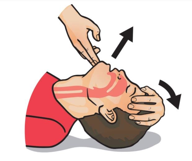
Opening Airway
Head Tilt/Chin Lift
Used For Non-Trauma Patients

Jaw Thrust
Suspected Spinal Injury (Prevents Moving/Hurting Spine Further)
Airway Adjuncts
Oropharyngeal (OPA)
Keeps Tongue From Blocking Airway
Inserting
select size (measure from earlobe to corner of mouth)
open mouth
hold upside down and insert
rotate 180 until flange rests on lips
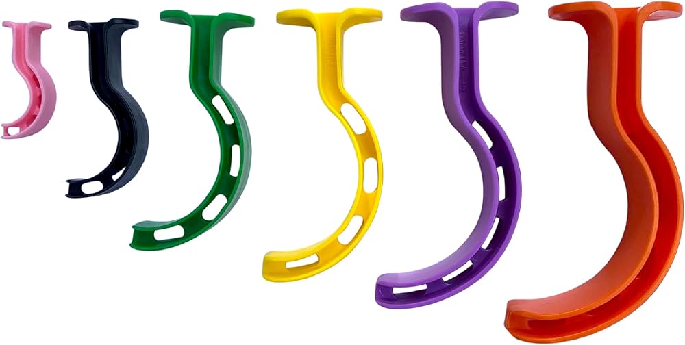
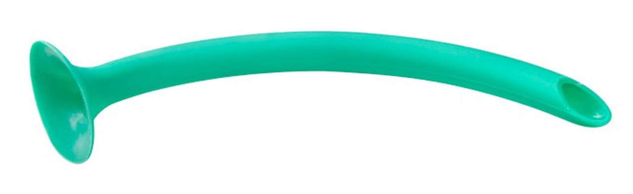
Nasopharyngeal Airway (NPA)
Used If The Patient Has A Gag Reflex
Inserting
select airway (measure from earlobe to nose)
lubricate
insert into right nostril with bevel toward septum (bridge of nose)
Oxygen
Given To Any Patient With Respiratory/Cardiac Issues
Oxygen Delivery
Non-Rebreather (NRB)
Provides 90% Oxygen
Set At 10-15 Liters/Min (LPM)
Unstable Patient

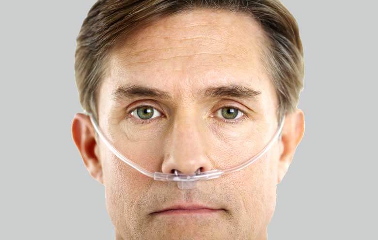
Nasal Cannula (NC)
35-50% Oxygen
2-6 LPM
Stable PT
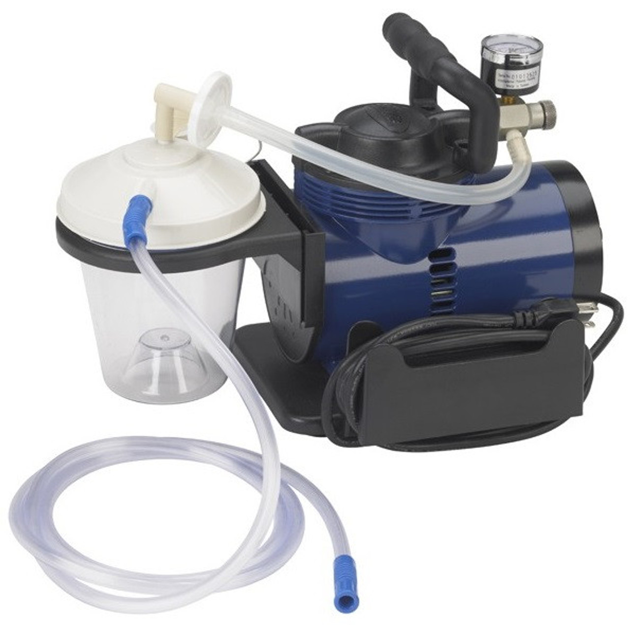
Suctioning
Never Suction For More Than 15 Seconds At A Time
Can Remove Patients Air Supply
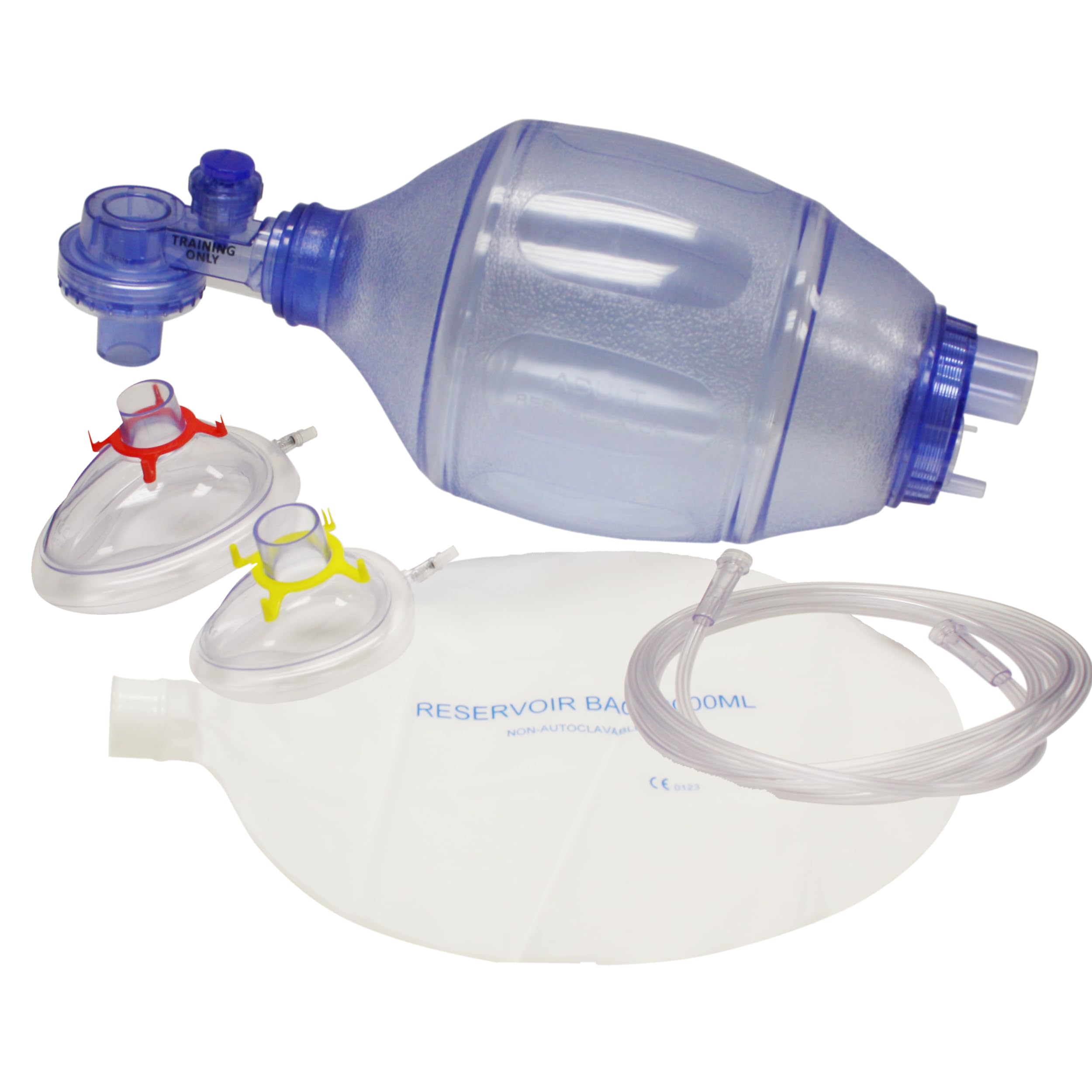
Artificial Ventilation
Bag Value Mask (BVM) - Manual Resuscitator That Helps Patients Who Are Not Breathing
Must Be Done Once Every 5-6 Seconds
Infants : 1-3 Seconds
Respiratory System
( IN ORDER )
Sinuses: Hallow spaces that warm and moisten air, produce mucus.
Nasal Cavity: Primary air entrance; warms, filters, and moistens air.
Nose: The external organ of smell and the primary physiological entry point for air into the respiratory system.
Larynx: The voice box, contains vocal cords for speech.
Pharynx: The throat; passageway for food and air.
Epiglottis: Covers trachea during swallowing to prevent aspiration.
Trachea: Windpipe; extends from larynx, supported by cartilage rings.
Bronchi: Main airways branching from trachea into lungs.
Bronchioles: Smaller branches leading to alveoli; regulate airflow.
Alveoli: Tiny air sacs where gas exchange occurs.
Diaphragm: Primary muscle of respiration; separates chest and abdomen.

Choking
Reasons For Choking (Ranking)
The Tongue Is Obstructing The Unconscious Victim’s Airway
Vomit → May Choke On Vomit Due To Age Or Drinking Heavily And Throwing Up While Passed Out
Foreign Body → Balloons & Food
Swelling → Allergic Reactions/Irritants
Spasm → Water Is Suddenly Inhaled
Chemicals & Fumes
NOTE
Majority Are Extremely Young Or Old
Recognizing
Audible Coughing/Breathing Sounds?
Are They High-Pitched?
Strong Or Weak Cough?
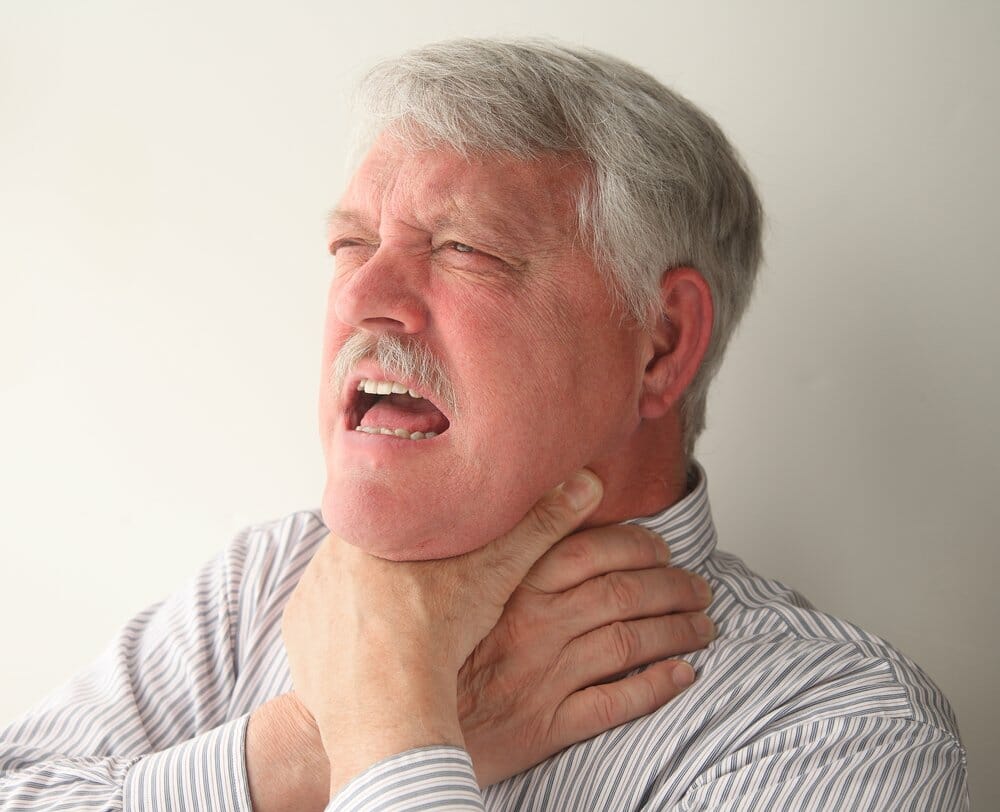
Can’t Speak, Breathe, Or Cough (Full Obstruction)
Universal Distress Signal (Clutching Neck)
Turning Blue (Cyanosis→ Lack Of O2)
Partial Obstructions With Bad Air Exchange Should Be Treated The Same As Complete Airway Blockages
If Strongly Coughing, Do NOT Intervene → Victim Is Still Breathing Strong
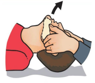
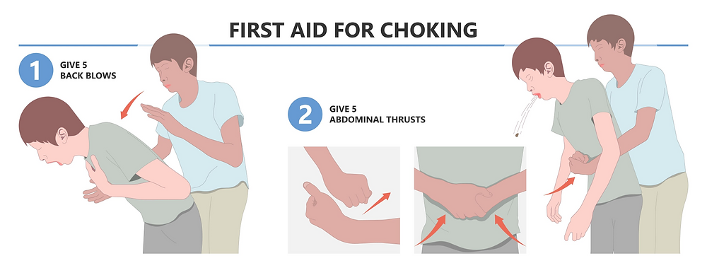
Conscious Choking
5 Abdominal Thrusts
Place Fist Above Umbilicus
5 Upward And Inward Thrusts
If Pregnant/Obese
5 Chest Thrusts
Fist On Sternum
If Unsuccessful, Support Chest With One Hand And Back Blows With Another
Continue Until Success Or Victim Is Unconscious
Choking While Alone
Use Fist
Use Corner Of Furniture
Be Creative
Call 911
Go To Large Area Where You Can Be Found
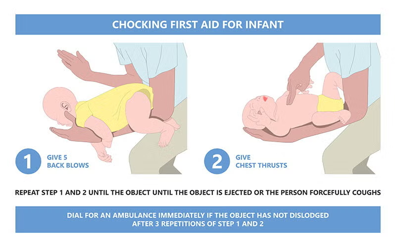
Conscious Choking Infants
Position With Head Downward (Usually Lie On Thigh)
5 Back Blows (Check For Expelled Object)
5 Chest Thrusts (Check For Expelled Object)
Repeat
Victim Becomes Unconscious
Call 911
Support Victim With Knees While Lowering To Floor
Assess Victim/Breathing
Begin CPR/Chest Compressions
Check For Object Before Giving Breaths
Empty Room With Victim Unconscious On Floor
Assess Victim
Give CPR If Needed
Compressions
Look For Object In Throat
Give Rescue Breaths
Order Of Unconscious Choking Rescue
A - Airway (HTCL)
B - Breathing
C - Compressions
Choking Unconscious Infant
If Breaths Don’t Go In Check For Objects And Continue
If Neither Go In Suspect Choking
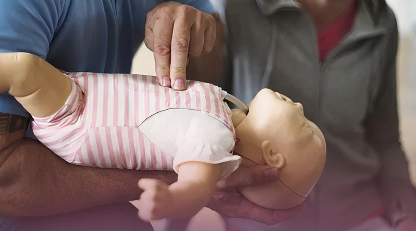
Begin 30 Compressions (2 Fingers)
Check For Object In Throat (NO BLIND FINGER SWEEP)
Give 2 Rescue Breaths
Notes
Start Compressions The Second Heart Rate Starts To Decrease, Don’t Start When Fully 0
SIDS (Sudden Infant Death Syndrome)
5000 Cases/Year
More Common In Males
No Known Cause
Babies Have Elevated Level Of CO2 In Blood During Autopsy
Believed That Airway Was Blocked Due To Plushies/Blankets Which Stopped Transfer Of CO2 And O2
No Indication Of Problems
Usually While Sleeping During First 6 Months
Prevention
Place Baby On Back (Now, Side)
Avoid “Fluffy” Blankets, ETC
Extra
Higher CPR Success Rate For Choking/Drowning Compared To Heart Attacks BC Body Was Somewhat Healthy In The First Place
Defibrillation
Sends An Electric Shock To The Heart To “Restart” It
Doesn’t Improve Heart Health, Just Saves Time
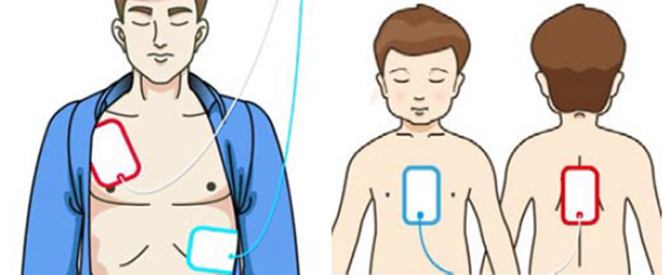
Location
Adults
Below Right Clavicle To Under Left Armpit (Diagonal)
Child
Right Shoulder To Under Left Armpit (Diagonal)
Baby
One Pad In Front Other Pad In Back
Note
Shocking Diagonally Increases Chances That The Heart Is Actually Shocked
Still Give Pregnant Woman Defib → Living Life Over Future Life
Extra
Do Not Touch Defibrillator When Shock Is Delivered
Remove All Metal And Piercings From The Body
Will Burn Skin Due To Metal Ions
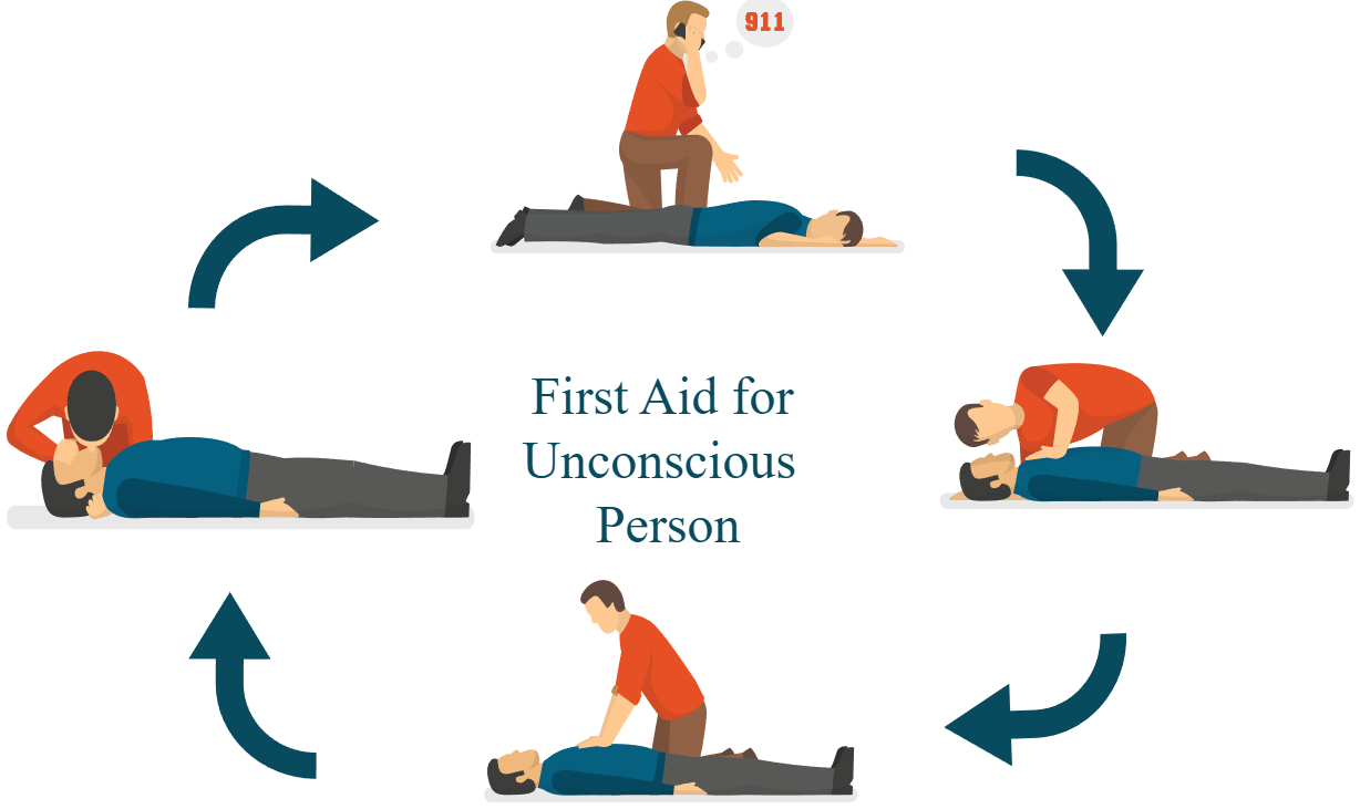
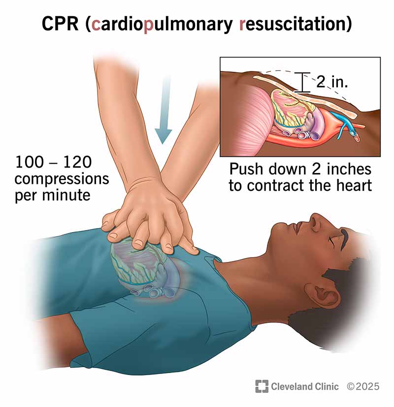
Cardiopulmonary Resuscitation
If Effective Can Provide ¼ - ⅓ Normal Blood Flow
Rescue Breaths Contain 16-21% O2
CPR Process
Start Immediately
Increases Chances Of Survival
Brain Damage Starts After 4-6 Minutes
Permanent Brain Damage After 10 Minutes W/O CPR
Do Not Move/Stop Until Qualified Help Arrives
Exceptions - Threat Of Fire, Victim Not On Hard Surface, Victims Head Not Level Or Above Body
Checking Vitals
Airway
Open Through Head-Tilt Chin-Lift
Jaw Thrust If Spinal Injury
Breathing
Look, Listen, And Feel For 5-10 Seconds
If Victim Not Breathing Give 2 Rescue Breaths
If They Don’t Go In Fix Airway And Try Again
If This Fails Again Suspect Choking
Compressions
Find Proper Hand Position (Between Nipples, 2 IN Above Sternum)
Stopping CPR
Victim Revives
Qualified Help Arrives
Too Exhausted To Continue
Unsafe Scene
Do Not Resuscitate Order
Circulatory System
Passes Nutrients, Gases, Hormones, And Blood Cells To And From Cells In The Body
Done To Maintain Body Temperature And Homeostasis + Fight Disease
Blood Vessels
Arteries
Carry Blood Away From The Heart
Usually Oxygenated Blood
Exceptions = Pulmonary (Going To Lungs) + Umbilical (Going To Placenta)
Major Arteries
Aorta - Largest Artery, Distributes Oxygenated Blood To All Parts Of The Body, Originates At Left Ventricle And Extends To Abdomen
Pulmonary - Carries Deoxygenated Blood From Heart To Lungs
Brachial - Inside Upper Arm, Where Blood Pressure Is Measured
Radial - Wrist, Where Pulse Is Measured
Carotid - Located In The Neck
Femoral - Medial Portion Of The Femur, Near Groin, Never Used
Veins
Carry Blood Towards The Heart
Majority Of Blood Carried Is Deoxygenated And Back To The Heart
Exceptions = Pulmonary (Leaving The Lungs) + Umbilical (Leaving The Placenta)
Capillaries
Only 1 Cell Thick
Allow For Exchange Of H2O, CO2, O2, ETC + Waste Materials
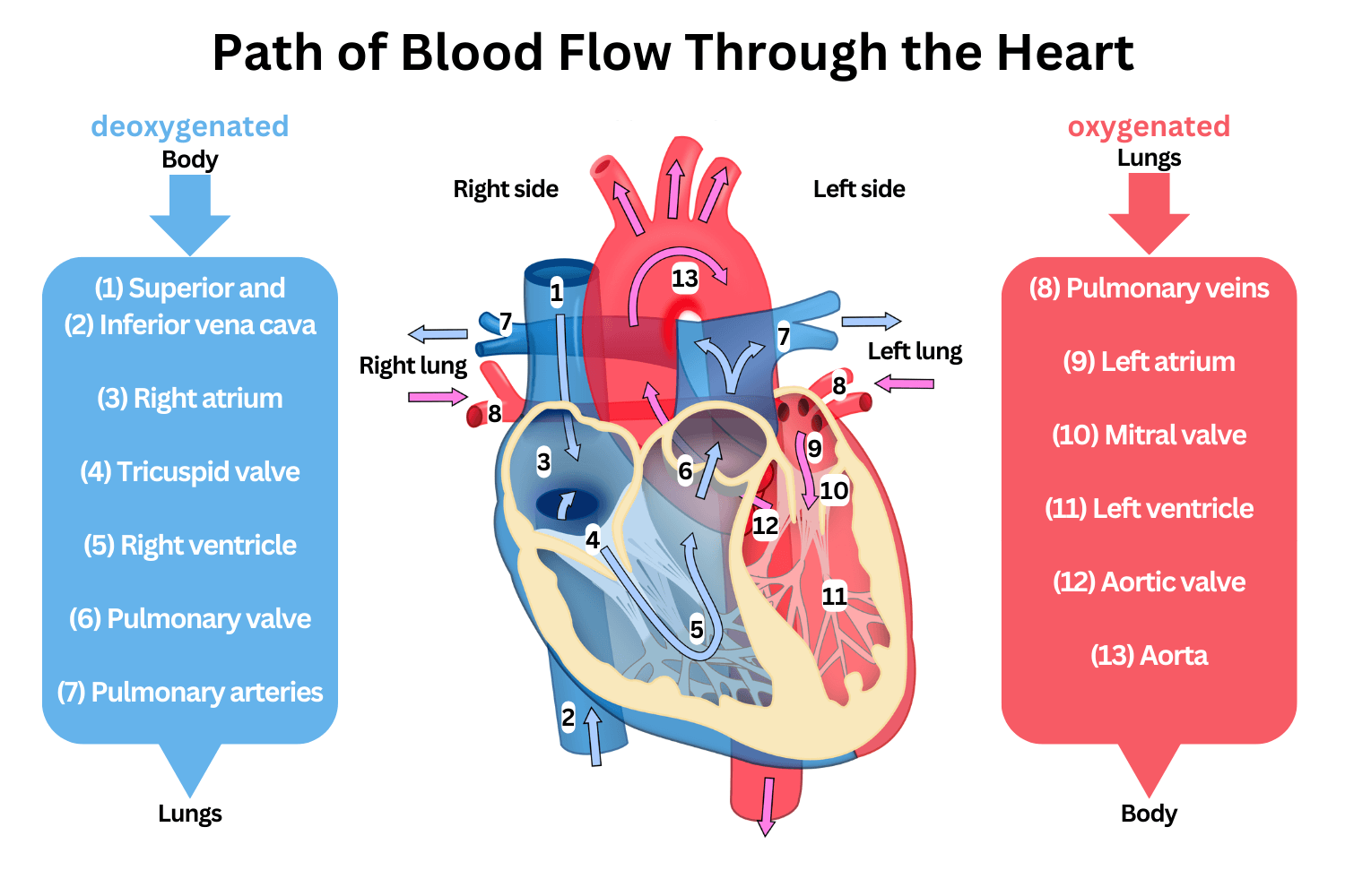
Blood Flow Order
1. Right Atrium
2. Right Ventricle
3. Pulmonary Artery
4. Lungs
5. Pulmonary Vein
6. Left Atrium
7. Left Ventricle
8. Aorta
9. Arteries, Capillaries, Veins
Blood
Perfusion
Circulation Within Tissues In Adequate Amount To Meet Cells Need For O2
Shock (Hypoperfusion)
Failure To Provide Adequate Circulation (BLOOD IS NOT CIRCULATING)
Cardiogenic
The Heart Loses Ability To Pump Blood/Not Circulating Blood And O2
Septic
Infection (Usually Bacterial) Of Blood
EX. The Appendix (Appendicitis) → The Appendix Isn’t Moved And Does Not Receive Blood Or Flush Out Bacteria, Causes Inflammation
Anaphylaxis
Severe Allergic Reactions
Hypovolemic
Decreased Water Volume → Blood Gets Thicker And Slower
Hemorrhage
Bleeding
Average Adult = 6 Liters Of Blood
Cannot Tolerate Greater Than 20% Blood Loss
Hemorrhagic Shock
Low Blood Volume Results In Inadequate Perfusion
Signs And Symptoms
Rapid, Weak (Thready) Pulse
Clammy (Moist, Sticky, Cold) Skin
Rapid & Shallow Respirations
Hypothermia (Due To Decreased Perfusion And Evaporation Of Sweat)
Enzymes Cannot Function Under 94°F
Thirst And Dry Mouth
Characteristics Of Bleeding
1. Arterial → Bright Red Spurting Blood (Oxygenated)
2. Venous → Dark Red Non-Spurting Blood (Deoxygenated)
3. Capillary → Easily Controlled Oozing Blood (Most Common)\
__ = More Dangerous Due To High Volume Of Blood Loss
EMS Treatment
Control Bleeding
Elevate Feet (If No Suspected Neck Injury)
Cover With Blanket
If Body Is Too Cold, Patient Will Become Unconscious
Oxygen (15 LPM Via NRB)
Controlling Bleeding
Direct Pressure With Bandage
Elevate Injury Above Heart
Blood Has To Go Uphill In Order To Get To Injury
If Bleeding Continues, Add More Bandages (NEVER REMOVE)
If Patient Is Punctured, Leave Object Inside
Object Is A Plug For The Injury
If Object Is Removed, Will Cut Patient Further
Tourniquet
Wrap Bandage Around Injury And Twist
Write “TK” And Time Applied On Patients Forehead
If Bleeding Does Not Stop, Patients Limb Might Be Amputated
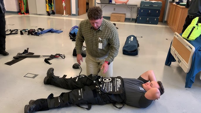
MAST Pants
Military Anti-Shock Trousers, Or Pneumatic Anti-Shock Garments (PASG)
Created In Vietnam
Stabilizes And Controls Blood Loss In Fractures Of Pelvis And Femurs
Puts Pressure On The Wound Which Limits Blood Flow
When Being Removed, Pressure Has To Be Removed Slowly So That Patient Does Not Pass Out
Limits Amount Of Pain
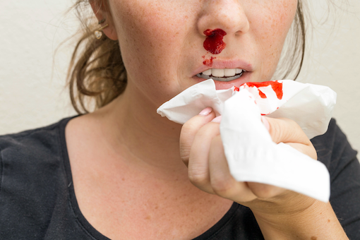
Nose Bleed
Have Patient Lean Forward And Pinch Nostrils
Apply Gauze Under Patients Upper Lip
Apply Ice Over Nose
Internal Bleeding
Hematoma
Bruise/Contusion
Capillaries Are Damaged Allowing Blood To Seep Into Tissues
Hematemesis
Blood In Vomit
Blood Pools Up In Stomach, Usually Thick/Clotted And Dark When Coming Out
Hemoptysis
Coughing Blood
Usually Sign Of Organ Failure, Respiratory Issues, ETC
Signs
Pain
Tenderness (Hurts To Touch)
Bruising
Swelling
Broken Ribs
Bruises On Chest
Distended Abs (Bloated)