Peripheral and Central Anatomy and Physiology
Anatomy and physiology of the peripheral vestibular system
The vestibular system allows us to maintain optimal balance and posture.
1st taught as a sensory system by Charles Darwin’s father
when someone moves, the vision system responds to that movement
more heavily considered as a system in the 1800s
The vestibular system works in tandem with the vision system and the proprioceptive system.
The triad
vestibular
vision
proprioception
The cochlea is anterior and medial to the vestibular system
The purpose of the vestibular system is to maintain balance to protect our bodies and ensure we do not fall and get hurt
Amniotes (not water bodied creatures) -
Relationship between rate of movement and involved system
At lower rates of movement → vision system dominates
At higher rate of movement → the vestibular system takes over
Rigid Body Motion
any rigid body can move in 6 different directions:
up and down
side to side
rotation (yaw)
tothe front or back (pitch)
rotation from
Otolith organs help records linear accelerations
utricle → front and back / horizontal
saccule → up and down /vertical
*Utricle and saccule both play a role in linear/translational movement
Semicircular canals (All 90 degrees to each other)
Horizontal/lateral semicircular canal → side to side/lateral movement
Superior/anterior/vertical semicircular canal
Posterior/ inferior semicircular canal →
The tail of the semicircular canals are connected to the utricle
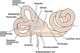
Codes movements into electrical energy
Ampulla are sensory structures that are kinda like a barrier between the semicircular canals and the utricle
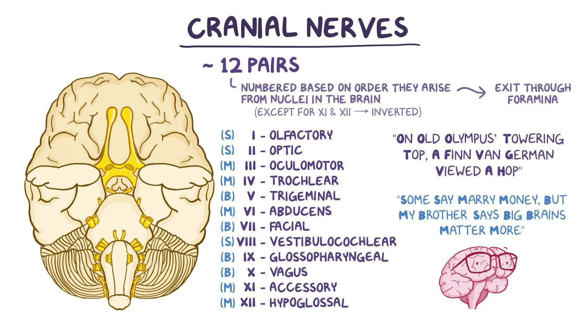
3, 4, and 6 are critical in vestibular awareness
responsible for ocular movements
Vestibular ocular pathway: helps to maintain stable vision
Vestibularospinal pathway: helps to maintain stable posterior
Structure of the Vestibular apparatus
semicircular canal’s sensory structure → crista (singular) cristae (plural)
cupula is a gelatinous mass that the hair bundles of the crista attach to
the density of the cupula and endolymph is the same at rest
otolith organs sensory structures → macula
type 1 cells are rounder than type 2 cells
there are more type 1 cells in the otolith organs
otoconia: calcium carbonate crystals are embedded on the otolithic membrane
the movement of the otoconia pulls the otolithic membrane, which causes hyperpolarization or depolarization
oval window opens on the saccular wall
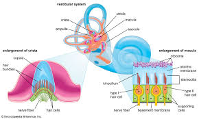
Type 1 and type 2 hair cells
Type 1
have a calyx terminal - allows for a very fast transmission of signals
there is a need for faster transmission
important for sudden, transient, jerk-related motion
higher concentration in otolith
more centrally located in the otolith
have shorter kinocillium which is just in the gel layer of the otolithic membrane which makes them more sensitive to movement
Type 2
code ongoing movement/signals
farther away from the striola (midline of the otolith organ and where kinocilium orientation changes)
taller kinocillium that is embedded in the mesh-layer
Mechanoreceptors: a sense organ or cell that responds to mechanical stimuli such as touch or sound.
The tallest hair cell is the kinocilium
Excitation versus inhibition in each plane
Resting firing rate (70-100 spikes/second) and the there is an excitation the firing rate goes up (400 spikes/second) and the opposite corresponding canal firing rate decreases (to 0 spikes/second)
the anterior or one and posterior of the other function together
They function in conjunction!
Dr. Raghav Jha
RALP (right anterior, left posterior)
LARP (left anterior, right posterior)
when one is excited the other is inhibited
the direction of movement is typically the side of excitation
Ewald’s Law
Stimulation of the semicircular canal generate eye movements in the place of the canal
excitation creates larger movement then inhibition (why?)
1) when the fluid moves the kinocilllium to the stereocillia
the plane of eye movement is the same as the plane of
ampullopetal flow causes depolarization of the semicircular canals
Every direction of movement must be encoded by the vestibular system
How are angular and linear movements encoded
Angular movements are coded in pairs
linear movements are coded in the same structure
Vestibular afferents
Regular
at the level of the type 2 hair cells
Irregular
found with type 1 hair cells
Otoliths vs. SCC VESITBULAR
Blood supply Related Disorders
vertebro-basiliar insufficiency
posteroinferior cerebellar
Anterior cerebellular artery
TIA of isolated cochlear artery
sudden SNHL
TIA isolated vestibular artery lesions
Central Vestibular Structures
Vestibular nucleus
SVN
IVN
LVN
MVN
Parteoinsular vestibular cortex