Connective Tissue (CT) Proper and Cartilage
Things to Know
CT is made of cells + fibers + ground substance
ECM = fibers + ground substance
Ground substance is a hydrated gel
Fibroblasts are the most common CT proper cell and make Type I collagen
Collagen = tensile strength
Type I collagen is coarse; Type II is fine and often not visible in cartilage; Type III reticular fibers
Loose CT generally has more cells/ground substance and thinner collagen bundles
Dense CT has thicker/more abundant fibers
Cartilage is mostly avascular, so nutrients diffuse through matrix
Lacunae are a major clue for cartilage
Perichondrium surrounds most cartilage, but there are exceptions
Isogenous groups/cell nests = groups of daughter cartilage cells
“Blasts” generally = building/growing, “cytes” = maintaining
Embryonic CT
Mesenchyme → the CT of the developing embryo
What are the two kinds of dense regular CT?
Dense Regular → 2 kinds: collagenous and elastic
Always regular → the collagen bundles tend to be oriented in the same direction
Cells → primarily fibroblasts
Collagenous
Most visible → are large bundles of Type I collagen
Typical of collagenous ligaments and tendons
Elastic
Predominantly large bundles of elastic fibers
Reduced amount of Type I collagen fibers
What are the two types of adipose tissue?
Adipose tissue
Cells → adipocytes
White adipose tissue → white fat
Unilocular → each cell has one large lipid vacuole
Brown adipose tissue → brown fat
Multilocular → each cell has multiple small lipid vacuoles
Distinguish between loose irregular CT, dense regular & irregular CT, reticular CT, and adipose tissue.
Loose irregular CT (areolar CT)
Orientation of collagen bundles varies
More ground substance and cells than dense irregular CT
Highly cellular and fewer fibers
Most visible fibers are small/thin bundles of Type I collagen
Image ID → lots of open/ground substance + scattered cells + thin collagen fibers
Dense irregular CT
Orientation of collagen bundles varies
Has less ground substance and cells than loose irregular CT
Has “wavy collagen”
Cells → primarily fibroblasts
Most visible fibers are larger/thicker bundles of Type I collagen
Image ID → thick, wavy collagen bundles running in different directions
Dense regular CT
Fibers tend to be oriented in the same direction
Cells → primarily fibroblasts
Large bundles of Type I collagen or elastic fibers
Image ID → fibers are parallel/all running in the same direction
Reticular CT
Cells → reticular cells
Fibers → reticular fibers
Form a mesh-like supportive framework
Image ID → looks like a mesh/network
Adipose Tissue
Cells → adipocytes
Image ID
White adipose → very large clear-looking cells with one large lipid vacuole

Brown adipose → cells have multiple small lipid vacuoles
What is the name of the “hydrated gel” polymer that binds to the fibers of the extracellular matrix in cartilage?
Hyaluronic acid/hyaluronan
Binds multiple aggrecan monomers to form large aggregates
Forms a “hydrated gel” with unique compressive properties
List the two cell types of cartilage. Which is actively secreting matrix? (note that visual distinction will not be required, as can be arbitrary)
Chondroblasts
Actively secrete cartilage matrix
Capable of division and matrix production
Produce mostly Type II collagen
Blasts = growing
Chondrocytes
Not actively dividing and secreting
Suspended in cartilage matrix
Maintain the matrix
Cytes = maintain
Which fiber is visible in fibrocartilage?
Type I collagen fibers are visible
Which fiber is visible in elastic cartilage?
Elastic fibers are visible
Which fiber is invisible in hyaline cartilage?
Type II collagen fibers are invisible
It blends into the matrix and cannot be seen
Distinguish between the 3 types of cartilage.
Hyaline cartilage
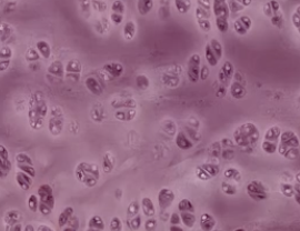
Chondrocytes reside within lacunae
Looks “glassy”
Type II collagen blends into the matrix and cannot be seen
Image ID → glassy/smooth matrix + lacunae + no visible fibers
Elastic cartilage
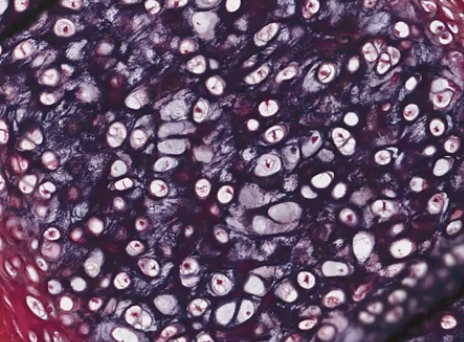
Identical to hyaline except it has elastic fibers
Chondrocytes located in frequent isogenous groups
Matrix is similar to hyaline cartilage with addition of elastic fibers
Image ID → lacunae + visible elastic fibers

Fibrocartilage
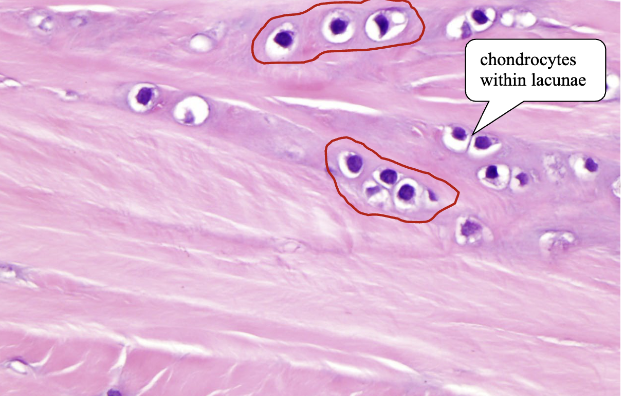
Small chondrocytes housed within lacunae
Chondrocytes inserted between large bundles of collagen fibers
Sometimes “stacked” in a row
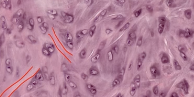
Type I collagen fibers
Often arranged in a “herring bone” pattern
Image ID → If you see lacunae + fibers = fibrocartilage
This can look like dense irregular CT
But in fibrocartilage, the fiber bundles are “too round: and the cells are located within lacunae
Mixed
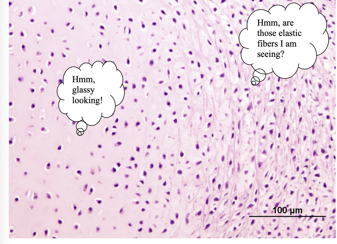
Hyaline (left) + elastic cartilage (right)
What is the difference between interstitial & appositional growth of cartilage?
Interstitial Growth
Growth within the inside
Chondroblasts divide and form multicellular lacunae → isogenous groups/cell nests
Daughter cells secrete matrix around and between each other
Causes expansion from within the cartilage
Think interstitial = inside
Appositional Growth
Growth upon the surface (outside)
Chondrocytes form from stem cells in the perichondrium
Secrete matrix onto the cartilage surface
Think appositional = adds onto the outside
Self-Quiz Image ID
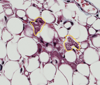
Brown adipose tissue cells circled in yellow
They are multilocular, meaning each cell has multiple small lipid vacuoles
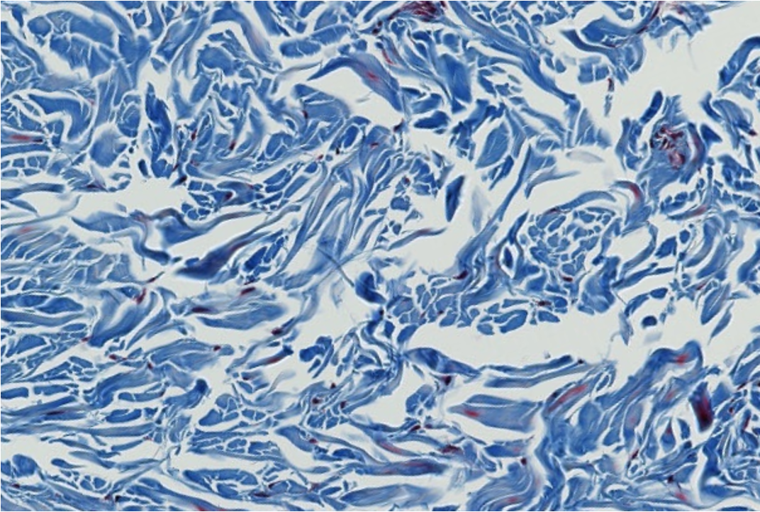
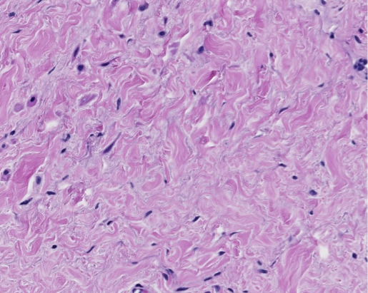
Dense CT has much more collagen and much less open ground substance than loose CT
There are relatively few cells
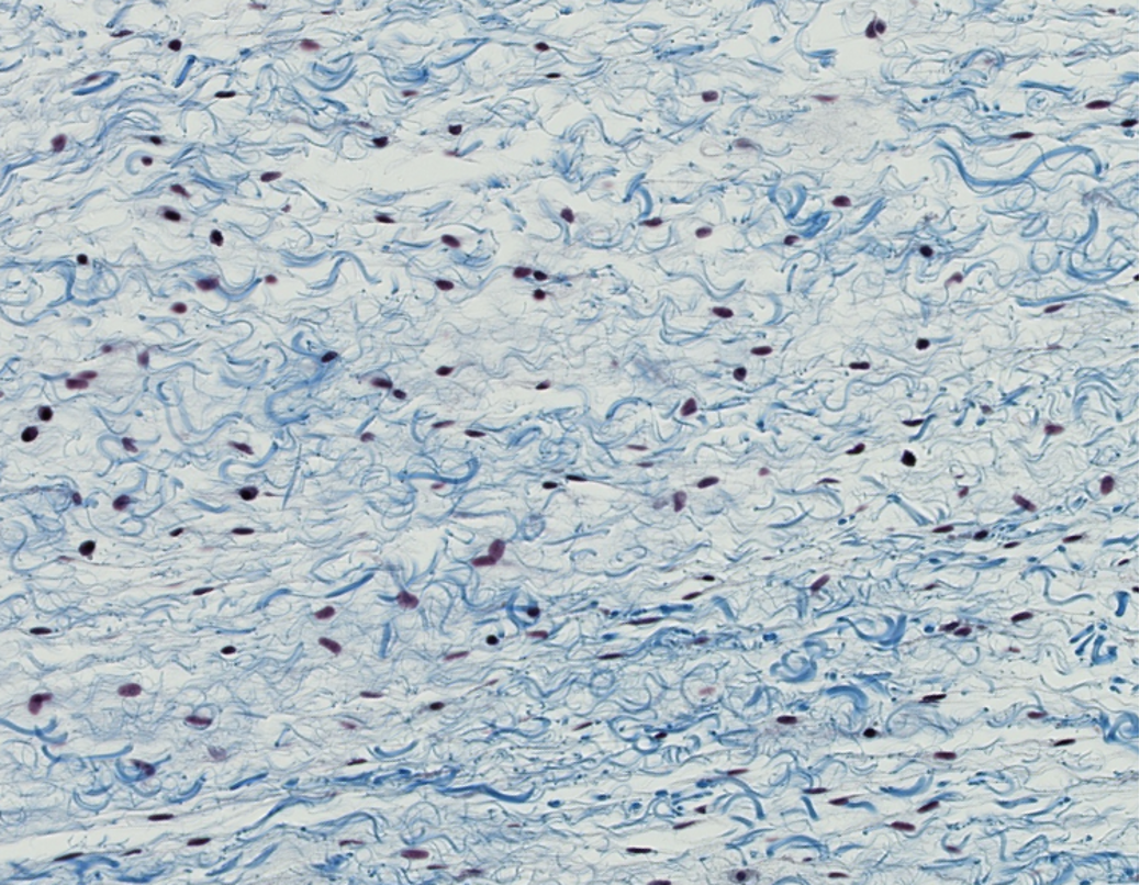
Loose CT
Aka areolar CT

Elastic ligaments classified as dense regular CT

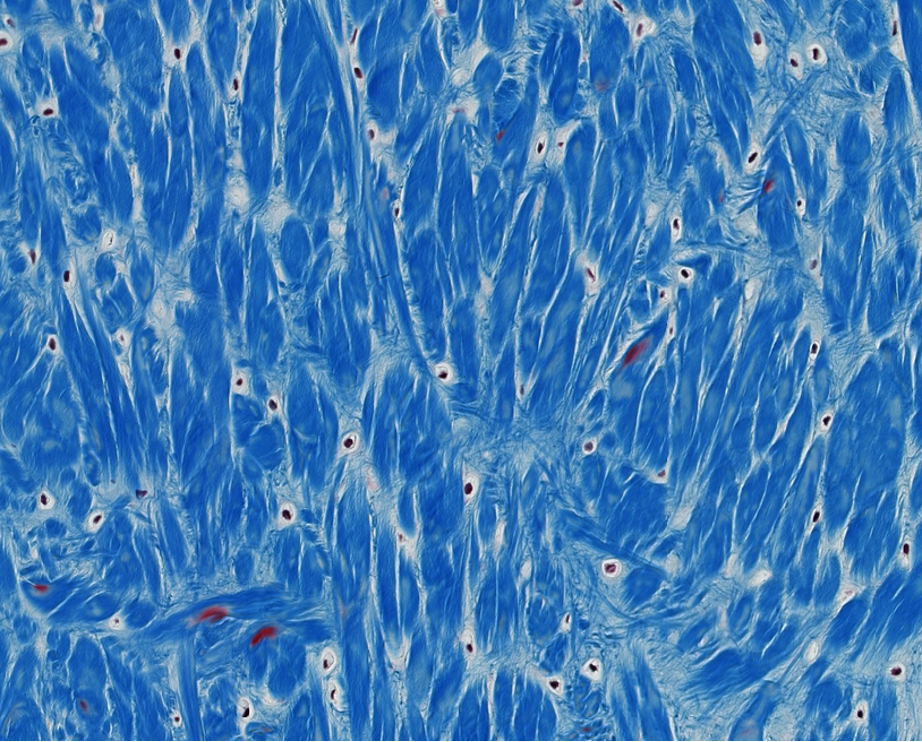
Fibrocartilage
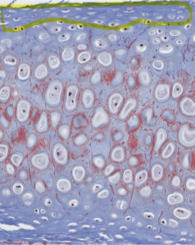
What do we call the CT capsule that surrounds hyaline & elastic cartilage?
Perichondrium