The Nervous System
: a system that controls all the activities of the body
→ NERVES are what allows you to feel, touch, and understand your feelings and your surroundings
homeostasis/equilibrium: balance. state of balance. keeping a constant internal environment
the main things we use our nervous system for:
maintaining blood glucose levels
maintaining temperature
systolic blood pressure
maintaining blood pH
the nervous system is made up of:
the brain
the nerves- talk to the spinal chord
the spinal chord- transmits info to the brain (like the highway)
senses- taste, sight, hearing
the main process of the nervous system:
sensory input
integration
motor output
sensory input: using senses/sensory receptors we receive information about our internal/external environments.
integration: interpreting what that information means, and what needs to happen consequentially. the information is often later integrated with stored information.
motor output: in situations where it is necessary, effector organs are told to respond and fix the issue.
effector organs: the part of the body that carries out the response. example: legs run, eyelids will blink, pancreas will produce insulin, so on.
the organization of the nervous system:
there are two main divisions:the central nervous system & the peripheral nervous system
CNS: the brain and spinal chord
coordinates the incoming and outgoing information
spinal chord acts as the message highway between the brain and the body
protecting the CNS:
bone coverings: the skull and the vertebrae
protective membranes surrounding brain: meninges
outer layer: dura mater
middle layer: arachnoid
inner layer: pia meter
shock absorber between the pia meter (inner) and arachnoid (middle layer) of the meninges and the central canal of the spinal chord: cerebral spinal fluid
the spinal chord: carries messages from receptor to brain and then back again from the brain to effector organs → receptor → up spine→ brain → down spine →effector organs
forum magnum: opening in skull for spinal chord
gray vs white:
gray: unmyelinated (no fat) neurons/axons (increased processing power)
white: myelinated (yes fat) inter neurons that connect spinal chord to brain (speed, message sending/receiving)
remember: smart think a lot people seem more grey. white counter tops fast fashion millennial- speedy
dorsal vs. ventral nerve:
dorsal: brings sensory info in (usually sensory neuron) (on the top)
ventral: carries motor info out to the effectors (usually motor neuron) (on the bottom)
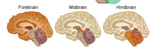
the brain: the brain has three regions
the forebrain: reason, intellect, memory, personality, language
the midbrain: relay center for eyes and ears
the hindbrain: muscles, balance, autonomic control
 the forebrain deep into it:
contains:
the cerebrum: what you think of when you think brain. the cerebral cortex is REALLY what you think of when you think brain. (gray matter- unmyelinated, processing power)
coordinating center for motor actions, speech, memory, personality
2 distinct hemispheres: right brain (visual/spatial awareness) and left brain (verbal skills and speech)
these two distinct hemispheres are linked by a communication bridge called the corpus callosum→ remember man with no corpus callosum
each hemisphere of the brain has 4 lobes: frontal, temporal, parietal, occipital
thalamus: interprets sensory information
hypothalamus: interprets internal environment and instructs the pituitary to produce hormones or the medulla Omblongota to send a nerve signal (autonomic)
olfactory bulbs: detect smell
how the cerebrum interprets speech:
DOES broca’s area: coordinates speech muscles and translates thoughts into speech (does the speech)
INTERPRETS wernickce’s area: language storage and comprehension (interprets the speech)
the four lobes of the forebrain:
frontal: voluntary movement (walking, speech), personality, memory, intellect, and conscious thought
temporal: interpreting sensory information (hearing, smell, vision)- receives auditory information → ear stuff sends it here
parietal: touch, pain, taste (temperature) awareness, interpreting speech, emotions
occipital: receives visual information
the mid brain
4 spheres of grey matter
relay center for eyes and ears reflexes
hindbrain:
contains:
cerebellum: controls limb movement, balance, and muscle tone
pons: relay station between cerebellum and medulla (like a bridge)
medulla oblongata: joins spinal chord to the cerebellum, the sight of autonomic nerve control
PNS: nerve and senses
carries information between the effectors/organs and the CNS
within the PNS there are two divisions: the somatic NS and the autonomic NS
→ the somatic (body) NS: consists of the nerves connected to sensory receptors and skeletal muscles, and is what permits voluntary action
controls all the nerves involved in body movement: eyes, mouth, ears muscles, and the nerves that create their actions: blink now, move your finger here- exception: reflexes
composed of:
12 paired cranial nerves: controls vision, hearing, balance, taste, smell, facial, tongue, head and neck movement
31 paired spinal nerves: controls skeletal muscles
→ the autonomic NS: permits the involuntary function of the organs inside of our body. additionally, the autonomic NS consists of the sympathetic and parasympathetic systems.
autonomic nervus system is involuntary internal homeostatic control
two types of ANS nerves:
preganglionic: CNS-ganglion (sends message from brain)
post ganglionic: ganglion-target organs (delivers to effectors)
autonomic nervous system halves:
the parasympathetic system (the off switch, rest and digest)
restores balance
long preganglionic nerve
preganglionic and post ganglionic release acetylcholine
master “off nerve”: vagus nerve (has control over heart, bronchi, liver, and digestive tract)
the sympathetic system (on switch, fight or flight)
prepares the body for stress
SHORT (fast) preganglionic nerve
pre ganglionic nerves release acetylcholine
post ganglionic nerves release norepinephrine (adrenaline)
cells of the nervous system: glial cells and neurons
glial cells are meant for support
neurons are to simple and basic to keep themselves alive, so glial cells act as a mother and hold them in place, provide them with nutrients, defend against infection and clean up after them if a cell dies
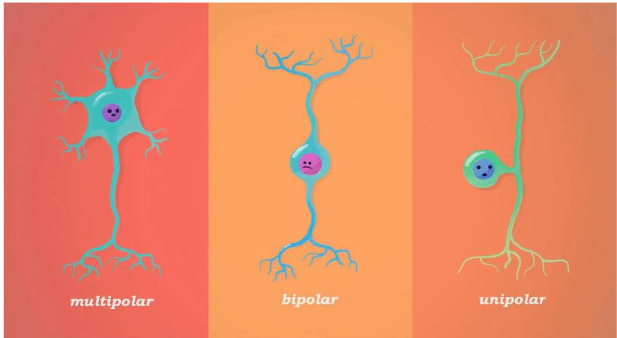
neurons (3 types)
sensory neurons (afferent, 90% are found only in PNS)
they relay information about the bodies internal/external environment to the CNS (example: from the hand to the spinal chord)
receive the first information in the process of the nervous system
a comes first in the alphabet- so afferent comes first here
→ afferent means that the cells carry information towards the CNS
inter neurons (100% are found in CNS)
links neurons in the spinal chord to neurons in the brain
motor neurons (efferent, everywhere)
carries impulses from the CNS to the effectors
efferent which means away from the CNS
→ efferent heads towards effectors
 motor neuron intern neuron sensory neuron
the anatomy of a nerve cell:
neurons: vary in size, shape, and appearance. work three main jobs: input (sensory neurons) integration (inter neurons), and output (motor neurons).
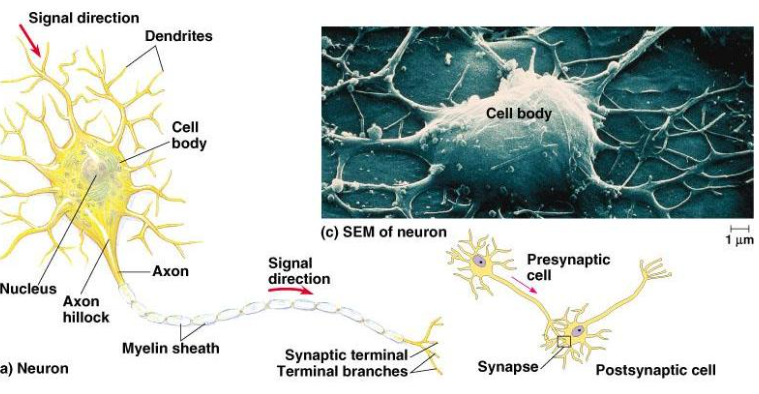
cell body: functional portion of a cell
→ when inside CNS called cell body, when inside PNS called ganglion
dendrites: looks like they are the arms of the cell body, but actually they are short extensions off the cell body that receive signals from other neurons or the environment
→ can connect to another cell body
axon: long extension off of the cell body that transmits impulses away to other neurons or effectors
the neurilemma: the membrane surrounding the axon that promotes cell regeneration
myelinated cells: cells with myelin sheath
Schwann cells are responsible for making myelin sheath → myelin sheath is essentially FAT. technically speaking: myelin sheath is the white insulation that surrounds the axon
myelin sheath acts as an insulator which helps prevent the loss of charged ions as they move through the axon. additionally myelin sheath allows for the nerve impulses to transmit across the axon MUCH faster.
people with MS end up with damaged myelin sheath- which makes it difficult and a very long process to transmit nerve impulses. the result of this is that eventually everything will slow down/degenerate.
the areas between myelin sheath are known as: nodes of Ranvier (essentially these are just gaps in the fat along the axon)
reflexes: involuntary actions that initially surpass interpretation by the brain
the message still transmits through all three neurons before there is a reaction, however the brain does not process what has happened (no thought)
→ this is called reflex arc and was developed in humans evolutionarily
→reflex arc: autonomic response controlled primarily by the spinal chord. the process of a reflex:
stimulus
receptors
sensory neuron
inter neuron
motor neuron
effector organ
= a response (leg has kicked or whatever)
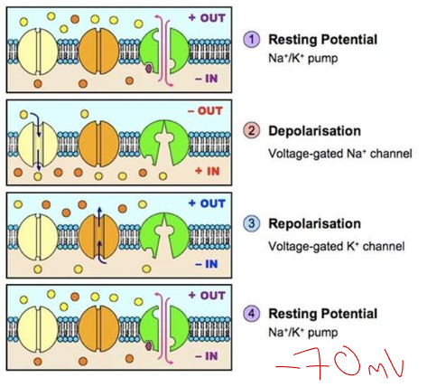
nerve impulses: nerves have two states
resting potential
nerves are polarized: they are off and just resting
internal charge is -70 mV
action potential
nerves are depolarized: turned on
charge jumps to +40 mV
how do we keep it -70 mV inside a neuron?
three main methods
sodium potassium pumps: uses ATP to pump 3 sodium ions in and 2 potassium ions out. this is an unequal distribution and leads to a polarized membrane
the presence of negatively charged plasma proteins and ions that never move: example chlorine (Cl-)
k+ (potassium) channels that allow potassium to leak out of the inside of the axon
action potential:
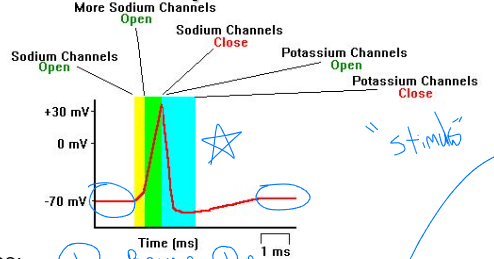
depolarization: stimulus bypasses threshold, causing -70mV to jump to +40mV
you cant feel something just a little bit- you either feel it or you don’t. which explains the all or nothing response
repolarization: Na+ channels close, and K+ gates open causing potassium to diffuse out of the cell which restores the original level of polarization (-70mV)
refractory period: the recovery time required before a neuron can return to its resting potential
saltatory conduction (in myelinated axons): HOP SCOTCH→ action potential jumps over myelin sheath and from node or Ranvier to node of Ranvier
this leads to faster conduction of action potential than on an unmyelinated axon
threshold level: the minimum amount of stimulus required to get a response (example >2mv)
all or nothing response: nerves and muscles respond completely or not at all
a charge that is greater than the minimum amount of stimulus required will not create an increased response
an increased response: more neurons are being activated
message priority is determined by:
more frequency of impulses
some neurons have higher threshold levels only set of with increased stimulus
therefore: the greater the number of impulses reaching the brain the greater the intensity of the response
THE SYNAPSE: the space between
a synapse is what divides neurons
presynaptic neurons: releases neurotransmitters into the synapse
postsynaptic neurons: receives neurotransmitters from the synapse
neurotransmitter: chemical messenger for neurons
IN ORDER FOR action potential to bridge the gab between two cells (the synapse) it must be converted into chemical energy
→ here is how it is done
the presynaptic membrane is depolarized (with the aid of calcium- Ca2+)
synaptic vesicles release neurotransmitters (ACETYLCHOLINE) from the axon bulbs/end plate
neurotransmitters diffuse and bind to receptors on post synaptic dendrites
post synaptic membrane opens either ion channel
Na+ flows in= excitatory
K+ flows out = inhibitory
- neurotransmitters are broken down by enzyme (cholinesterase) and ion gates close
- neurotransmitters are absorbed by pre-synaptic neurons for rebuilding
the role of neurotransmitters:
common neurotransmitters: dopamine, serotonin, endorphins
excitatory (when Na+ flows in): triggers receptors in post synaptic cleft to allow positive ions (sodium) in→ this leads to depolarization
inhibitory (when potassium goes out): triggers potassium channels to open→ leads to hyperpolarization
summation: the effect of the accumulation of neurotransmitters from two or more neurons, can be inhibitory or excitatory
neurotransmitter disorders:
parkinson’s disease: inadequate dopamine (inhibitory) causes involuntary muscle contraction
alzheimer’s disease: inadequate acetylcholine (excitatory) deterioration of memory and mental capacity
brain imaging technology
PET scan: track activity and usage- radioactive glucose is consumed in certain parts of the brain and when that part of the brain is used it will light up with different colors on the scan.
MRI: giant magnets that detect changed in H+ that emit radio signals
the structure of the eye:
composed of three separate layers: sclera, choroid layer, retina
sclera: outer most layer of the eye- protection. “whites”- no blood vessels, gets O2 from dissolved tears
covered by the cornea (transparent tissue that refracts light towards pupil)
gets nutrients from the aqueous humor- clear liquidly fluid in the front of the eye
lack of nutrients and O2: glaucoma
choroid layer: middle layer that has pigments that prevent scattering by absorbing stray light- has many blood vessels
iris: colored muscle that regulates the amount of light that enters the eye
pupil: opening for light to go in
lens: focuses image on the retina by action of dorsal and ventral ciliary muscles
vitreous humor: cloudy, jellylike material that maintains eye shape and lets light through
retina: innermost layer, composed of three layers of cells:
light sensitive rods and cones: c=color
bipolar cells: pass messages from rods and cones to cells of the optic nerve
optic nerve cells: ganglion cells (cell in a cluster of cells found in the PNS)
HOW IT WORKS:
light depolarizes bipolar cells
bipolar cells deliver messages to rod and cones
bipolar cells send color shade/information to optic nerve
rods and cones:
rods: used in dim light
cones: used for identifying colors- red, blue, and green cones
retina continued:
fovea centralis: closely packed with cones at the center of the retina; most sensitive area of the eye
blind spot: lack of rods and cones, where the optic nerve comes in contact with the retina
focusing an image: light entering the eye is bent/focused by cornea towards the pupil→ the lens further bends (refracts) light towards a focal point (fovea centralis). The image is inverted but the brain corrects it.
accommodations: adjustments made to lens and pupil to view near or far objects
focusing for close vision: thickening the lens shape by flexing ciliary muscles allows tendons to relax
focusing for distant vision: lens becomes thin by ciliary muscles relaxing and tendons pulling- pupils may also dilate to allow for as much light in as possible
vision defects:
cataracts: lens or cornea becomes clouded
solutions: replace cloudy lens with a plastic one or remove lens and use glasses instead
astigmatism: abnormal curvature of the cornea or surface of the lens
solution: unevenly ground lenses
nearsightedness (myopia): image is focused in front of the retina
solution: glasses with concave lenses
farsightedness (hyperopia): image is focused beyond the retina (the eyeball is too small)
solution: glasses with a convex lens
color blindness: inherited condition where one lacks certain cones (r. b, or g)
after images: caused by the fatigue of a cone in an area of the retina: can be positive (strobe light) or negative (green and red cross)
the muscles of the eye:
extrinsic muscles: OUTSIDE, move the eyeball in the socket
rectus muscle: up and down, in and out
oblique muscles: rotation, left and right
intrinsic muscles: INSIDE, controls lens and iris and ciliary muscle body
ciliary body muscles: involuntary muscles that contract, causing the body to move forward and the lens shape to change.
ciliary body is a part of the choroid (middle) layer
contains ligaments that attach the lens to smooth muscles
the structure of the ear
ear is crucial in hearing and balance (equilibrium): both mechanoreceptors
essentially: the inner ear cells have tiny cilia which respond to mechanical stimuli (movement) which causes the nerve cell to generate an impulse
the ear can be divided into three sections: inner, middle, and outer
the outer ear:
the pinna: outer flap, acts as a sound funnel, directing it into the auditory canal (what we think of when we think of ear.
shape is very intentionally designed for the purpose of directing it into the auditory canal
auditory canal: channel that carries sound waves towards the ear drum
specialized sweat glands within the auditory canal produce ear wax which is intended to trap invading particles
the middle ear: begins at the ear drum ends at the vestibule
tympanic membrane: the eardrum
ossicles: amplifies/passes sound waves from the eardrum to the oval window by using three small bones:
malleus (hammer)
incus (anvil)
stapes (stirrup) -smallest bone in the body
oval window: smaller than the eardrum, amplifies sound- connects vestibule to middle ear, flap like bone
eustachian tube: air filled tube that equalizes pressure between internal and external ear (the reason your ears pop on airplanes)
also drains excessive fluid to nasal cavity
the inner ear:
vestibule: connected to middle ear by the oval window
inside of the vestibule there are two small sacs that are involved in maintaining head balance- the utricle and saccule
semi circular canals: three fluid filled canals that help body balance
COCHLEA: the snail. coiled snail shell lined with hair cells that respond to different sound waves and frequencies and then convert them into electrical impulses
auditory nerve: transmits sound signals to the CNS
hearing and sound HOW IT WORKS:
sound energy waves (mechanical energy) get converted into chemical energy (Na+, K+)
ossicles move a shorter distance then eardrum =amplification of sound
intense noise causes the muscles around the ossicle to contract = restricting movement, and pulling stapes from the oval window which means there will be less sound amplified and transmitted
oval window is moved inwards, which moves the round window outwards
triggers waves in fluid within the inner ear to the cochlea- this converts the waves to electrical impulses
organ of chorti: primary sound receptor in the cochlea
one inner row and three outer rows of hair
basilar membrane: anchors the receptor hair cells to the organ of chorti
fluid vibrations move the basilar membrane and the hair cells bend, which stimulates the sensory nerves that send the auditory signal to the brain (temporal lobe)
tectorial membrane: non mobile top layer
stereocilia: “the ear hairs”,
differ in width and lengths for pitch and volume perception
→ sound wave → tympanic membrane → ossicles → oval window →organ of chorti (cochlea) → round window → temporal lobe
hearing loss:
conductive (vibration) hearing loss: caused by wax buildup, middle ear infection, punctured ear drum
treatment: hearing aid: amplifies sound and transmits to ear drum
sensorineural hearing loss: auditory nerve is severed, or cochlear hair cells are damaged
treatments: cochlear implants
the ear and balance:
the liquid in your inner ear is what is responsible for balance: this liquid moves when we move and sends information to the brain to indicate how we are moving
in the body: balance is maintained by three semi-circular canals
in each fluid filled canal there is a pocket (ampulla) that contains hair (cilia) that moves with the gel like (cupulla).
cupula is pulled by forces (example gravity)
movement causes the cupula to bend the cilia, initiating a nerve impulse that is carried by the CNS
in the head: maintained by two fluid filled sacks in the vestibule: saccule and utricle
in each sac there are hair like receptors that are suspended in a jelly like material that contains tiny carbon stones called otoliths
when the hair is bent sensory receptors are stimulated and send a message to the brain