Lab Exam 2:
Lab 7: CNS Anatomy
Cerebral Cortex:
Frontal lobe: voluntary motor control, personality, intellectual processing, verbal communication
Pre-Central Gyrus: conscious motor control
Parietal Lobe: understanding words and formulating words to express thoughts
Post Central Gyrus: somatosensory cortex, sensation from skin, joints muscles, and tendons
Temporal Lobe: hearing center
Occipital lobe: visual center
Insula: takes sensation from visceral and keeps it in memory
example: food poisoning results in never eating that food or at that restaurant again
Cerebellum:
Cerebellum: posture, balance, smooths and directs movement
example: walk white line, touch finger to nose, drunk driving test
Diencephalon:
Thalamus: release station for all sensory information, except smell, as it passes on the cerebrum
Hypothalamus: control center for hunger, thirst, body temperature, hormone secretion
Brain Stem:
midbrain: relay center for auditory information, motor coordination, reward behavior
Pons: respiratory center
Medulla Oblongata: respiratory center, vasomotor center, cardiac control center
Spinal Cord:
Gray horns: contains sensory neuron axon terminals, interneurons, motor neuron cell bodies
White Columns: contains myelinated axons of interneurons
Dorsal Root: contains myelinated axons of sensory neurons
Dorsal Root Ganglia: contains cell bodies of sensory neurons
Ventral Root: myelinated axons of motor neurons
Spinal Nerve: contains myelinated axons of both sensory and motor neurons
Neuron:
Dendrite: site of graded potential generation
Cell body: site of graded potential generation, integration of incoming signals
Axon: propagation of action potential, release of neurotransmitters
Lab 8: Reflex Arc
Reflex: simple motor response to a particular stimulus
A short neural pathway not under conscious control
Clinical Significance:
Specific simple reflex arcs are associated with specific spinal cord segments, making tests for reflexes useful in diagnosing neurological disorders
Reflex arc: consists of sensor, sensory neuron, interneuron (not present in all reflex arcs) , motor neuron, and effector
sensory receptor: receptor that detects changes in the environment and generates impulses that are transmitted to the central nervous system
sensory neuron: communicates information from PNS to CNS with axon potential
interneuron: integration, decision maker neuron
motor neuron: extends out of CNS to effector organ
effector: muscle or gland
Knee Jerk Reflex: stretch reflex, that is monosynaptic and involves a sensory receptor that is a muscle spindle
contains sensory neuron (muscle spindle), sensory neuron, motor neuron, effector (skeletal muscle)
NO interneuron
sensory neuron and motor neuron synapse directly, in the lumbar region of the spinal cord
stretch reflex: protect muscles from over stretch and play role in subconscious postural adjustments
Muscle Spindles: stretch receptors that respond to changes in muscle length
sensory organ within skeletal muscle that is sensitive to muscle stretch and provides a length detector within muscles.
provide major source of proprioceptive sensory information
Monosynaptic Reflex: Simple reflex involving a sensory neuron synapsing directly with a motor neuron.
No interneuron in between
sensory neuron and motor neuron are next to each other on same muscle
muscle maintained in optimal length
allows for maintaining adequate overlap of thick and thin filaments
prevents overstretching
if patient has damage to cervical spinal cord, there would be no effect in knee jerk reflex because information does not have to travel to travel to and from the cerebral cortex
Latent period: time between the the onset of the stimulus and the onset of the effector muscle contraction
the time it takes for neural message to go from sensor to effector
reflects the number of synapses in a particular pathway
Experiment comparing Latent periods:
testing the knee jerk reflex with voluntary response of raising leg
result: voluntary latent period was longer
due to synaptic transmission taking longer than propagation of action potentials
neural pathway for voluntary response contains many synapses, taking longer than the reflex response that has only one synapse
Plantar Reflex: neurological test that is elicited by cutaneous receptors on the sole of the foot
cutaneous sensory receptor in foot transmits information to lumbar region of spinal cord to sensory cortex of the brain, then through the pyramidal motor tracts descending from cerebral cortex to lower motor neurons in the spinal cord
Normal response: adduction and plantar flexion of toes
abnormal response: abduction and dorsiflexion of toes
also known as Babinski reflex
Clinical Significance:
abnormal response infers the patient has damage along the pyramidal motor tract
infants exhibit babinski’s reflex normally because neural control is not fully developed yet
if patient has damage to cervical spinal cord, it would prevent sensory input from reaching the brain and motor output from reaching the foot
Complex Neurological Pathway:
Disynaptic Reflex: A type of polysynaptic reflex that involves only two synapses, where a sensory neuron synapses with an interneuron, which synapses with a motor neuron
Lab 9:
Sensory receptors: receive stimuli (changes in the environment) and change (transduce) incoming signals into action potentials that are sent to specific parts the of the brain for interpretation
Adaptation: ability that some sensory (phasic receptors) receptors have to reduce the singal sent in response to a continued stimulus
general senses: found body wide
general senses are somesthetic and proprioceptive
somesthetic: information about body feelings (touch, pressure, temperature, pain)
proprioceptive: information about body position (muscle strength and contraction, joint position)
special senses: localized in the head
Law of Specific Nerve Energies:
Law of Specific Nerve Energies: the nature of perception is defined by the pathway over which the sensory information is carried
sensation produced by the “adequate” stimulus (normal stimulus) is the one the brain will perceive, even if the stimulus is different than normal nature
the mind directly perceives the activity of sensory nerves, not the external stimuli themselves. Each sensory nerve produces its own characteristic sensation regardless of how it is stimulated
adequate stimulus: type of stimulus to which the receptor is most sensitive to
adequate stimulus for cones of the retina: bright light and color
adequate stimulus for hair cells of cochlea: sound waves
adequate stimulus for Pacinian corpuscles: deep pressure
adequate stimulus for Meissner’s corpuscles: light touch
Testing Law of Specific Nerve Energies:
Experiment: pressing on the eye creates a sensation of light ("seeing stars") because the optic nerve is activated, even though the stimulus was physical pressure, not light.
Testing Perception:
cutaneous receptors are rarely stimulated singly, often different types of receptors are stimulated simultaneously
when placing gloved hand into beaker of water, hand remains dry but sensation of wetness is felt
due to sensation of temperature and pressure
Receptor Fields and Sensory Acuity
Acuity: ability of sensory receptors to discriminate between two different points of stimulation
Receptor Fields:The area of skin associated with a single sensory receptor.
They are often overlapping.
different parts of the body have different sizes of receptor fields
when stimulated, causes action potential on axon of single neuron
density of sensory receptors/receptor size are inversely related to the size of somatosensory cortex devoted to a specific body part
receptor fields are smaller in the finger tips (than the back of the neck), meaning there are a higher density of sensory receptors in the fingers and a larger area of the somatosensory cortex for interpretation for the fingers
greater number of sensory receptors serving an area of the body = the smaller the receptor field = the better the sharpness of sensation of that area
Two-Point Discrimination Test (two point touch threshold test): measurement of approximate size of receptive fields
take calipers and lightly touching skin at same time, measuring the minimum distance two points of touch can be perceived as separate
two point touch threshold: minimum distance at which two points can be felt as two
measures the distance between receptor fields

Adaptation:
Adaptation: ability that some sensory receptors have to reduce the singal sent in response to a continued stimulus
meissner’s corpuscles adapt rapidly to presence of stimuli
taste receptors adapt slowly to presence of stimuli
cutaneous pain receptors do not adapt at all
experiment: place a coin on forearm and let it remain there, and place a rubber band around head and see which adaptation occurs faster
result: sensation of the coin adapted quicker
Equilibrium and Hearing
Inner Ear Anatomy
Vestibular Apparatus: part of the inner ear responsible for equilibrium
includes the utricle and saccule that detect linear acceleration and the semicircular canals, that detect rotational move ments
Cochlea: part of the inner ear Responsible for hearing, where sound vibrations are converted to neural signals.
Bony Labyrinths: contain the membranous labyrinths within a body cavity, filled with fluid (perilymph)
membranous labyrinths: tubular structure within the bony labyrinth that contains fluid (endolymph)
endolymph in membranous labyrinths and perilymph in bony labyrinths contain different concentrations of potassium

Sensory Hair Cells
Hair Cells: type of mechanoreceptor, that depolarizes based off mechanical deformation of cell
contain sterocilia that bend
sterocilia: modified microvilli arranged by height
kinocilium: tallest cilium
if bend in one direction, opens potassium channels causing depolarization, causing an increase in frequency of action potentials
opens potassium channels, allowing potassium from endolymph to enter cell and depolarization occurs
when stereocilia is bent towards the kinocilium, the plasma membrane is depressed and opens potassium channels, allowing potassium to enter, causing depolarization of the hair cells
if bend in other direction, causes hyper-polarization, reducing frequency of action potentials
when bent away from kinocilium, cause potassium channels to close

Hearing Tests
endolymph in inner hear vibrate in response to sound waves conducted through skull bones, bypassing the middle ear ossicles
makes it possible to differentiate between conduction deafness and sensory deafness
conduction deafness: deafness resulting from middle ear damage such as damage to the external auditory canal or middle ear
caused by: external auditory canal impairment, damage to tympanic membrane, middle ear damage such as otosclerosis ( bone spur on ear ossicles), or otitis media (inflamed and fluid filled middle ear cavity)
Detecting for conductive deafness:
strike tuning fork and place handle on mastoid process behind ear
sound will transmit to ears through vibrations in skull bones
vibrating skull bone makes fluid in cochlea move, causing basilar membrane move, bending stereocilia into tectorial membrane, creating actions potential that are sent to auditory cortex
bone conduction: bypassing inner ear and transmitting sound through the skull
when sound is no longer heard, place tuning fork next to external auditory canal
if no damage to middle or inner ear, sound waves will follow normal pathway of sound
air conduction: sound heard through normal pathway
if patient does NOT hear tuning fork when placed next to the external auditory canal (sound perceived longer by bone conduction), indicates conductive deafness
sensory deafness: deafness resulting from inner ear damage to the cochlea or vestibulocochlear nerve
caused by: prolonged exposure to loud noises, inflammation to inner ear, tumor, trauma to head, autoimmune disease, aging, circulation problems, use of drugs that damage inner ear, damage to neural pathways
Detecting whether hearing loss is sensory or conductive deafness:
strike tuning fork and place base at center of forehead
determine if sound is louder in left ear, right ear, or equally loud in both ears
sound is equally louder in both ears, indicates normal hearing
sound is louder in damaged ear indicates conductive deafness
damaged ear is more upregulated (sensitive to sound) relative to normal ear
sound is louder in normal ear indicates sensory deafness
cochlea in damaged ear has diminished ability to generate action potentials
Vestibule: Utricle and Saccule
vestibule: consists of utricle and saccule
utricle: senses horizontal acceleration
saccule: senses vertical acceleration
utricle and saccule contain a macula
macula: consist of hair cells with stereocilia that embedded in a gelatinous otolithic membrane
otolithic membrane contain otoliths (crystals of calcium carbonate) that weigh down the gelatinous otolithic membrane
when head moves in horizontal direction, gelatinous otolithic membrane in the utricle bend in that direction, causing stereocilia to bend towards kinocilium, opening potassium channels to cause depolarization
when head moves in vertical direction, gelatinous otolithic membrane in the saccule bend in that direction, causing stereocilia to bend towards kinocilium, opening potassium channels to cause depolarization
Semicircular canals: detect angular or rotational acceleration
3 different semicircular canals, at 3 different planes (anterior, posterior, lateral)
crista ampullaris: where sensory hair cells are located
stereocilia of hair cells are embedded in a gelatinous structure called the cupula
when rotational movements occur, endolymph in semicircular canals move, causing cupula to bend, causing stereocilia to bend towards kinocilium, opening potassium channels to cause depolarization creating action potentials to be sent to the brain


Neural Pathways for Equilibrium and Balance
integration center for vestibular apparatus goes to the cerebellum and vestibular nuclei (in brain stem)
neurons for utricle, saccule, and semicircular canal go to the cerebellum and vestibular nuclei (in brain stem)
The Middle Ear
consists of the tympanic membrane, ossicles (malleus, incus, and stapes) and the oval window
tympanic membrane (ear drum): detects vibrations, moves in concert with the vibrations
ossicles (malleus, incus, and stapes): amplify vibrations
oval window: entrance into the cochlea
sound: vibrations of the air
Cochlea: part of the inner ear responsible for hearing, where sound vibrations are converted to neural signals.
contiains the organ of corti
organ of corti (spiral organ): sits on the basilar membrane in the cochlear duct, containing hair cells and stereocilia
structure within the cochlea that constitutes the functional unit of hearing. It consists of hair cells on the basilar membrane that help to transduce sound waves into nerve impulses
basilar membrane: contain sensory receptors
tectorial membrane: gelatinous membrane that cause hair cells to get bent

sound waves vibrate tympanic membrane, causing ossicles amplify vibrations, sending vibrations to oval window, then into tubes of cochlea causing fluid in cochlea to vibrate, vibrating the basilar membrane, pushing stereocilia on hair cells against the tectorial membrane, opens potassium channels causing depolarization

Pitch (frequency) Transduction
vibrations are larger in amplitude, they stimulate more hair cells, resulting in a higher frequency of action potentials, which the brain interprets as a louder sound.
basilar membrane is triangular in shape, getting wider the further you get down, which allows different frequencies of sound waves to resonate at specific locations along its length
hair cells stimulated on the basilar membrane closer to the oval window respond to higher frequencies.
hair cells stimulated on the basilar membrane further from the oval window respond to lower frequencies

Neural Pathways of Hearing
hearing pathways go to the auditory cortex of the temporal lobe
specific frequencies go to different locations in the auditory cortex
The Eyes and Vision
Photoreceptors: stimulated by electromagnetic radiation
amount of energy in electromagnetic radiation varies depending on how long the wavelength is
wavelength is inversely proportional to frequency
higher frequency, the smaller the wavelength

Eye Anatomy
cornea: focus light onto retina
cornea and lens function together to focus and refract (bend) light rays onto photoreceptors of the retina
pupil: opening for light to go through
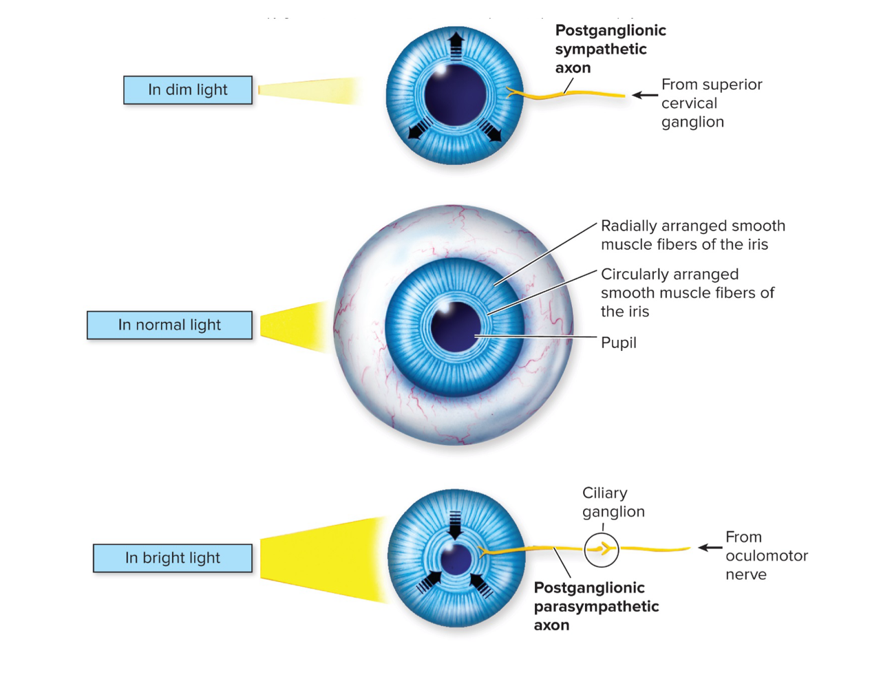
iris: regulate size of the pupil
post ganglionic sympathetic neuron causes dilation the pupil in dim light
post ganglionic parasympathetic neuron causes constriction of the pupil in bright light
lens: provides accommodation (seeing close or seeing far away)
thick lens - allows seeing close up
thin lens - allows seeing far away
lens are attached to ciliary muscle that contract or relax to change the shape of the lens
when ciliary muscle relaxes, causes tension of lens making it thinner, allowing focusing on distant objects

when ciliary muscle contracts, causes lens to get bulgy, allowing focusing on closer objects

Visual Acuity and Vision Pathology
visual acuity: the sharpness of vision, the power to discriminate details
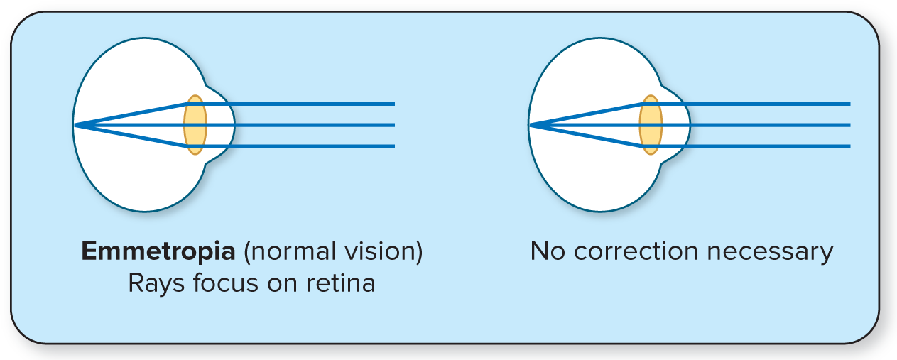
Emmetropia: normal vision, rays focus on retina
Hyperopia: farsightedness, rays focus behind retina
convex lens correct farsightedness
image is brought to focus in behind of retina, rather than on the retina
due to eyeball that is egg shaped, too short

Myopia: nearsightedness, rays focus in front of retina
concave lens correct nearsightedness
image is brought to focus in front of retina, rather than on the retina
due to eyeball that is too long

Astigmatism: rays do not focus
uneven lens correct astigmatism
corrected by cylindrical lenses that compensate for asymmetrical lens/cornea
Unequal curvature of the refractive surfaces of the cornea and/or lens, so the light that enters the eye along certain meridians does not focus on the retina.

Testing for Visual Acuity
standing 20 feet from snellen eye chart, cover one eye and read the line with the smallest letters you can see
line 1 should be easily read at 200 feet
line 8 should be easily read at 20 feet
measured as d/D
d: distance at which person can read letters
D: distance at which a normal eye can read letters
Testing for Astigmatism
standing 20 feet from astigmatism chart and cover one eye
positive for astigmatism: some spokes are sharp and dark, while others are blurred and lighter
Testing Accommodation
using a meter stick and pin, hold meter stick under one eye, extending outward and close other eye
hold pin arms length away and gradually move pin closer until pin first appears blurred
near point: closest distance you can see an object in sharp focus
Testing Blindspot
hold card with + and o symbol about 20 inches away from face, with one eye covered
other eye must be covered because the eyes compensate for each other
keep open eye focused on circle
slowly bring card closer to face until cross disappears
continue to move card towards face until cross reappears
The Retina
optic disc: where optic nerve forms, blindspot
does NOT have retina, causing blindspot
Fovea Centralis: Area of the highest visual acuity, containing only cones
located directly behind the pupil
Retina: contains photoreceptors
contains pigment which dissociates when absorbs or transduce light energy
photoreceptor cells: rods and cones
when photons hit photoreceptor cells, causes cells to depolarize and cause action potentials
transduce light energy into action potentials
rods: sensitive to low light and dark light
responsible for black-and-white vision under low illumination.
cones: sensitive to bright light and color
provides color vision and high visual acuity.
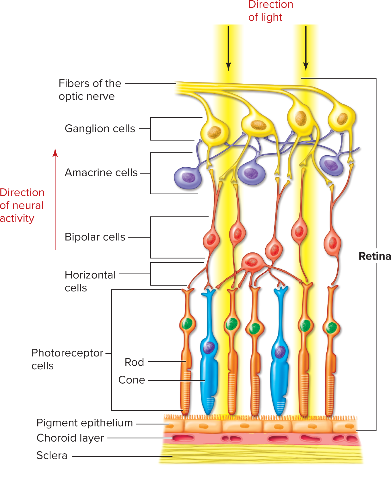
retina is inverted - incoming light passes various nerve cells to reach cones and rods of the retina
once reaches retina, light passing through must go through bipolar cells
bipolar cells synpase with ganglion cells
ganglion cells synapse with fibers of the optic nerve that transmit information to the brain
light passes through one way, then action potentials travel in the same way it came from
Light Pathway (Front to Back): Light travels through the eye, passes through the ganglion cell layer and the bipolar cell layer, and finally hits the photoreceptors (rods and cones) located at the back of the retina.
Signal Pathway (Back to Front): When the photoreceptors detect light, they change their electrical activity and send signals to the bipolar cells, which in turn signal the ganglion cells.
Action Potential Direction: The ganglion cells generate action potentials that travel back towards the front of the retina, join together to form the optic nerve, and exit the eye to go to the brain
Trichromatic vision
3 types of cones:
S (red) cone
M (green) cone
L (blue) cone
cones overlap, allowing for a wider range of colors to be perceived due to the combination of signals sent to the brain.
screens contain red, green, and blue LEDs to stimulate red, green, and blue cones in our eyes
color blindness: when one or more of cones is defective

Retina Convergence and Light Sensitivity
generally, photoreceptors are convergent networks
multiple rods and cones synapse with a single ganglion cell
fovea centralis only contains cones and has NO convergent network
each cone synapses with its own ganglion cell, each creating its own action potential to the brain
allows for fovea centralis to have highest visual acuity
Neural Pathway for Vision
axons of the ganglion cells converge to form the optic nerve, landing in the Occipital Lobe
Lab 10:
Myofilaments: filamentous proteins involved in contraction.
smallest unit of muscle
Myofibrils: Rod-shaped structures composed of myofilaments, that extend and attach to each end of the muscle
myofilaments and myofibrils are part of cytoskeleton, inside of the sarcolemma
Myofibers: Skeletal muscle cells
Sarcomere: repeating segments, contractile subunit of a myofibril
when all contract together, shortens myofibril
extend from z disc to z disc
Sarcomere Structure:
Thick filaments: made from protein called myosin
possess a collection of myosin "heads" (motor proteins) for interaction with actin
A band: part of the sarcomere that contains thick filaments
always found in center of sarcomere
thick filaments give A bands the dark appearance
thin filaments: made of filamentous proteins called actin
I band: Region with only thin filaments and Z-discs, extends from one end of the thick filament to another
thin filaments give I bands the light appearance
contain tropomyosin and troponin, which are a collection of regulatory proteins that stimulate muscle contraction
Z disc: Ends of each sarcomere that anchors the thin filament
H band: space between the ends of the thin filaments, only containing thick filaments
Sarcoplasm: The cytoplasm of a muscle fiber
Transverse Tubules (T-tubules): Invaginations (holes) of the sarcolemma into the cell
narrow membranous tunnels formed from sarcolemma, directly attached to SR
open to the extracellular environment through pores in the cell surface
substances from extracellular matrix can enter cell via t-tubules
able to conduct action potentials inward adjacent to SR (into the cell)
voltage (action potential) travels into t-tubules
Neuromuscular Junction: synapse between neuron and effector (muscle)
contains 3 major components:
Synaptic Knob: End of the axon that releases neurotransmitters
Synaptic Cleft: Space between the neuron and muscle fiber where neurotransmitters are released.
Motor End Plate: Folded portion of the sarcolemma that contains receptors for neurotransmitters
folded to increase surface area
Sliding Filament Theory:
When a muscle contacts, all sarcomeres shorten simultaneously:
A bands DO NOT shorten, but move closer together
thick filaments DO NOT shorten
thin filaments DO NOT shorten
thin filaments slide towards the H band
I bands shorten
H bands shortens or disappears
during contraction, thin filaments slide past thick filaments toward the center
thin filaments are attached to Z discs, so when it slides past the thick filaments, the Z discs move closer together
causes H zone to get smaller and smaller as tips of thin filaments get closer
also cause I bands to get smaller, since the I bands are comprised solely of thin filaments and their distance from each other reduces during contractions.
Stimulating a Muscle Fiber
Complete Steps of Muscle Contraction:
(ACh) is released from the lower motor neuron
Acetylcholine binds to Nicotinic ACh receptors on the motor end plate
Na+ channels open, allowing Na+ to diffuse into the fiber, creating an action potential at the neuromuscular junction
Action potential propagates down the scarolemma and into transverse tubules, entering the interior of the muscle fiber
Action potential causes voltage-gated Ca2+ channels on the t-tubules to undergo a conformational change
conformational change of voltage-gated Ca2+ channels triggers opening of Ca+ release channels on the SR, allowing Ca2+ to moves down its concentration gradient
Ca2+ diffuses into the sarcoplasm and is released on the myofibrils, binding to troponin
troponin undergoes a conformational change, which moves the attached tropomyosin out of the way
exposes myosin binding site
allows for myosin head to bind to myosin binding site on actin
cross bridge action is initiated and continues while calcium concentration is high
release of phosphate causes conformational change in myosin head
allows for myosin head to pulled, pulling the attached actin filament towards the center of the sarcomere
Completion of the conformational change causes ADP to be released from the myosin head
A new ATP binds to myosin head causing conformational change that results in the myosin head to detach from the actin filament
Hydrolysis of ATP causes another conformational change, returning myosin head to its original orientation, preparing for the next contraction cycle.
Twitch, Summation, and Tetanus
Twitch: when electrical stimulus is applied to a muscle, it quickly contracts and relaxes
Summation of Twitches: occurs when a new stimulus arrives before the muscle fiber relaxes, twitches are summed together
summation of muscle twitch are produced in lab by increasing frequency of stimulus
summation of muscle twitch are produced normally in the body by increasing frequency of action potentials
summation of action potentials are not possible due to refractory period
Tetanus: maintained maximum contraction, due to elevated concentration of Ca2+ in the sarcoplasm
graded contraction of muscles occurs:
Summation of Twitches: increasing frequency of action potentials
Recruitment of Motor Unit: increasing the number of active motor units
Primary means of varying tension in a whole muscle
sustained contractions achieved by:
sustaining stimulus (action potentials)
asynchronous recruitment of motor units (prevent fatigue)
Lab: adding ATP, potassium, and magnesium ions to muscle fiber, causes length of muscle fiber to decrease
potassium and magnesium are cofactors