Comprehensive Nursing Notes for Mobility Problems
Learning Outcomes
Utilize the nursing process to administer care for clients with mobility issues, including:
Fractures
Surgical correction of herniated disks (lumbar/cervical)
Hip fractures (total hip replacement/open reduction/internal fixation)
Total knee replacement
Spinal cord injury (SCI)
Understand complications associated with fractures and provide nursing interventions to prevent these complications.
Explain nursing responsibilities regarding traction and casts.
Conduct neurovascular assessments to monitor blood flow and nerve function.
Collaborate with healthcare professionals for rehabilitation and home care needs, ensuring comprehensive patient support.
Structures and Functions of the Musculoskeletal System
Composition:
Voluntary muscle: enabled movement under conscious control.
Bone: provides structure, support, and houses bone marrow for blood cell production.
Cartilage: reduces friction between bones and joints, maintains their shape, and acts as a cushion.
Ligaments: strong connective tissue that connects bone to bone, providing joint stability.
Tendons: fibrous connective tissue that attaches muscle to bone, enabling movement.
Fascia: connective tissue that separates structures and allows movement (surrounding muscles, bones, and blood vessels).
Bursae: small fluid-filled sacs that reduce friction between tendons, muscles, and bones at joints.
Connective tissue: provides support and flexibility throughout the body.
Purposes:
Protects body organs from injury.
Provides support and stability to maintain posture.
Stores minerals (like calcium and phosphorus) crucial for various bodily functions.
Allows for coordinated movement necessary for daily activities.
Blood cell production (hematopoiesis) occurs in the bone marrow of bones.
Bone Types
The human skeleton contains 206 bones, classified into:
Long bones: provide support, enable movement, and are crucial for blood cell production (e.g., femur, humerus).
Short bones: cube-shaped and composed of spongy bone, providing stability (e.g., carpals in the wrist).
Flat bones: protect internal organs and serve as attachment points for muscles (e.g., skull, ribs).
Irregular bones: support and protect various body parts (e.g., vertebrae, pelvis).
Sesamoid bones: small, round bones embedded in tendons, aiding in joint function (e.g., patella).
Assessment of the Musculoskeletal System
Objective Data
Physical Examination:
Muscle strength testing (scale of 0-5) to evaluate functional ability:
0/5: No contraction detected.
1/5: Flicker or trace contraction observed.
2/5: Active movement with gravity eliminated.
3/5: Active movement against gravity.
4/5: Active movement against gravity with some resistance.
5/5: Active movement against full resistance (considered normal strength).
Other Measures:
Measure limb length and circumferential muscle mass to assess for asymmetry.
Assess for use of assistive devices, posture, and gait to identify mobility issues.
Inspect for scoliosis and perform straight-leg raises to gauge flexibility and strength.
Diagnostic Studies of the Musculoskeletal System
Common Diagnostic Studies Include:
Standard X-ray: the first-line imaging for fractures and bone abnormalities.
Bone scan: useful for detecting bone cancer that will light up like a hot spot on the scan.
CT scan: provides detailed cross-sectional images of bone and soft tissue.
Diskogram: evaluates pain originating from intervertebral discs.
DEXA (Dual energy X-ray absorptiometry): measures bone density to assess for osteopenia or osteoporosis.
EMG (Electromyogram): evaluates the health of muscles and the nerve cells that control them.
MRI (Magnetic Resonance Imaging): provides detailed views of soft tissues, including ligaments and discs.
Myelogram: uses contrast to look for issues in the spinal column, such as herniated discs.
SSEP: sensory-evoked potentials assess the functional integrity of pathways from the spinal cord to the brain.
Thermography: captures heat patterns in tissues, often used for muscular issues.
QUS (Quantitative Ultrasound): assesses bone density without ionizing radiation.
Interventional Studies:
Arthrocentesis: procedure to aspirate fluid from a joint for analysis.
Arthroscopy: minimally invasive procedure using a camera to visualize internal joint structures.
Intervertebral Disc Disease
Degenerative Disc Disease (DDD):
Characterized by loss of fluid in intervertebral discs leading to reduced elasticity and flexibility, resulting in potential pain and limited mobility.
Herniated Disc:
Occurs when disc material protrudes and compresses spinal nerves, often due to degeneration or trauma.
Surgical Interventions:
Spinal fusion (ankylosis) using bone graft from the patient’s fibula or iliac crest or donor tissue (allograft), which stabilizes the spine post-surgery.
May involve metal fixation to enhance stability of the spine post-operation.
Nursing Management: Vertebral Disc Surgery
Postoperative Care:
Maintain proper spinal alignment until healing is confirmed through X-ray or patient's functional recovery.
Use of pillows under thighs when supine and between legs when side-lying to minimize pressure on the surgical site.
Log roll the patient to reposition safely without twisting the spine.
Monitor pain levels regularly and implement pain management strategies per physician’s orders.
Avoid bending, twisting, or lifting weights over 10lbs to facilitate recovery.
Neurovascular Assessment:
Continuous assessment for signs of spinal cord edema, particularly after cervical surgery where loss of function may be imminent.
Assessment of Donor Site:
Regularly check the bone graft site for signs of complications like infection or graft failure.
Peripheral Neurological Status:
Assess movement and sensation in extremities as well as vital signs every 2-4 hours for the first 48 hours post-op.
Compare with pre-op neurological status to detect any deterioration or recovery.
Assess circulation through capillary refill time and pulses.
Spinal Cord Injuries (SCI)
Caused by trauma or damage leading to temporary or permanent changes in function.
Incidence:
Approximately 17,000 new cases annually in the U.S.; currently 282,000 individuals live with SCI, highlighting the need for proper management and rehabilitation.
High mortality risk within the first year is prevalent due to complications.
Approximately 30% rehospitalization rate due to secondary complications like infections or falls.
Types of Injuries:
Variance in function depending on injury level (e.g., C4 injury results in tetraplegia; T6 injury results in paraplegia).
Can be caused by blunt or penetrating trauma and damage that progresses after the initial injury (e.g., inflammation or swelling).
Conditions like spinal shock could occur immediately after injury leading to temporary loss of function.
Vasogenic shock may occur with injuries at T6 or higher, affecting vital signs and circulation.
Compression fractures may lead to chronic pain and complications if not treated promptly.
Common Fracture Types
Types of Fractures:
Closed (simple): does not break through the skin.
Open (compound): breaks through the skin, classified into:
Grade I (clean wound, minimal contamination).
Grade II (larger wound, more soft tissue damage).
Grade III (contaminated with significant soft tissue injury).
Transverse
Spiral
Greenstick - incomplete fracture
Comminuted
Oblique
Pathologic
Stress
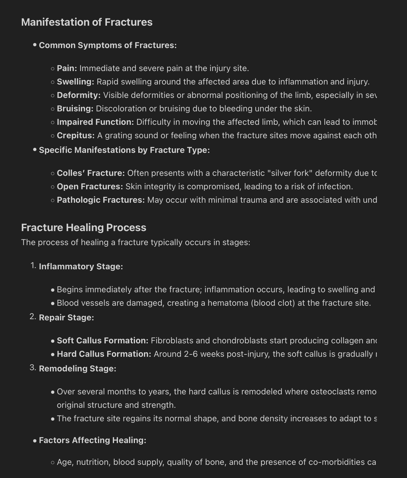
Colles’ Fracture
Characteristics:
Most common in older adults due to osteoporosis, often resulting from falls.
Presentation includes dorsal displacement of the distal fragment (known as silver-fork deformity).
Assessment:
Movement
Capillary refill
Pulses
Color
Temperature
Management:
Closed reduction followed by casting or splinting; neurovascular assessment should be done to check for complications.
Reduce edema (ice and splinting)
Move fingers and shoulder to reduce edema, increase venous return, and prevent stiffness and contracture
Nursing Process: The 6 P's for Neurovascular Assessment
Key Indicators:
Pain: assess and manage to facilitate comfort.
Pressure: monitor for swelling or compartment syndrome.
Pallor: check for color changes indicating compromised circulation.
Pulselessness: assess for weak or absent pulses.
Paresthesia: monitor for sensations of numbness or tingling.
Paralysis: assess for loss of movement indicating neurological compromise.

Complications of Fractures
Early Complications:
Shock: a life-threatening condition due to blood loss or other factors.
Fat embolism: fat droplets may enter the bloodstream, causing blockages.
Compartment syndrome: pressure within muscles builds to dangerous levels.
Venous thromboembolism (VTE), pulmonary embolism (PE): blood clots forming due to immobilization or injury.
Delayed Complications:
Avascular necrosis: death of bone tissue due to insufficient blood supply.
Complex regional pain syndrome (CRPS): chronic pain condition that can occur after injury.
Nonunion or malunion: improperly healed fractures leading to further complications.
Emergency Management of Fractures
Initial Actions:
Immobilization of the affected body part to prevent further injury.
Ensure splinting above and below the fracture site for stabilization.
Cover open fractures with sterile dressings to prevent infection.
Medical Management of Fractures
Fracture Reduction:
Aim to restore anatomic alignment through either closed (manipulation without surgery) or open (surgical intervention) methods.
Use of fixation devices (like pins, plates, or screws) as needed to maintain alignment during healing.
External fixation requires pin care and constant infection monitoring
Caring for Patients with Casts or Traction
Assessment:
Regularly check neurovascular status to monitor for circulation and nerve function.
Monitor for signs of complications: pressure ulcers, deep vein thrombosis (DVT), and skin integrity issues.
Patient Education:
Instruct patients on care techniques and warning signs to report, including increased pain, swelling, or skin changes.
Traction Management
Used to align fractures and reduce muscle spasms effectively.
Key Principles:
Continuous force must be applied to maintain alignment and promote healing.
Monitor for complications such as skin breakdown, nerve damage, or blood flow issues.
Rehabilitation Considerations
Pelvic Fractures:
Management varies by type of fracture; may involve bed rest and active symptom management.
Hip Fractures:
Likely to require surgical fixation, with care approaches similar to other orthopedic surgeries for optimal recovery.
Nursing Priorities Post-Operative Care for Hip Replacement
Prevent dislocation and enhance mobility through proper positioning and support devices.
Regularly assess for complications and manage pain effectively, involving patient education regarding activity limitations.
Schedule rehabilitation therapies, including physical therapy (PT) and occupational therapy (OT), for comprehensive recovery support.
Knee Replacement Needs
Postoperative assessment includes monitoring for complications and the strategic use of continuous passive motion (CPM) devices to enhance recovery.
Education:
Discuss the importance of activity restrictions and signs of potential complications such as swelling or infection post-surgery.
Osteomyelitis Management
Treatment includes IV antibiotics and thorough wound assessments to prevent complications, including chronic infections.
Patient education focuses on understanding signs of infection, proper hygiene practices, and the importance of follow-up appointments.
Amputation Care
Focus on stump care routines, managing phantom limb sensations, and promoting mobility through the utilization of prosthetics.
Regular monitoring for skin integrity and circulation to prevent complications is critical in post-amputation care.