DIGESTIVE SYSTEM OF THE FROG
DIGESTIVE SYSTEM OF THE FROG
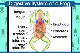
The digestive system of frogs comprises the alimentary canal or digestive tract along with the related digestive glands.
In the alimentary canal, processes such as digestion, mastication, and absorption occur while the digestive glands produce some enzymes that cause digestion of the food that is ingested.
➢ The function of the frog digestive system is digestion and absorption
➢ Frog digestive system consists of the alimentary canal along with digestive glands
Alimentary canal
➢ long, complete and coiled tube
➢ consist of mouth, buccal cavity, pharynx, oesophagus, stomach, intestine, rectum, cloaca.
Mouth
➢ The alimentary canal starts with the mouth
➢ very wide gap extending from one side of the snout to the other.
➢ two bony jaws are found in the mouth, and the jaws are covered by the immovable lips.
The upper jaw is fixed, while the lower jaw is flexible – it can move up and down to open or close the mouth.
Buccal Cavity
➢ The mouth opens into the buccal cavity
➢ wide, large, and shallow.
➢ comprises a ciliated columnar epithelial lining, which has mucous glands, and these glands secrete mucus which aids in food lubrication. The frogs do not have salivary glands.
Teeth
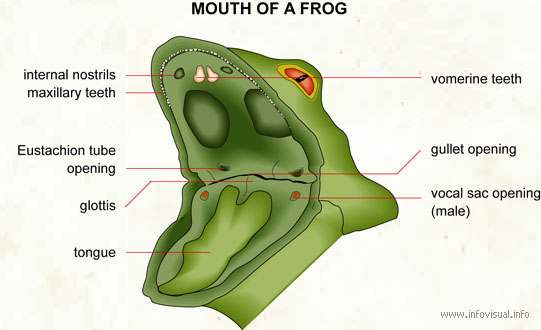
➢ The lower jaw does not have teeth. But it occurs in a row on either side of the maxillae and premaxillae bones of the upper jaw. Teeth are pointed backward. There are two more patches of teeth seen on either side of the median line of the roof of the buccal cavity called vomerine teeth. The vomers also comprise two groups of vomerine teeth.
➢ These teeth are not utilized to chew but to check the escape of captured prey.
➢ The upper jaw has a row of closely set, uniform, small, and hook-like pointed teeth.
Internal Nostrils
➢ In its roof near the vomerine teeth, the buccal cavity comprises two openings – the posterior or internal nares associated with the nasal cavities through which respiratory gases move to and from the buccal cavity at the time of respiration.
Tongue
➢ The tongue in frogs is large, sticky, muscular, and protrusible.
➢ It is found at the base of the mouth cavity.
➢ The anterior end of it is attached to the inner border of the lower jaw while the posterior end is bifid and free.
➢ The upper surface has taste buds forming small papillae and mucous glands of which the secretions cause the tongue to be sticky.
➢ Digestive enzymes are neither produced by the mucous glands nor the taste buds.
Orbit-Bulging
➢ Behind the vomerine teeth, the roof of the buccal cavity has two oval and large pale areas, the bulging of the eyeballs.
➢ At the time of swallowing food, eyes are pressed down into the buccal cavity, pushing the food into the pharynx.
Pharynx
➢ The buccal cavity tapers behind the pharynx.
➢ It opens through the gullet into the oesophagus.
➢ The pharynx and the buccal cavity at times are referred to as the buccopharyngeal cavity.
➢ At the roof of the pharynx on each of the lateral sides, a wide Eustachian tube can be found with an opening that communicates with the middle ear.
➢ The glottis is a median slit in the pharynx behind the tongue, that shields the entry into the lungs. During breathing, it is always open and closes at the time of swallowing. In the angle of the lower jaw on the floor of the pharynx in male frogs, two openings of the vocal sacs are formed too. They serve as resonators during croaking.
Oesophagus
➢ Gullet directs into a broad, short and muscular section of the alimentary canal referred to as the oesophagus.
➢ This section of the alimentary canal is extremely short as a result of the absence of the neck however, highly distensible as their inner lining into numerous longitudinal folds that enable enough expansion of the oesophagus at the time of passage of ingested food via it to the stomach.
➢ They open into the stomach, wherein no demarcation line forms between the stomach and the oesophagus.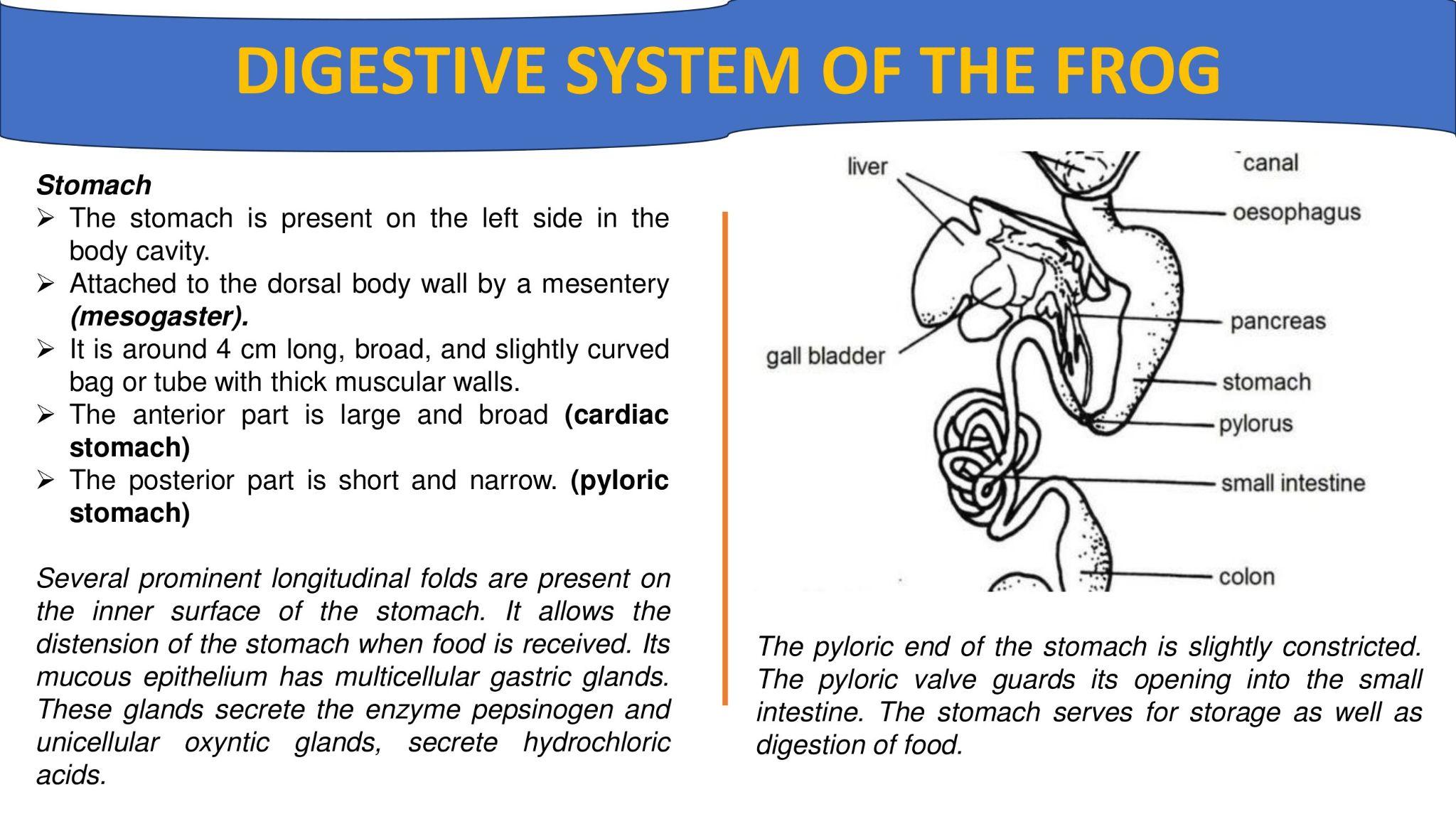
Stomach
➢ The stomach is present on the left side in the body cavity.
➢ Attached to the dorsal body wall by a mesentery (mesogaster).
➢ It is around 4 cm long, broad, and slightly curved bag or tube with thick muscular walls.
➢ The anterior part is large and broad (cardiac stomach)
➢ The posterior part is short and narrow. (pyloric
stomach)
Several prominent longitudinal folds are present on the inner surface of the stomach. It allows the distension of the stomach when food is received. Its mucous epithelium has multicellular gastric glands. These glands secrete the enzyme pepsinogen and unicellular oxyntic glands, secrete hydrochloric acids.
The pyloric end of the stomach is slightly constricted. The pyloric valve guards its opening into the small intestine. The stomach serves for storage as well as digestion of food.
Small intestine
➢ Small intestine is a long, coiled, and narrow tube
➢ It comprises of two parts: anterior duodenum and posterior ileum
➢ The ducts from the liver and pancreas open into the duodenum
➢ Bile juice emulsifies fat and pancreatic juices digest carbohydrates and proteins
➢ Digested food is absorbed by the ileum The mucosal lining of the small intestine apart from the intestinal glands comprises two types of cells
Goblet cells – large cells possessing granular substances and oval vacuoles producing mucus. The nucleus is found near the base of the cell
Absorbing cells – these are distinguished as small cells with nuclei found near the base.
Rectum
➢ Large intestine is a short and wide tube
➢ Its inner lining forms numerous folds
➢ In the rectum undigested food material is stored which is ready to expel from the cloaca
Cloaca
➢ The anus and the urinogenital apertures open into the cloaca
➢ Cloaca opens to outside by the vent or cloacal aperture, lying at the hind end of the body.
➢ Cloaca remove undigested food material.
(2)DIGESTIVE GLANDS OF THE FROG
Liver
❑ The largest gland of the body
❑ Bile is a greenish alkaline fluid secreted by the liver
❑ Bile is stored in a sac called the gallbladder
❑ It changes the PH of food from acidic to alkaline
❑ Hepatic ducts join the pancreatic duct to form a hepatopancreatic duct
❑ Ultimately opens into duodenum
Pancreas
❑ Pancreas is a long and irregularly lobed gland
❑ It secretes pancreatic juice
❑ Pancreatic juice poured into the duodenum through the hepatopancreatic duct
❑ Pancreatic juice helps digestion of ingested food
ACTIVITY OF THE ORGANS IN DIGESTION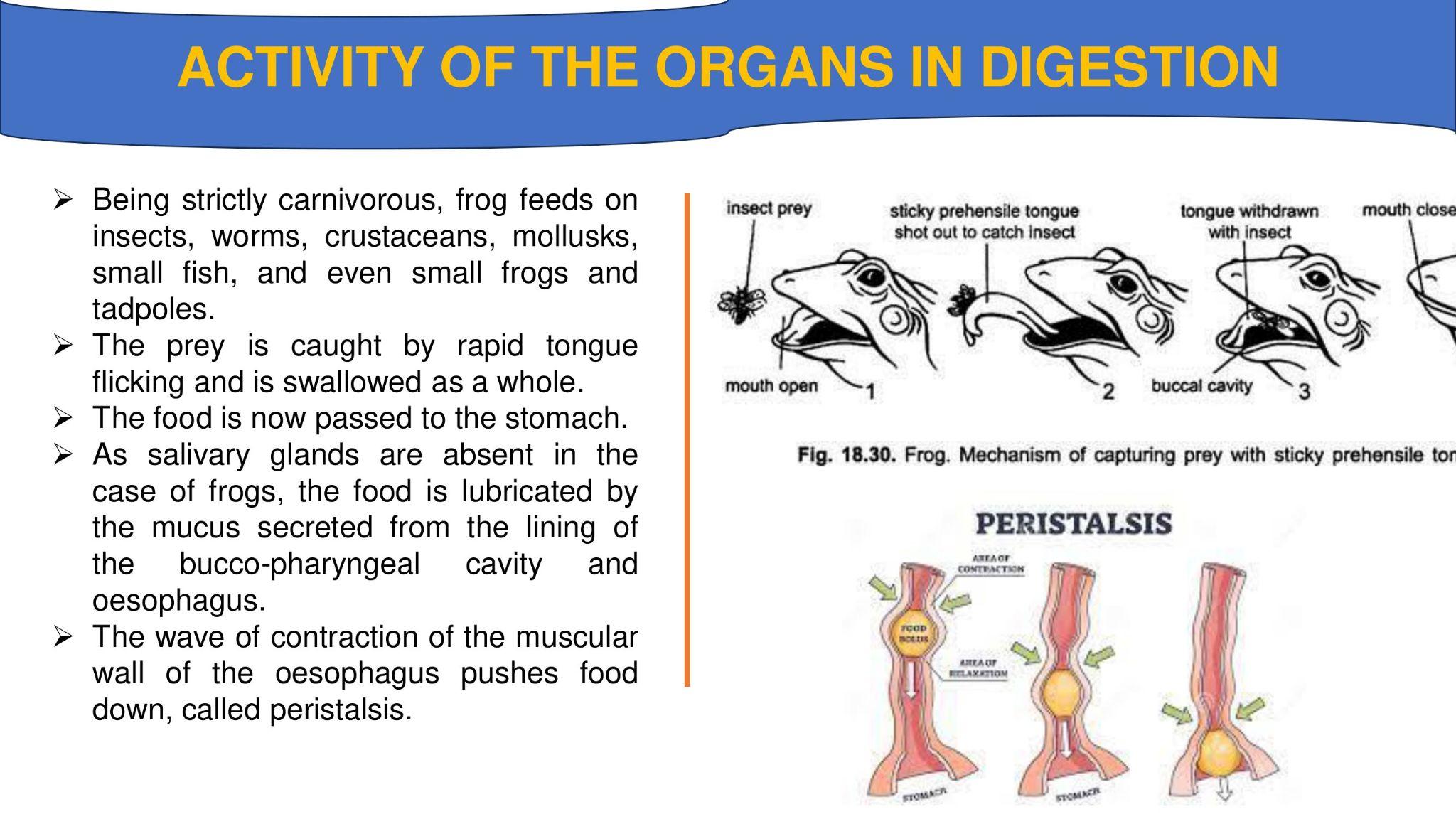
➢ Being strictly carnivorous, frog feeds on insects, worms, crustaceans, mollusks,
small fish, and even small frogs and tadpoles.
➢ The prey is caught by rapid tongue flicking and is swallowed as a whole.
➢ The food is now passed to the stomach.
➢ As salivary glands are absent in the case of frogs, the food is lubricated by the mucus secreted from the lining of the bucco-pharyngeal cavity and oesophagus.
➢ The wave of contraction of the muscular wall of the oesophagus pushes food down, called peristalsis.
Buccal Digestion:
In frog, the captured prey is neither subjected to any physical change (mastication) nor any chemical action in the buccal cavity as the buccal epithelium does not have any digestive gland.
From the buccal cavity the prey is directly pushed into oesophagus where it undergoes
physical changes due to the constant peristaltic movement of its wall. Besides mucus, the glands of oesophagus also secrete an enzyme called pepsin but no digestion occurs as it does not become active till it reaches the stomach, while the mucus simply makes the active food inactive and soft and, thus, makes the passage easier.
Gastric Digestion:
Due to oesophageal contractions and relaxations, the food comes down to the stomach.
The stomach performs the following three main functions:
(a) Storage;
(b) Mechanical mixing; and
(c) Chemical modifications.
➢ The gastric juice secreted by the gastric glands contains a large amount of water, inactive pepsinogen enzyme, and free hydrochloric acid.
➢ Inactive pepsinogen changes into active pepsin on being mixed with hydrochloric acid.
➢ The acid also prevents bacterial decomposition and dissolves the inorganic salts as well as makes the food soft.
➢ The pepsin of the stomach along with the pepsin of oesophagus acts on proteins of food and changes them into peptones and proteoses.
Inactive Pepsinogen + HCl → Active pepsin
Pepsin + Proteins → Peptones + Proteoses
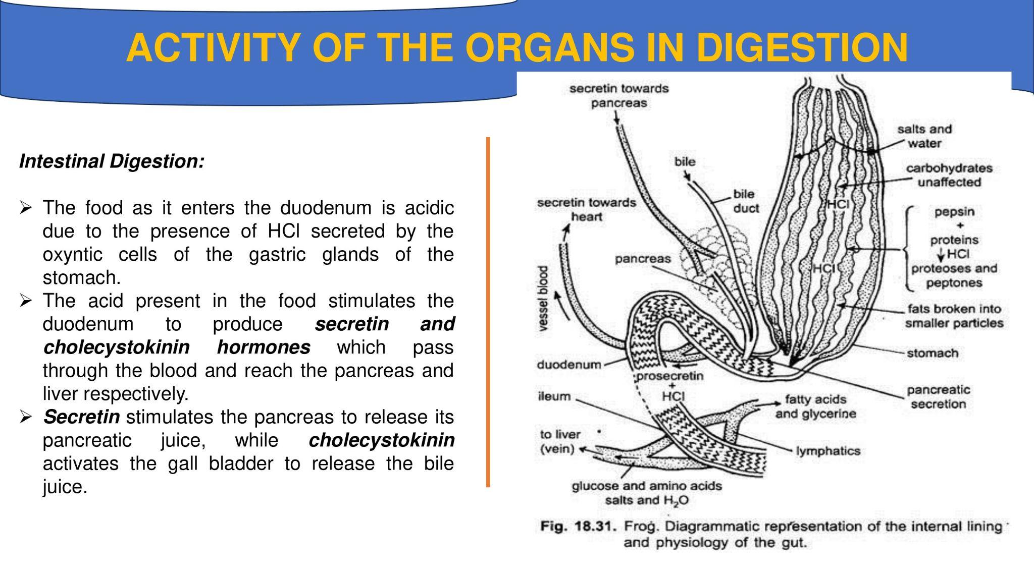
Intestinal Digestion:
➢ The food as it enters the duodenum is acidic due to the presence of HCl secreted by the oxyntic cells of the gastric glands of the stomach.
➢ The acid present in the food stimulates the duodenum to produce secretin and cholecystokinin hormones which pass through the blood and reach the pancreas and liver respectively.
➢ Secretin stimulates the pancreas to release its pancreatic juice, while cholecystokinin activates the gall bladder to release the bile juice.
Bile:
➢ It is a greenish alkaline fluid which contains no digestive enzymes so it does not take any part in the digestion of food.
➢ It is alkaline because it contains certain inorganic salts like sodium bicarbonate, sodium glycocholate, and sodium torocholate which not only neutralize the acidity of the semi- digested food, chyme but also prepare it for the activity of enzymes of pancreatic juice which can act only in the alkaline medium.
➢ They also break down fats to form small globules which can be emulsified. Bile juice also activates the fat-digesting enzyme of the pancreas, the lipase.
Pancreatic Juice:
➢ It is also a watery alkaline fluid containing three powerful to liver enzymes called trypsinogen, amylopsin (amylase), and steapsin (lipase).
(i) Trypsinogen is an inactive enzyme unless it is mixed with the enterokinase enzyme of succus entericus. Enterokinase, thus, activates the trypsinogen to form active trypsin. Trypsin acts on proteins, peptones, and proteoses changing them into simple amino acids.
(ii) Amylopsin (amylase) acts on starches reducing them into maltose,
(iii) Steapsin (lipase) acts on emulsified fat to form fatty acids and glycerol.
(c) Succus Entericus:
It is also an alkaline fluid contains many enzymes known as enterokinase, peptidases (erepsin), lipase, maltase, invertase, lactase, ribonuclease, and deoxyribonuclease.
(i) Enterokinase activates the inactive trypsinogen of pancreatic juice to form active trypsin.
(ii) Peptidases includes proteolytic enzymes like erepsin which act on polypeptides breaking them into amino acids.
(iii) Lipase and maltase along with the same enzyme of the pancreatic juice act on emulsified fats and maltose and convert them into fatty acids and sugars, respectively.
(iv) Invertase and lactase act on sucrose and lactose reducing them to glucose,
(v) Ribo- and deoxyribonuclease change the nucleic acids into nucleotides which are used in the synthesis of DNA, RNA and ATP.
Thus, the food is thoroughly digested with the help of these enzymes. As a result, all the proteins are reduced to amino acids, the carbohydrates to glucose and similar sugars and fats to glycerol and fatty acids.
Egestion, Absorption and Assimilation of Digested Food
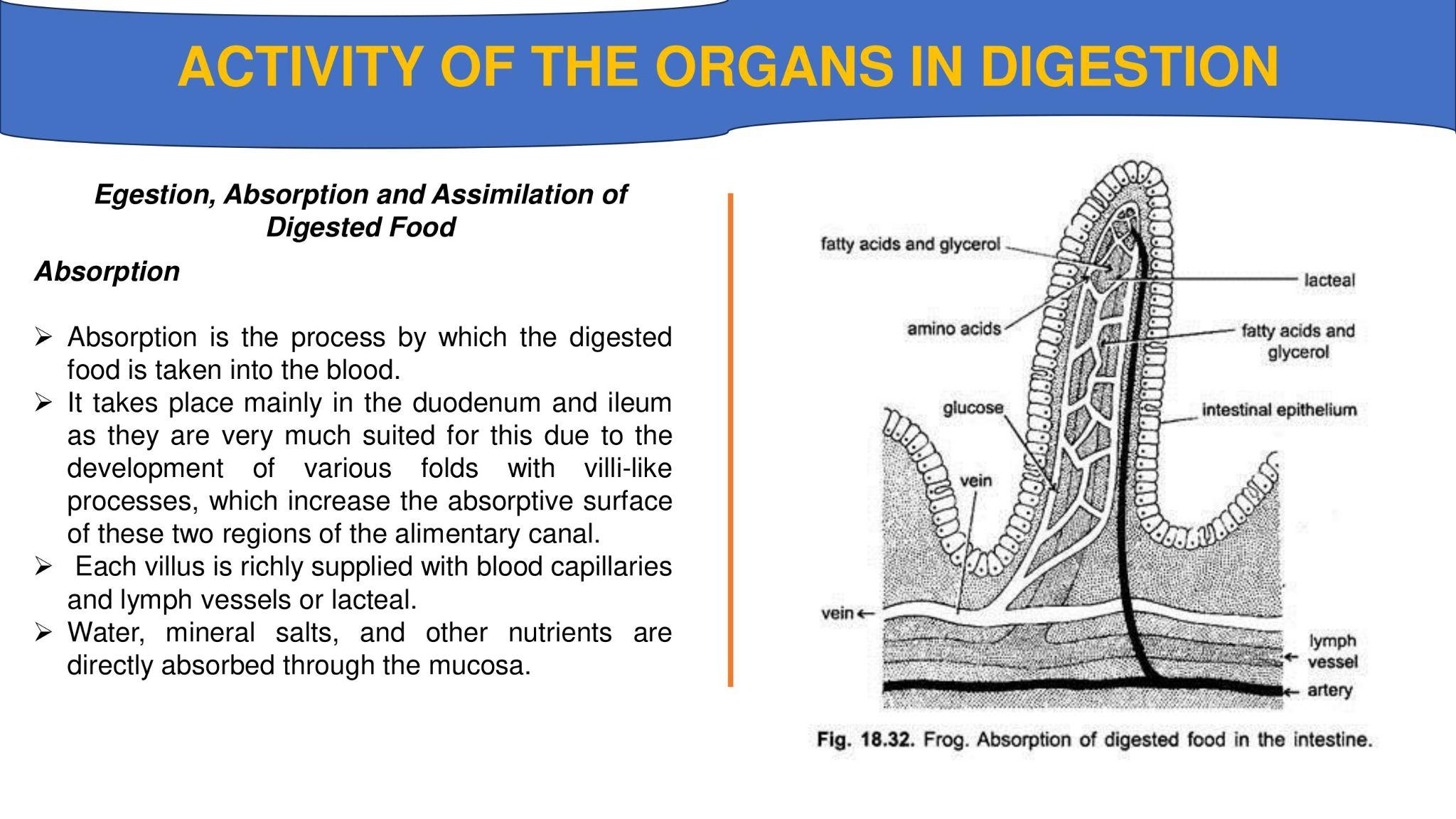
Absorption
➢ Absorption is the process by which the digested food is taken into the blood.
➢ It takes place mainly in the duodenum and ileum as they are very much suited for this due to the development of various folds with villi-like processes, which increase the absorptive surface of these two regions of the alimentary canal.
➢ Each villus is richly supplied with blood capillaries and lymph vessels or lacteal.
➢ Water, mineral salts, and other nutrients are directly absorbed through the mucosa.
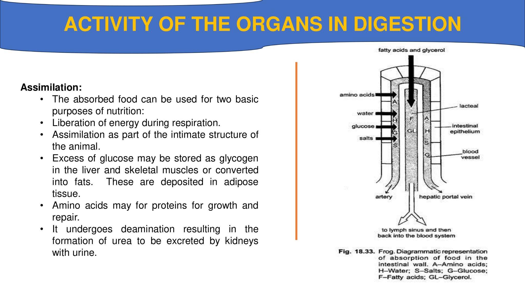
Assimilation:
• The absorbed food can be used for two basic purposes of nutrition:
• Liberation of energy during respiration.
• Assimilation as part of the intimate structure of the animal.
• Excess of glucose may be stored as glycogen in the liver and skeletal muscles or converted into fats. These are deposited in adipose tissue.
• Amino acids may for proteins for growth and repair.
• It undergoes deamination resulting in the formation of urea to be excreted by kidneys with urine.
Egestion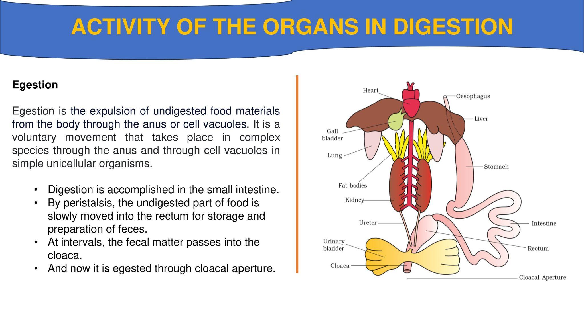
Egestion is the expulsion of undigested food materials from the body through the anus or cell vacuoles. It is a voluntary movement that takes place in complex species through the anus and through cell vacuoles in simple unicellular organisms.
• Digestion is accomplished in the small intestine.
• By peristalsis, the undigested part of food is slowly moved into the rectum for storage and preparation of feces.
• At intervals, the fecal matter passes into the cloaca.
• And now it is egested through cloacal aperture.
(3)HISTOLOGY OF THE ORGANS IN DIGESTION
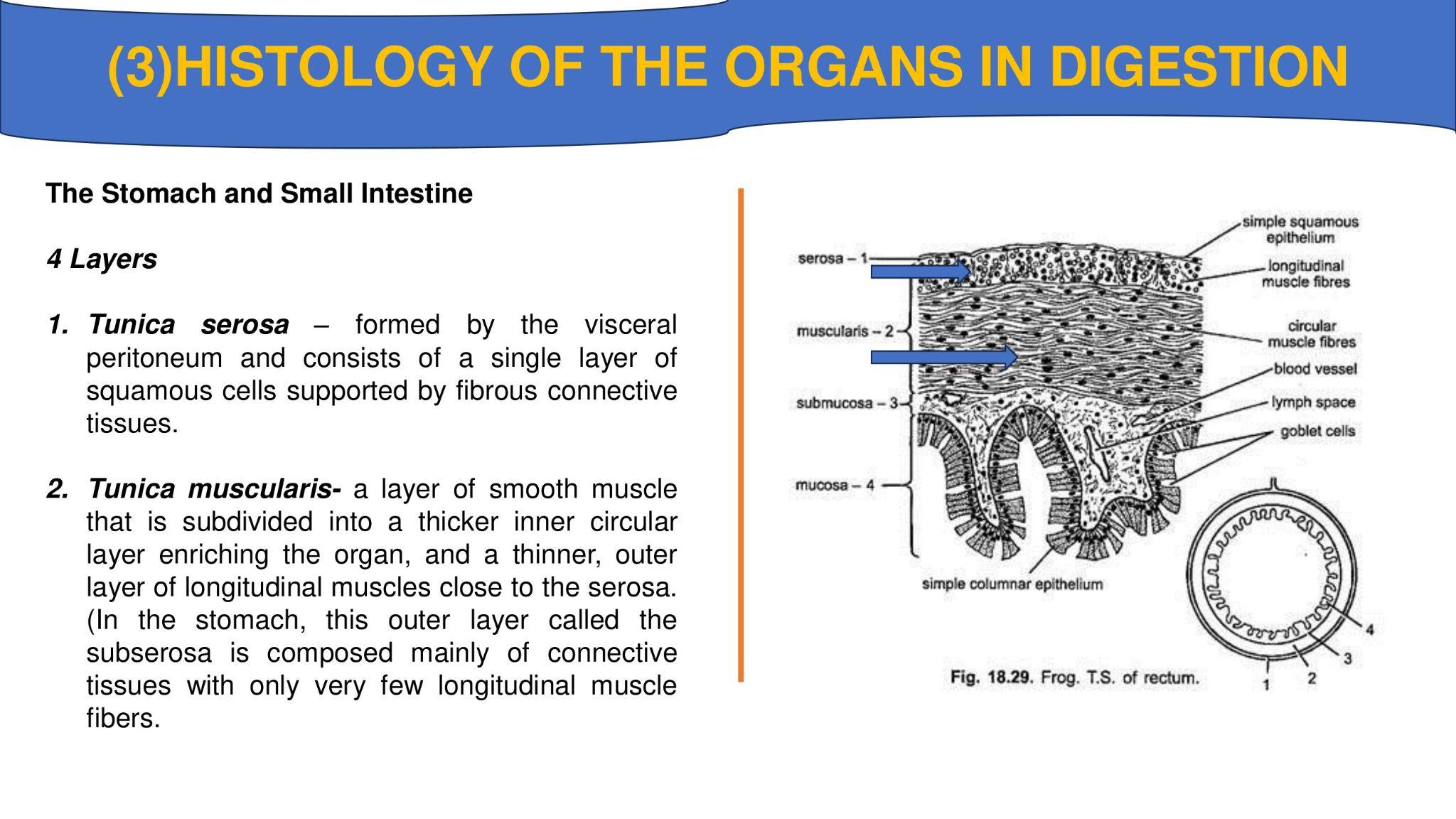
The Stomach and Small Intestine
4 Layers
1. Tunica serosa – formed by the visceral peritoneum and consists of a single layer of squamous cells supported by fibrous connective tissues.
2. Tunica muscularis- a layer of smooth muscle that is subdivided into a thicker inner circular layer enriching the organ, and a thinner, outer layer of longitudinal muscles close to the serosa. (In the stomach, this outer layer called the subserosa is composed mainly of connective tissues with only very few longitudinal muscle fibers.
3. Tunica submucosa – an internal layer of connective tissues with numerous blood vessels and nerves. This tissue extends into the folds of the inner coat.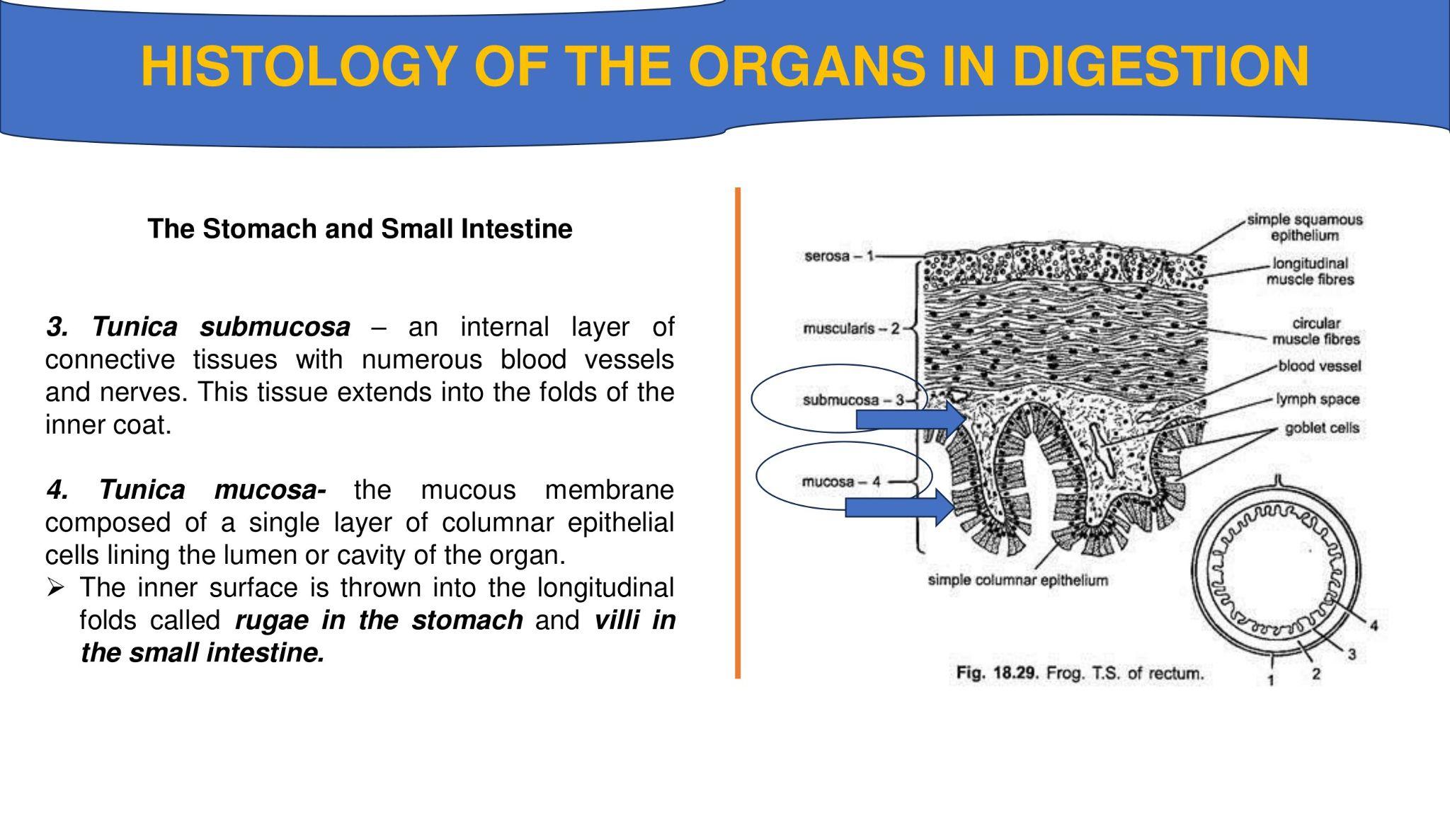
4. Tunica mucosa- the mucous membrane composed of a single layer of columnar epithelial cells lining the lumen or cavity of the organ.
➢ The inner surface is thrown into the longitudinal folds called rugae in the stomach and villi in the small intestine.
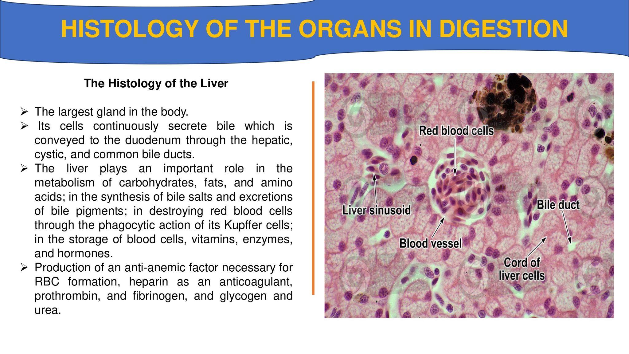
The Histology of the Liver
➢ The largest gland in the body.
➢ Its cells continuously secrete bile which is conveyed to the duodenum through the hepatic, cystic, and common bile ducts.
➢ The liver plays an important role in the metabolism of carbohydrates, fats, and amino acids; in the synthesis of bile salts and excretions of bile pigments; in destroying red blood cells through the phagocytic action of its Kupffer cells; in the storage of blood cells, vitamins, enzymes, and hormones.
➢ Production of an anti-anemic factor necessary for RBC formation, heparin as an anticoagulant, prothrombin, and fibrinogen, and glycogen and urea.
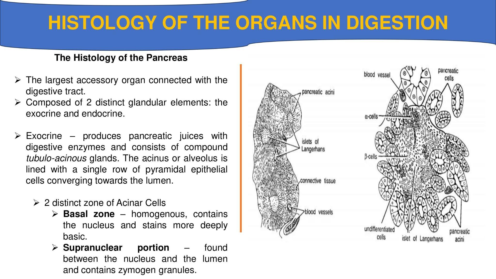
The Histology of the Pancreas
➢ The largest accessory organ connected with the digestive tract.
➢ Composed of 2 distinct glandular elements: the exocrine and endocrine.
➢ Exocrine – produces pancreatic juices with digestive enzymes and consists of compound tubulo-acinous glands. The acinus or alveolus is lined with a single row of pyramidal epithelial cells converging towards the lumen.
2 distinct zone of Acinar Cells
➢ Basal zone – homogenous, contains the nucleus and stains more deeply basic.
➢ Supranuclear portion – found between the nucleus and the lumen and contains zymogen granules.
➢ Islets of Langerhans – The islets vary in size and are irregularly clustered, scattered among the acini.
➢ When stained, the irregular prismatic cells appeared lighter than the acinar cells.
➢ The islets are delimited from the acini by a thin reticular membrane and supplied with numerous blood capillaries.
➢ The close relationship between the islets and the blood capillaries provides a rapid transfer of cell secretions to the bloodstream.
COMMON DISORDERS RELATED TO DIGESTION
1. Gastric hyperacidity - due to excessive secretion of hydrochloric acid often associated with peptic ulcer and gastritis. Hyposecretion of mucin causes ulcer formation; hyposecretion of HCI, other forms of gastritis and carcinoma
2. Vomiting - a reflex act brought by the mechanical irritation of the throat, or by irritating substances in the stomach and duodenum. This is also effected by pain, motion sickness and certain emotions.
3. Indigestion or dyspepsia - disturbed digestion or failure to digest food
4. Peptic ulcer - loss of tissues in areas of the digestive tract like lower esophagus, stomach and duodenum.
5. Colic - acute abdominal pain usually due to smooth muscle contraction, obstruction, or twisting.
6. Gallstones - precipitation of the concentrated bile constituents in the gall bladder or bile ducts. Gallstones cause obstructive jaundice and obstruction of the bile ducts causes severe unbearable pain known as biliary colic
7. Jaundice - the yellowness of the skin and the whites of the eyes due to obstruction of bile flow that bilirubin (bile pigments) is carried by the blood to all parts.
8. Constipation - a condition in which bowels are evacuated at long interval or with difficulty; feces are retained in the rectum due to decreased lumen, relaxed state of the colon's muscles.
9. Diarrhea - increased frequency and loose bowel movement -melini brought about by gastro-intestinal diseases like bacterial or viral infection.
10. Flatulence - the presence or sensation of excessive gas in the stomach and intestine