Detailed reference
Overview of the Integumentary System
Structure and Function of the Skin
The skin is the largest organ of the body, serving as a barrier to protect underlying tissues from external harm.
It consists of three main layers: the epidermis, dermis, and hypodermis, each with distinct functions and structures.
The epidermis is primarily responsible for protection and is composed of keratinized stratified squamous epithelium.
The dermis provides structural support and elasticity, containing connective tissue, blood vessels, and nerve endings.
The hypodermis anchors the skin to underlying structures and provides insulation and fat storage.
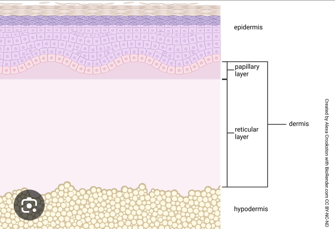
Layers of the Epidermis
The epidermis has five distinct layers: stratum corneum, stratum lucidum, stratum granulosum, stratum spinosum, and stratum basale.
The stratum corneum is the outermost layer, composed of dead keratinocytes that protect against environmental damage.
The stratum lucidum is found only in thick skin areas, providing an additional layer of protection.
The stratum granulosum contains keratinocytes that begin to flatten and lose their nuclei, contributing to the waterproof barrier.
The stratum spinosum contains Langerhans cells that play a role in immune response.
Structure and Function
The integumentary system is composed of the skin and its accessory structures, including hair, nails, and glands, providing overall protection to the body.
The skin is a complex organ made of multiple layers of cells and tissues, held together by connective tissue, which supports its structure and function.
Accessory structures such as hair and nails serve various functions, including protection, sensory input, and thermoregulation.
The skin is divided into two main layers: the epidermis (outer layer) and the dermis (inner layer), with the subcutaneous tissue lying beneath.
The epidermis is avascular, meaning it lacks blood vessels, relying on diffusion from the dermis for nutrient supply.
The integumentary system plays a crucial role in homeostasis, protecting against environmental hazards and regulating body temperature.
The Dermis and Its Layers
Composition of the Dermis
The dermis is divided into two layers: the papillary layer and the reticular layer, both composed of connective tissue.
The papillary layer contains loose areolar connective tissue, providing nutrients to the epidermis and containing sensory receptors.
The reticular layer is thicker and composed of dense irregular connective tissue, providing strength and elasticity to the skin.
Fibroblasts in the dermis produce collagen and elastin fibers, essential for skin structure and resilience.
The dermis houses blood vessels, lymphatic vessels, hair follicles, and glands, playing a crucial role in thermoregulation and sensation.
Functions of Dermal Components
Blood vessels in the dermis regulate temperature through vasodilation and vasoconstriction.
Nerve endings in the dermis allow for the sensation of touch, pressure, and pain, enhancing the body's response to the environment.
Sweat glands in the dermis help regulate body temperature through perspiration, while sebaceous glands secrete oils to keep the skin moisturized.
Hair follicles in the dermis are responsible for hair growth and are associated with sensory functions.
The Hypodermis and Accessory Structures
The Hypodermis
The hypodermis, or subcutaneous layer, is composed of loose connective tissue and adipose tissue, providing insulation and cushioning.
It connects the skin to underlying structures such as muscles and bones, allowing for flexibility and movement.
The hypodermis also serves as a fat storage site, which can be utilized for energy and thermal insulation.
Accessory Structures of the Integumentary System
Hair is a keratinous filament that grows from hair follicles in the dermis, providing protection and sensory functions.
Nails protect the tips of fingers and toes and enhance the ability to grasp objects.
Glands such as sebaceous and sweat glands play vital roles in skin health and thermoregulation.
The presence of sensory receptors in the skin allows for the detection of environmental stimuli, contributing to the body's overall sensory experience.
Hair Structure and Growth
Anatomy of Hair
The hair shaft is the visible part of the hair, while the hair root lies beneath the skin's surface, ending in the hair bulb.
The hair bulb contains the hair matrix, a layer of basal cells that divide and push hair cells outward as they grow.
The hair papilla, located within the hair bulb, is made of connective tissue and contains blood vessels and nerve endings, providing nutrients and sensation.
Hair is composed of three main layers: the medulla (central core), cortex (outer layer of keratinized cells), and cuticle (outermost protective layer).
Hair texture is determined by the shape of the cortex and the presence of the medulla, influencing whether hair is straight or curly.
Hair serves multiple functions, including protection from UV radiation, sensory input, thermoregulation, and communication (e.g., goosebumps).
Hair Growth Process
Hair growth begins with the production of keratinocytes in the hair bulb, which are pushed outward as new cells are formed.
Keratinization occurs as cells move toward the skin surface, resulting in the formation of the hair shaft, which is composed entirely of dead keratinized cells.
The arrector pili muscle, connected to each hair root, contracts in response to nerve signals, causing hair to stand up and trap air for insulation.
The average hair growth rate is approximately 1.25 cm per month, influenced by factors such as genetics and health.
Hair loss can occur due to various factors, including hormonal changes, stress, and nutritional deficiencies.
Understanding hair structure and growth is essential for addressing hair-related conditions and treatments.
The Epidermis and Its Layers
Composition of the Epidermis
The epidermis is primarily composed of keratinized, stratified squamous epithelium, providing a protective barrier against environmental damage.
Keratinocytes produce keratin, a fibrous protein that contributes to the hardness and water-resistant properties of skin, hair, and nails.
The epidermis is divided into four or five layers, depending on the thickness of the skin: stratum basale, stratum spinosum, stratum granulosum, and stratum corneum.
Thin skin, which covers most of the body, has four layers, while thick skin, found on palms and soles, includes an additional layer called the stratum lucidum.
The stratum basale is the deepest layer, consisting of a single row of cuboidal or columnar keratinocytes that attach to the basal lamina.
The epidermis is avascular, relying on the dermis for blood supply and nutrient exchange.
Functions of the Epidermis
The primary function of the epidermis is to act as a barrier, protecting underlying tissues from pathogens, chemicals, and physical damage.
The epidermis plays a role in regulating water loss, preventing dehydration through its keratinized outer layer.
Melanocytes, located in the epidermis, produce melanin, which protects against UV radiation and contributes to skin pigmentation.
The epidermis contains sensory receptors, such as Meissner's corpuscles, which detect touch and pressure, enhancing sensory perception.
The process of keratinization ensures that the outermost layer of the epidermis is continuously renewed, maintaining skin integrity.
The epidermis also participates in immune responses, with Langerhans cells acting as antigen-presenting cells.
Nails and Sweat Glands
Anatomy of Nails
Nails are specialized structures of the epidermis found at the tips of fingers and toes, providing protection and enhancing dexterity.
The nail body is formed on the nail bed, which is rich in blood vessels, and is composed of densely packed dead keratinocytes.
The nail root contains a matrix of proliferating cells that enable continuous nail growth, while the cuticle (eponychium) protects the nail root.
The lunula is the crescent-shaped region at the base of the nail, where the nail bed is thickened, and the hyponychium is the area beneath the free edge.
Nails serve functional purposes, such as picking up small objects and providing support to the fingertips during mechanical stress.
Nail health can be indicative of overall health, with changes in color or texture signaling potential health issues.
Sweat Glands and Their Functions
Sudoriferous (sweat) glands are responsible for producing sweat to regulate body temperature through evaporative cooling.
Sweat glands develop from epidermal projections into the dermis and are classified as merocrine glands, releasing secretions via exocytosis.
There are two main types of sweat glands: eccrine glands, which are distributed throughout the body, and apocrine glands, which are concentrated in specific areas (e.g., armpits, groin).
Eccrine glands are involved in thermoregulation, while apocrine glands are associated with emotional responses and body odor.
The secretion of sweat contains water, electrolytes, and metabolic waste, playing a role in excretion and maintaining electrolyte balance.
Understanding the function of sweat glands is essential for addressing conditions such as hyperhidrosis (excessive sweating) and skin hydration.
Overview of the Integumentary System
Structure and Function of the Skin
The skin is the largest organ of the body, serving as a barrier to protect underlying tissues from external harm.
It consists of three main layers: the epidermis, dermis, and hypodermis, each with distinct functions and structures.
The epidermis is primarily responsible for protection and is composed of keratinized stratified squamous epithelium.
The dermis provides structural support and elasticity, containing connective tissue, blood vessels, and nerve endings.
The hypodermis anchors the skin to underlying structures and provides insulation and fat storage.
Layers of the Epidermis
The epidermis has five distinct layers: stratum corneum, stratum lucidum, stratum granulosum, stratum spinosum, and stratum basale.
The stratum corneum is the outermost layer, composed of dead keratinocytes that protect against environmental damage.
The stratum lucidum is found only in thick skin areas, providing an additional layer of protection.
The stratum granulosum contains keratinocytes that begin to flatten and lose their nuclei, contributing to the waterproof barrier.
The stratum spinosum contains Langerhans cells that play a role in immune response.
Structure and Function
The integumentary system is composed of the skin and its accessory structures, including hair, nails, and glands, providing overall protection to the body.
The skin is a complex organ made of multiple layers of cells and tissues, held together by connective tissue, which supports its structure and function.
Accessory structures such as hair and nails serve various functions, including protection, sensory input, and thermoregulation.
The skin is divided into two main layers: the epidermis (outer layer) and the dermis (inner layer), with the subcutaneous tissue lying beneath.
The epidermis is avascular, meaning it lacks blood vessels, relying on diffusion from the dermis for nutrient supply.
The integumentary system plays a crucial role in homeostasis, protecting against environmental hazards and regulating body temperature.
The Dermis and Its Layers
Composition of the Dermis
The dermis is divided into two layers: the papillary layer and the reticular layer, both composed of connective tissue.
The papillary layer contains loose areolar connective tissue, providing nutrients to the epidermis and containing sensory receptors.
The reticular layer is thicker and composed of dense irregular connective tissue, providing strength and elasticity to the skin.
Fibroblasts in the dermis produce collagen and elastin fibers, essential for skin structure and resilience.
The dermis houses blood vessels, lymphatic vessels, hair follicles, and glands, playing a crucial role in thermoregulation and sensation.
Functions of Dermal Components
Blood vessels in the dermis regulate temperature through vasodilation and vasoconstriction.
Nerve endings in the dermis allow for the sensation of touch, pressure, and pain, enhancing the body's response to the environment.
Sweat glands in the dermis help regulate body temperature through perspiration, while sebaceous glands secrete oils to keep the skin moisturized.
Hair follicles in the dermis are responsible for hair growth and are associated with sensory functions.
The Hypodermis and Accessory Structures
The Hypodermis
The hypodermis, or subcutaneous layer, is composed of loose connective tissue and adipose tissue, providing insulation and cushioning.
It connects the skin to underlying structures such as muscles and bones, allowing for flexibility and movement.
The hypodermis also serves as a fat storage site, which can be utilized for energy and thermal insulation.
Accessory Structures of the Integumentary System
Hair is a keratinous filament that grows from hair follicles in the dermis, providing protection and sensory functions.
Nails protect the tips of fingers and toes and enhance the ability to grasp objects.
Glands such as sebaceous and sweat glands play vital roles in skin health and thermoregulation.
The presence of sensory receptors in the skin allows for the detection of environmental stimuli, contributing to the body's overall sensory experience.
Hair Structure and Growth
Anatomy of Hair
The hair shaft is the visible part of the hair, while the hair root lies beneath the skin's surface, ending in the hair bulb.
The hair bulb contains the hair matrix, a layer of basal cells that divide and push hair cells outward as they grow.
The hair papilla, located within the hair bulb, is made of connective tissue and contains blood vessels and nerve endings, providing nutrients and sensation.
Hair is composed of three main layers: the medulla (central core), cortex (outer layer of keratinized cells), and cuticle (outermost protective layer).
Hair texture is determined by the shape of the cortex and the presence of the medulla, influencing whether hair is straight or curly.
Hair serves multiple functions, including protection from UV radiation, sensory input, thermoregulation, and communication (e.g., goosebumps).
Hair Growth Process
Hair growth begins with the production of keratinocytes in the hair bulb, which are pushed outward as new cells are formed.
Keratinization occurs as cells move toward the skin surface, resulting in the formation of the hair shaft, which is composed entirely of dead keratinized cells.
The arrector pili muscle, connected to each hair root, contracts in response to nerve signals, causing hair to stand up and trap air for insulation.
The average hair growth rate is approximately 1.25 cm per month, influenced by factors such as genetics and health.
Hair loss can occur due to various factors, including hormonal changes, stress, and nutritional deficiencies.
Understanding hair structure and growth is essential for addressing hair-related conditions and treatments.
The Epidermis and Its Layers
Composition of the Epidermis
The epidermis is primarily composed of keratinized, stratified squamous epithelium, providing a protective barrier against environmental damage.
Keratinocytes produce keratin, a fibrous protein that contributes to the hardness and water-resistant properties of skin, hair, and nails.
The epidermis is divided into four or five layers, depending on the thickness of the skin: stratum basale, stratum spinosum, stratum granulosum, and stratum corneum.
Thin skin, which covers most of the body, has four layers, while thick skin, found on palms and soles, includes an additional layer called the stratum lucidum.
The stratum basale is the deepest layer, consisting of a single row of cuboidal or columnar keratinocytes that attach to the basal lamina.
The epidermis is avascular, relying on the dermis for blood supply and nutrient exchange.
Functions of the Epidermis
The primary function of the epidermis is to act as a barrier, protecting underlying tissues from pathogens, chemicals, and physical damage.
The epidermis plays a role in regulating water loss, preventing dehydration through its keratinized outer layer.
Melanocytes, located in the epidermis, produce melanin, which protects against UV radiation and contributes to skin pigmentation.
The epidermis contains sensory receptors, such as Meissner's corpuscles, which detect touch and pressure, enhancing sensory perception.
The process of keratinization ensures that the outermost layer of the epidermis is continuously renewed, maintaining skin integrity.
The epidermis also participates in immune responses, with Langerhans cells acting as antigen-presenting cells.
Nails and Sweat Glands
Anatomy of Nails
Nails are specialized structures of the epidermis found at the tips of fingers and toes, providing protection and enhancing dexterity.
The nail body is formed on the nail bed, which is rich in blood vessels, and is composed of densely packed dead keratinocytes.
The nail root contains a matrix of proliferating cells that enable continuous nail growth, while the cuticle (eponychium) protects the nail root.
The lunula is the crescent-shaped region at the base of the nail, where the nail bed is thickened, and the hyponychium is the area beneath the free edge.
Nails serve functional purposes, such as picking up small objects and providing support to the fingertips during mechanical stress.
Nail health can be indicative of overall health, with changes in color or texture signaling potential health issues.
Sweat Glands and Their Functions
Sudoriferous (sweat) glands are responsible for producing sweat to regulate body temperature through evaporative cooling.
Sweat glands develop from epidermal projections into the dermis and are classified as merocrine glands, releasing secretions via exocytosis.
There are two main types of sweat glands: eccrine glands, which are distributed throughout the body, and apocrine glands, which are concentrated in specific areas (e.g., armpits, groin).
Eccrine glands are involved in thermoregulation, while apocrine glands are associated with emotional responses and body odor.
The secretion of sweat contains water, electrolytes, and metabolic waste, playing a role in excretion and maintaining electrolyte balance.
Understanding the function of sweat glands is essential for addressing conditions such as hyperhidrosis (excessive sweating) and skin hydration.