Chapter 23: The Digestive System
23.1 Overview Of the Digestive System
The function of the digestive system:
Breaks down foods, releases nutrients, and absorbs them into the body.
The small intestine is the primary site for digestion and nutrient absorption into the blood or lymph.
Each digestive organ contributes to the nutrient absorption process.
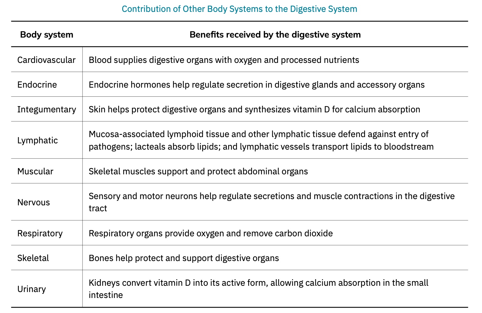
Interrelation with other body systems:
The digestive system works cooperatively with other systems (e.g., cardiovascular and endocrine systems).
Ex. Arteries supply organs with oxygen and processed nutrients.
While intestinal veins (the hepatic portal system) drain the digestive tract and lead to the liver for nutrient processing.
the hepatic portal system, which directs blood from the digestive organs to the liver instead of back to the heart.
These nutrients are off-loaded for processing before blood completes its circuit back to the heart.
The digestive system provides nutrients and support to muscle and vascular tissues.
Hormones from endocrine glands ( and endocrine cells of the pancreas, the stomach, and the small intestine) contribute to control of digestion and nutrient metabolism.
The digestive system supports overall homeostasis by providing nutrients for endocrine function.
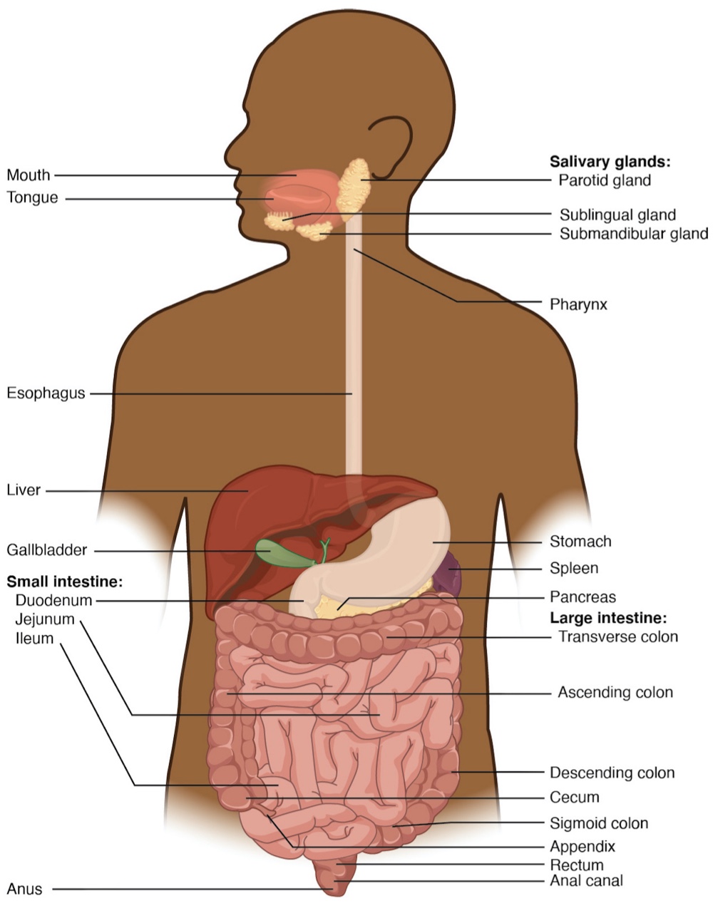
Digestive System Organs
The digestive system is divided into two main categories:
Alimentary canal organs
Accessory digestive organs
Critical for orchestrating the breakdown of food and the assimilation of its nutrients into the body
Alimentary Canal Organs:
The alimentary canal is a one-way tube, approximately:
7.62 meters (25 feet) in length during life
10.67 meters (35 feet) in length after death (once smooth muscle tone is lost)
Also called the gastrointestinal (GI) tract or gut.
Main function: nourish the body.
Begins at the mouth and terminates at the anus.
Between these points, the canal is modified as:
Pharynx
Esophagus
Stomach
Small and large intestines
Both mouth and anus are open to the external environment.
so food and wastes within the canal are considered outside the body until absorbed.
Nutrients enter into and nourish the body’s "inner space."
Accessory Structures:
Each accessory digestive organ aids in the breakdown of food.
Mechanical digestion begins in the mouth with the teeth and tongue.
Salivary glands begin chemical digestion.
Once foods enter the small intestine, the gallbladder, liver, and pancreas release essential secretions (bile, enzymes) for digestion.
These structures are called accessory organs as they develop from the lining cells of the gut and augment its function.
Even after development they maintain connection to the gut by way of ducts.
Vital contributions from these organs are necessary for life.
Malfunctions can lead to significant diseases.
Histology of the Alimentary Canal
The 4 tissue layers of the alimentary tract have details that vary to fit their specific functions.
The alimentary tract is composed of four tissue layers throughout its length:
Mucosa
Submucosa
Muscularis
Serosa (continuous with the mesentery)
Starting from the lumen and moving outwards
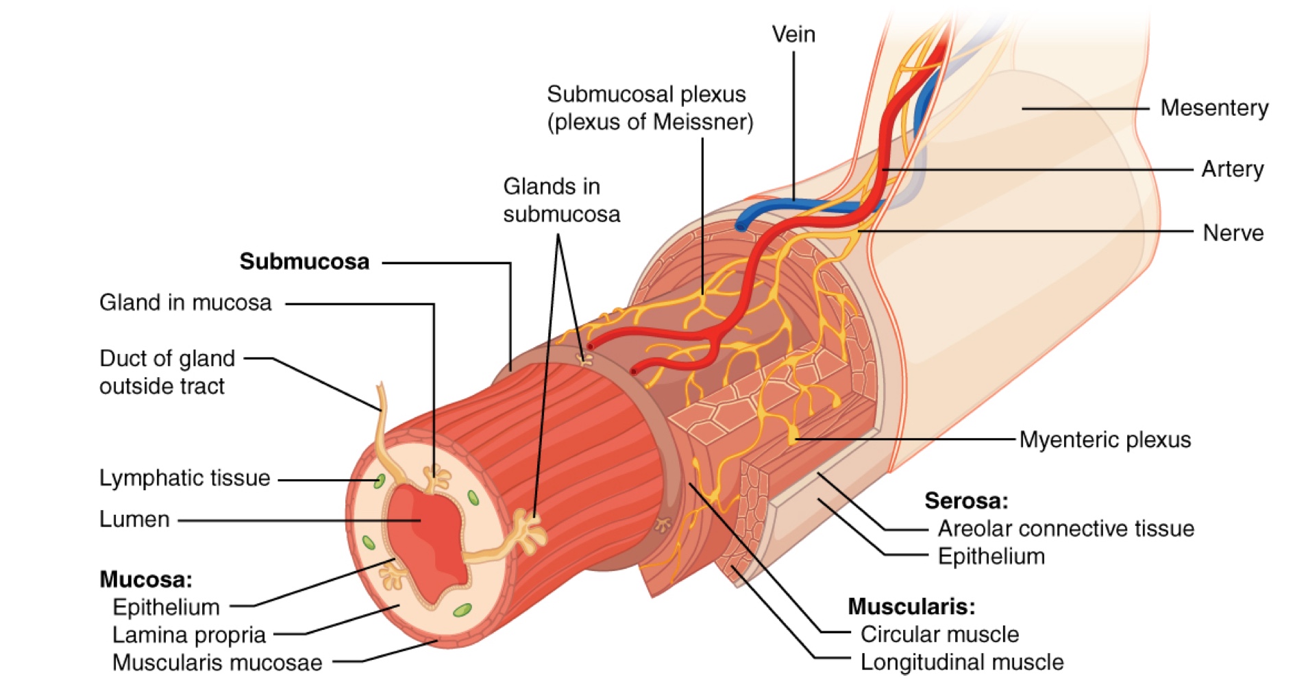
Mucosa:
Referred to as a mucous membrane due to mucus production, characteristic of gut epithelium.
Composed of:
Epithelium (in direct contact with ingested food)
Lamina propria ( layer of connective tissue similar to the dermis)
Muscularis mucosa (thin, smooth muscle layer)
Epithelium:
In the mouth, pharynx, esophagus, and anal canal: primarily a non-keratinized, stratified squamous epithelium.
In the stomach and intestines: simple columnar epithelium.
The epithelium is in direct contact with the lumen.
Epithelial cells:
Goblet cells secrete mucus and fluid into the lumen;
enterendocrine cells secrete hormones into interstitial spaces between cells.
Lifespan of epithelial cells:
Mouth: a couple of days
Gut: about a week
Process of rapid cell renewal preserves health despite wear from food contact.
Lamina Propria:
Loose connective tissue with numerous blood and lymphatic vessels.
These vessels and blood transports nutrients absorbed through the alimentary canal to other parts of the body.
Serves an immune function by:
Housing clusters of lymphocytes (mucosa-associated lymphoid tissue - MALT).
Lymphocyte clusters substantial in distal ileum (Peyer's patches).
Plays a role in defending against pathogens encountered in the alimentary canal.
Muscularis mucosae:
Thin layer of smooth muscle in a constant state of tension, pulling the mucosa into undulating folds.
These folds increases surface area for digestion and absorption.
Submucosa:
Lies immediately beneath the mucosa.
A broad layer of dense connective tissue connects the mucosa to the underlying muscularis.
Contains blood and lymphatic vessels (transport absorbed nutrients).
Houses submucosal glands that release digestive secretions.
Serves as a conduit for the submucosal plexus (a dense nerve network).
Muscularis (also called the muscularis externa):
Composed of a double layer of smooth muscle (inner circular and outer longitudinal layers).
Contractions of layers promote mechanical digestion, exposes more food to digestive chemicals, and moves food along the canal.
Involves skeletal muscle in some regions for voluntary control in the most proximal and distal regions(e.g., esophagus, mouth).
This includes the mouth, pharynx, anterior part of the esophagus, and the external anal sphincter.
Basic two-layer structure found in the small intestine, modified in other organs.
Stomach provides churning function through the additional oblique muscle layer.
Colon has two layers but longitudinal layer appears as three narrow parallel bands (tenia coli).
Serosa:
Portion superficial to the muscularis in the abdominal cavity.
Consists of a layer of visceral peritoneum and loose connective tissue.
Differs from organs like the stomach, esophagus which have dense collagen sheaths (adventitia).
Adventitia helps stabilize the alimentary canal in place near the vertebral column.
Nerve Supply
As soon as food enters the mouth, it is detected by receptors that send impulses along the sensory neurons of cranial nerves.
Without these nerves:
Food would lack taste.
Individuals would be unable to feel the structures of their mouth.
Individuals would be unable to avoid biting themselves while chewing, an action enabled by motor branches of cranial nerves.
Intrinsic innervation of much of the alimentary canal:
Provided by the enteric nervous system, which runs from the esophagus to the anus.
Contains approximately 100 million motor, sensory, and interneurons (unique to this system compared to all other parts of the peripheral nervous system).
Enteric neurons are grouped into two plexuses:
Myenteric plexus (plexus of Auerbach):
Located in the muscularis layer of the alimentary canal.
Responsible for motility, especially the rhythm and force of contractions of the muscularis.
Submucosal plexus (plexus of Meissner):
Lies in the submucosal layer.
Responsible for regulating digestive secretions and reacting to the presence of food.
Extrinsic innervations of the alimentary canal:
Provided by the autonomic nervous system, which includes both sympathetic and parasympathetic nerves.
Sympathetic activation (fight-or-flight response):
Restricts the activity of enteric neurons, thereby decreasing GI secretion and motility.
Parasympathetic activation (rest-and-digest response):
Increases GI secretion and motility by stimulating neurons of the enteric nervous system.
Blood Supply
The blood vessels serving the digestive system have two functions:
Transport the protein and carbohydrate nutrients absorbed by mucosal cells after food is digested in the lumen.
Supply the organs of the alimentary canal with the nutrients and oxygen needed for their cellular processes.
Specifically,:
The more anterior parts of the alimentary canal are supplied with blood by arteries branching off the aortic arch and thoracic aorta.
Below this point, the alimentary canal is supplied with blood by arteries branching from the abdominal aorta.
The celiac trunk services the liver, stomach, and duodenum, whereas the superior and inferior mesenteric arteries supply blood to the remaining small and large intestines.
The veins that collect nutrient-rich blood from the small intestine (where most absorption occurs) empty into the hepatic portal system.
This venous network takes the blood into the liver where the nutrients are either processed or stored for later use.
Only then does the blood drained from the alimentary canal viscera circulate back to the heart.
To appreciate how demanding the digestive process is on the cardiovascular system, consider that while you are "resting and digesting," about one-fourth of the blood pumped with each heartbeat enters arteries serving the intestines.