Hearing
Chapter 7: Audition, the Body Senses, and the Chemical Senses
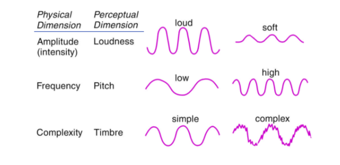
7.1 Sound Waves
Sound waves are vibrations that travel through air and other mediums.
7.2 Divisions of the Ear
Outer Ear: Contains the channel to the tympanic membrane.
Middle Ear: Houses the ossicles (small bones that transmit sound).
Inner Ear: Contains the cochlea, crucial for auditory processing.
7.3 The Cochlea
Composed of three chambers:
Scala Vestibuli and Scala Media (separated by a membrane).
Scala Tympani and Scala Media (separated by the basilar membrane).
Organ of Corti: Contains hair cells that transduce sound waves into nerve impulses.
Components include:
Basilar membrane (base).
Tectorial membrane (roof).
Hair cells in between.
7.4 Auditory Hair Cells
Inner Hair Cells (approximately 3500): Form a singular line, destruction leads to hearing loss.
Outer Hair Cells (approximately 12,000): Arranged in three rows, mainly structural.
Cilia on top project into the tectorial membrane; movement due to sound waves creates receptor potentials.
7.5 Auditory Transduction
The cilia tips are joined by fiber links that open ion channels when they move, leading to depolarization and ion flow (Calcium and Potassium).
7.6 Auditory Pathways
Afferent Pathways:
Cochlear nuclei → Superior olivary nuclei → Inferior colliculus → Medial geniculate → Auditory cortex.
Efferent Pathways:
Olivocochlear bundle controls inner ear feedback.
7.7 Place Coding of Pitch
Different sound frequencies cause maximal distortion in specific basilar membrane locations:
High Frequency: Near the base.
Moderate Frequency: Near the apex.
Tonotopic representation in auditory cortex where adjacent neurons correspond to adjacent membrane areas.
7.8 Support for Place Theory
Findings by von Bekesy demonstrate maximal displacement occurs at varied membrane locations for different frequencies.
Hair cell loss from antibiotics affects high frequency hearing first.
Cochlear implants can restore hearing by stimulating specific basilar membrane regions.
7.9 Analysis of the Auditory System
Auditory system components are responsible for sound detection, localization, and identity recognition.
Lesions at different auditory system levels produce distinct loss characteristics in pitch, frequency detection, and overall deafness.
Bilateral auditory cortex: animal can detect pitch, intensity diff, but not “tunes”
Brachium of inf. colliculus: animal cannot detect frequency or intensity difference
Lateral lemniscus: animal is deaf
7.10 Somatosenses
Provide sensory information regarding skin and body events:
Cutaneous Senses: Signals regarding Pressure, Vibration, Heating/Cooling, and Pain.
Kinesthesia: Signals from joints, tendons, and muscles about body position and movement.
7.11 Morphology of Skin
Comprised of two main layers:
Dermis: Inner layer housing blood vessels, nerves, and glands.
Epidermis: Outer protective layer composed of skin cells.
7.12 Cutaneous Senses
Primary sensations:
Touch: Perception through pressure & vibration (detected by Pacinian corpuscles).
Temperature: Interpreted through warmth and cold receptors located at varying skin depths.
Pain: Related to tissue damage, poorly localized.
7.13 Somatosensory Pathways
Dorsal Columns: Carry touch-related info, precise localization.
Spinothalamic Tract: Carries pain and temperature signals, less precise.
Somatosensory cortex is organized into numerous cortical maps of the body surface.
7.14 Pain
Pain serves as a survival function; lack of pain receptors greatly increases risk.
Induces escape and withdrawal responses and can motivate behavior.
Pain involves tissue destruction induced by:
Thermal Stimuli
Mechanical Force
Pain is poorly localized and features an emotional component affecting perception severity.
7.15 Pain Receptors
Nociceptors:
Free nerve endings responsive to intense mechanical, chemical, or thermal stimuli;
free nerve endings networks within the skin that respond to intense pressure
receptors that are sensitive to ATP
Found in skin, muscles, internal organs, cornea, teeth. Activated by tissue damage.
7.16 Analgesia
Refers to reduced pain perception.
Can be induced by various means including hypnosis, massage, acupuncture, placebo, attention shifts and medications (like opiates).
Pain stimuli activate somatosensory areas of the brain, influencing the emotional response to pain.
The anterior cingulate cortex is involved in the aversiveness of pain (hypnosis and PET scanner study)
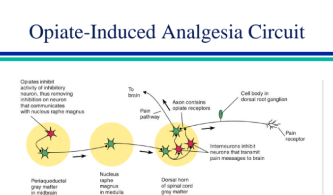
7.17 Opiates and Pain
Exogenous opiates decrease pain reactivity.
The brain naturally produces endorphins to modulate pain perception.
Naloxone serves as an antagonist, reversing opiate effects. (Naloxone reversibility is taken as an indication of opiate involvement)
Focal brain stimulation can reduce pain
PAG in particular is effectiveBrain stimulation activates a descending pathway that modulates pain (Basbaum and Fields model)
7.18 Gustation
Involves taste related to food and liquid intake.
Molecules from food activate receptors categorized by taste:
Sweet: Safe foods.
Salty: Sodium sources.
Bitter: Potentially poisonous foods.
Sour: Spoiled food.
7.19 Transduction of Taste
Taste molecule binding alters receptor potential, initiating signal transduction:
Saltiness responds to sodium ions. (Receptor for saltiness may be a simple sodium channel) (sodium chloride)
Sourness: interacts with hydrogen ions in acid solutions
Bitterness: typical stimulus is an alkaloid (e.g. quinine)
Receptors involve a hydrophobic residue
Sweetness: stimulus - sugar
Receptors have a hydrogen ion site
7.20 Gustatory Processing
Gustatory information is transmitted through cranial nerves 7 (anterior tongue), 9 (posterior tongue), and 10 (palate and epiglottis)
First relay station for taste information is the nucleus of the solitary tract (medulla)
Taste information is then transmitted to primary gustatory cortex, to the amygdala, and to the hypothalamus
Taste signals transmit via cranial nerves to the medulla's nucleus of the solitary tract, then to the primary gustatory cortex, amygdala, and hypothalamus.
Taste perception involves overlaps in taste quality response and temperature sensitivity.
Recordings from chorda tympani (7th cranial nerve) indicate that taste fibers respond to more than one taste quality and to temperature
cortex, the major groups of taste-sensitive neurons were salty and sweet