BIOL 2100 CH. 6
Concept 6.1: Biologists use microscopes and biochemistry to study cells
The development of the microscope in 1590 and later in the 1600’s helped humans to discover and study cells
Dead cells were first seen on oak bark by Robert Hooke in 1665
Living cells were first seen by Leeuwenhoek a few years later
The most common microscope known as the light microscope allows visible light to pass through the specimen and seen through a glass lens
The lens refracts(bends) light to project a magnified image into the eye or camera
In microscopy magnification, resolution, and contrast are most important
Magnification: is the ratio of an objects image to its relative size
Resolution: is a measure of clarity for an image being the minimum distance two points can be separated and distinguished as two separate points
Contrast: is the difference in brightness between light areas and dark areas of an image

Brightfield microscopes: are when light passes directly through the specimen, the can be stained or unstained, staining them kills cells
Phase-contrast microscope: allow variations in density within a specimen to amplified and enhance contrast in unstained cells
Differential interference contrast microscopes :allow variations in density within a specimen the only difference being the exaggerations in the differences in density
Fluorescence microscopes: reveal locations of speicific molecules through fluorescent dyes or antibodies which absorb UV radiation and in turn emit visible light
Confocal microscopes: use lasers to create a single plane of fluorescence, through this many different planes captured can create a 3D image
Deconvolution microscope: uses digitals to remove out of focus light and reassigns it to the desired source creating a sharper 3D image
Super-resolution microscopes: use UV light to excite fluorescent molecules allowing an image of where the position of the molecules are record to create a sharp image
Electron Microscopy
Focuses a beam of electrons through a specimen rather than focusing light, but this oftentimes kill cells
(SEM) Scanning electron microscopy :use a electron microscope to show a 3D image of the surface of a specimen where the beam excites electrons on the surface showing a black and white image but it can be colored artifically
Used mainly for detailed topography of a specimen
(TEM) Transmission electron microscopy: shows a thin section of a specimen revealing it’s internal structure
Both TEM and SEM use electromagnets to bend paths of electrons to get a focused image for viewing
(cryo-EM) Cryo-electron microscopy: uses tissues of specimens that are frozen rapidly to lock the molecule into a rigid state, this allows a beam of electrons to pass through and create a visual molecule through the microscope
These microscope allow the study or organelles in eukaryotic cells and in the study of cytology which is the study of cell structure
Organelles are membrane enclosed structures with specialized functions, these are suspended in cytosol of eukaryote cells
Cell fractionation: technique where cells are disrupted and separated by centrifugation at high speeds (differential centrifugation)
Allows viewing of organelles and subcellular structures individually

Concept 6.2: Eukaryotic cells have internal membranes that compartmentalize their functions
In cells there Eukaryotic and Prokaryotic cells
Bacteria and Archea = Prokaryotic Cells
Animals, Fungi, Protists, Plants = Eukaryotic Cells
The difference between the two is that eukaryotes keep DNA in the nucleus which is double membrane enclosed while prokaryotes keep DNA in a nucleoid which is non-membrane enclosed

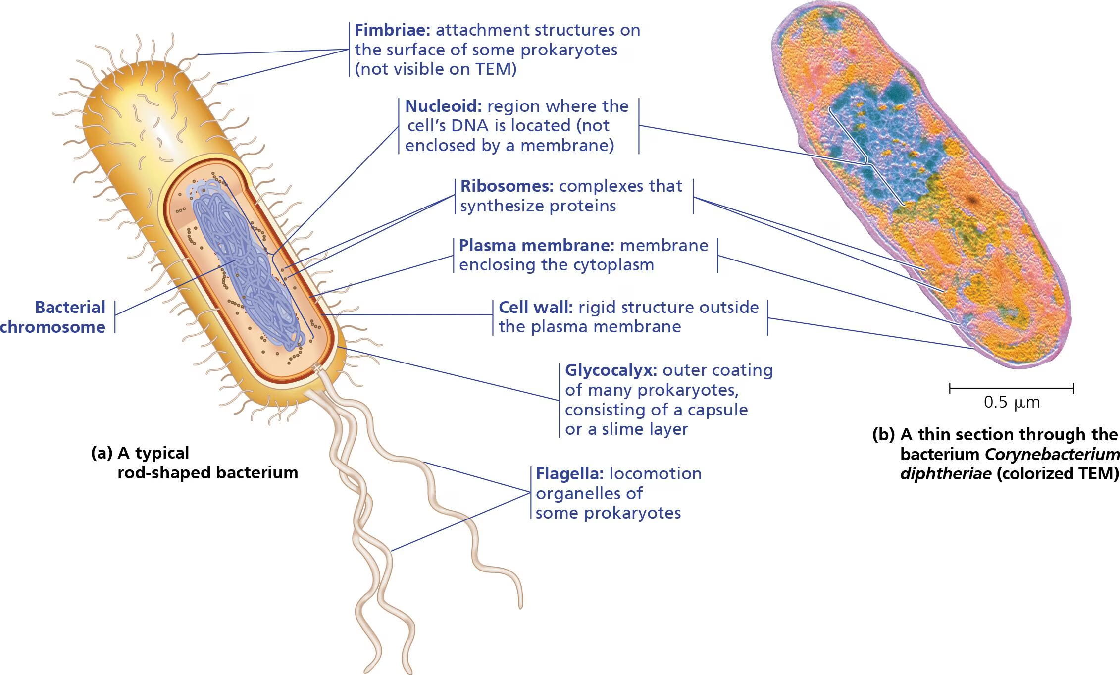
In both cells cytoplasm is the interior but function in different ways
In eukaryotes it’s the reigon btw. the nucleus and plasma membrane that consist of organelles of specialized form and function
In prokaryotes there is an absence of membrane bound structures, but some have regions surrounded by proteins where reactions take place
Eukaryotes are larger with a 10-100 micrometer diameter
Plasma membranes allow the passage of nutrients into cells due to eukaryotes size the surface area allows a smaller surface to volume ratio meaning they have smaller surface area

Having a higher ratio of surface area to volume allows more cell exchange
Eukaryotic Cells


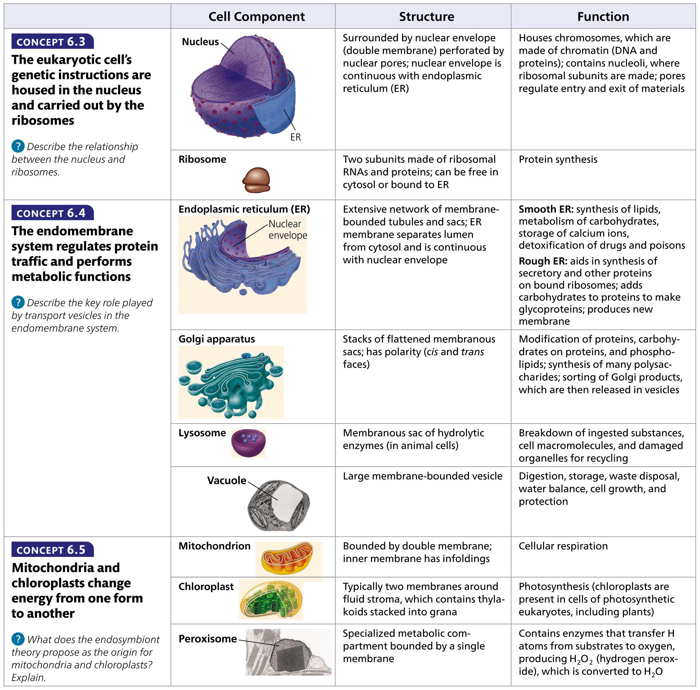
Concept 6.3: The eukaryotic cell’s genetic instructions are housed in the nucleus and carried out by the ribosomes
The nucleus contains most of the genes in eukaryotic cells, it’s made up of chromatin and is a cluster of neurons
The nuclear envelope is a double membrane that encloses the nucleus from cytoplasm
It consists of a pore complex that aids in the entry and exit of proteins, RNA’s, and large macromolecules
At the pores nuclear lamina which is a net of protein filaments help maintain the shape of the nucleus by supporting the envelope

Within DNA chromosomes consist of a long DNA molecules which are made up of small proteins known as histones
Histones help coil DNA molecules of each chromosome allowing it to fit into the nucleus
The complexes of DNA and proteins making up chromosomes is known as chromatin
When cell division is not being enacted stained chromatin appears as diffuse mass in micrographs oftentimes being confused for chromosomes
Chromosomes in human cells are numbered at 46 in the nucleus, sex cells have 23 chromosomes each
The nucleolus consists of rRNA genes where rRNA synthesis and ribosomal subunits assemble
The nucleus directs protein synthesis through synthesizing mRNA which carries info over to DNA
mRNA is transported to the cytoplasm through the nuclear pores where ribosomes translate mRNA’s genetic message into what we known as a primary structure of a specific polypeptide
Ribosomes carry out protein synthesis and are complexes made of rRNA
The more ribosomes a cell has the more protein synthesis will occur

They build proteins in two regions free and bound ribosomes
Free ribosomes: suspended in cytosol and function within such as enzymes that catalyze glucose breakdown
Bound ribosomes: attached to the outside of the ER or nuclear envelope, usually make proteins made for insertion into membranes or exportation from secretion
ex. the pancreas secretes digestive enzymes meaning there will be a higher proportion of bound ribosomes
Concept 6.4: The endomembrane system regulates protein traffic and performs metabolic functions
The endomembrane system: collection of membrance-bounded organelles inside a eukaryotic cell
Nuclear envolope, Endoplasmic reticulum(ER), Golgi apparatus, Lysosomes, Vesicles/Vacuoles, and Plasma Membrane
These organelles are linked through the use of vesicles that are membrane-bound sacs inside or outside of cells
The ER is. a network of tubules and sacs know as cisternae that separate the ER lumen/ cisternal space from cytosol

Smooth ER due to the lack of ribosomes on it’s outer surface
Rough Er due to the magnitude of ribosomes on it’s outer surface
It’s attached to the cytoplasmic side of the nuclear envelopes outer membrane that continues with rough ER
Smooth ER synthesizes lipids, metabolism of carbohydrates, detoxifing drugs/poisons, and storage of calcium ions
Enzymes of smooth ER are important of the synthesis of oils and steroids
Sex hormones are produced in smooth ER of animal cells and steroid hormones secreted by adrenal glands
Detoxification occurs through the addition of hydroxyl groups to drug molecules making them hydrophilic and easier to eradicate from your system
Due to this doses of alcohol, drugs, etc. may need to be taken in higher doses to achieve sedation or desired outcome due to to an increased tolerance after repeated used
Ex. alcohol tolerance and tolerance to certain drugs
Calcium ions such as in muscle cells are pumped from cytosol into ER lumen
When muscle cells are stimulated by nerve impulses calcium ions rush into cytosol across the ER membrane causing contractions of muscle cells
Rough ER function through the secretion and creation of secretory proteins such as pancreatic cells that synthesize insulin where rough ER secretes the hormone into the bloodstream
Most of these secretory proteins are glycoproteins meaning that they’re covalently attached to one or more carbohydrates
Secretory proteins can depart from rough ER through transport vesicles to it’s desired location
It also is a factory for the cell where added membrane proteins and phospholipids grow into it’s own membrane
The Golgi Apparatus is essentially the mail center of the cell
Products of the ER are modified and stored to be sent to other destinations
It consists of flattened membranous sacs that separate the internal space of the GA from cytosol

The GA stack has two sides known as the cis face and trans face
The cis face recieve vesicles that bud from Er to add it’s membrane and contents of its lumen through fusing with GA
The trans face ships vesicles that travel to other sites
In the GA products from the ER are modified
Ex.sugar monomers in carbohydrates may be removed creating a larger variety of said product
The GA also creates macromolecules where most polysaccharides secreted by cells are Golgi products
nonprotein Golgi products depart from the trans face that fuse with the plasma membrane which can add to surfce area when contents of said product are released
The GA manufactures and refines products in stages using different and unique enzymes to do so
Lysosomes are membranous sacs of hydrolytic enzymes often times used for digestion of macromolecules
they thrive in acidic enviornments such as the digestive tract
they can break open or leak which is fine due to cytosol almost neutral pH but excessive leakage can cause cell destruction due to cell digestion
The hydrolytic enzymes are made in rough ER then sent to GA for more processing and refinement
Lysosomes carry out intracellular digestion through processes such as Phagocytosis where small substances or organisims are engulfed by a cell where enzymes will then digest the food
In human cells macrophages (type of white blood cell) engulf harmful bacteria and invaders

Lysosomes also use their hydrolytic enzymes to recule organic material in a cell called autophagy
This process consists of damaged organelles or small amounts of cytosol being surrounded by a double membrane where lysosomes will then fuse with the outer membrane
The enzymes will destroy the inner membrane allowing organic compounds inside to be released in the cytosol for reuse
Vacuoles are large vesicles from the ER/ GA that is a selective in solute transportation
Food vacuoles: membranous sacs waiting to be used as food for the cell
In unicellular protists contractile vacuoles pump excess water out to maintain a stable concentration of ions and molecules in cells
In plants/fungi certain vacuoles use enzymatic hydrolysis to store/hold organic compounds for future use
Also in mature plant cells a central vacuole plays a role in growth, storage, and holding of toxic compounds

Endomembrane Transportation

Concept 6.5: Mitochondria and chloroplasts change energy from one form to another
In eukaryotes the mitochondria and chloroplasts are energy converting organelles
Mitochondria are the sites of cellular respiration using O2 to generate ATP from extracting energy from glucose, fats, etc.
Chloroplasts work in a similar way but for solely plant cells where photosynthesis is used to generate chemical energy (oxygen, glucose) through the use of sunlight, co2, and water
With these two organelles the Endosymbiont theory states that the ancestor of eukaryotic cells engulfed a prokaryotic cell forming the relationship known as endosymbiont which is essentially a cell living within another cell
Over time the host cell and endosymbiont became a single organism, the eukaryotes having the endosymbiont becoming the mitochondrion and prokaryotes having chloroplasts

Mitochondria
Factors such as the double membranes of these two organelles support the theory and the addition of ribosomes as well as DNA molecules that program the synthesis of the proteins on ribosomes, and lastly their autonomy with independent reproduction within each respective organelle
Mitochondria are made up of two membranes enclosing the mitochondrion which is a phospholipid bilayer harboring embedded proteins
The outer membrane is smooth while the inner membrane is convoluted with cristae which are infoldings
Cristae allows a larger surface area due to infoldings thus increasing the frequency and productivity of cellular respiration
The inner membrane has two compartments the mitochondrial matrix and intermembrane space
The intermembrane space is the reigion btw. the inner and outer membranes
The matrix has enzymes, mitochondrial DNA, and ribosomes that may help catalyze certain steps of cellular respiration and enzymes that make ATP

Chloroplast
They contain the green pigment known as chlorophyll that function in the production of sugar and are often found in leaves and other green organs of plants
Inside thylakoids which are innerconnected sacs are stacked on top of one another, these stacks are called granum
The outside of thylakoids is stroma which harbors chloroplast DNA, enzymes, and ribosomoes
This organization allows the chemical process of photosynthesis to occur

Peroxisomes:Oxidation
These are metabolic compartments bounded by single membranes consisting of enzymes that remove hydrogen atoms and transfer them to O2 creating hydrogen peroxide H2O2 which is very toxic itself but has an enzyme that converts it to water
Peroxisomes can be used to break down fatty acids into smaller molecules that can be transported to to mitoch. and fuel cellular respiration
They function in liver detoxification and other harmful/poisinous substances through transfering hydrogen from poisinous compounds to oxygen
Specialized peroxisomes known as glyoxysomes contain enzymes intiating the conversion of fatty acids to sugar
This can be used as a source of energy in the tissues of plant seeds until it can produce glucose by photosynthesis
Concept 6.6: The cytoskeleton is a network of fibers that organizes structures and activities in the cell
In organelles it’s been revealed that they consist of cytoskeletons
They’re networks of fibers extending throughout the cytoplasm
They function in giving mechanical support to the cell and maintaining its shape as well as anchoring organelles
They’re very fluid meaning they can be easily dismantaled and put back up
Cell motility is also vital to the cytoskeleton where interactions with motor proteins will take place allowing cells to move along fibers outside the cell
Vesicles utilize motor proteins as feet to walk to their intended destinations along a path provided by the cytoskeleton
The cytoskeleton manipulates the plasma membrane by bending it inward forming food vacuoles

Components of Cytoskeletons

Microtubules are hollow tubes that grow out from centrosomes and are made up of dimers which are molecules made up of two components
Within centrosomes centrioles are cylinders of microtubule triplets that give it it’s ring shape
They help maintain cell shape and cell motility through the disassembley and building of tubulins elsewhere in the cell
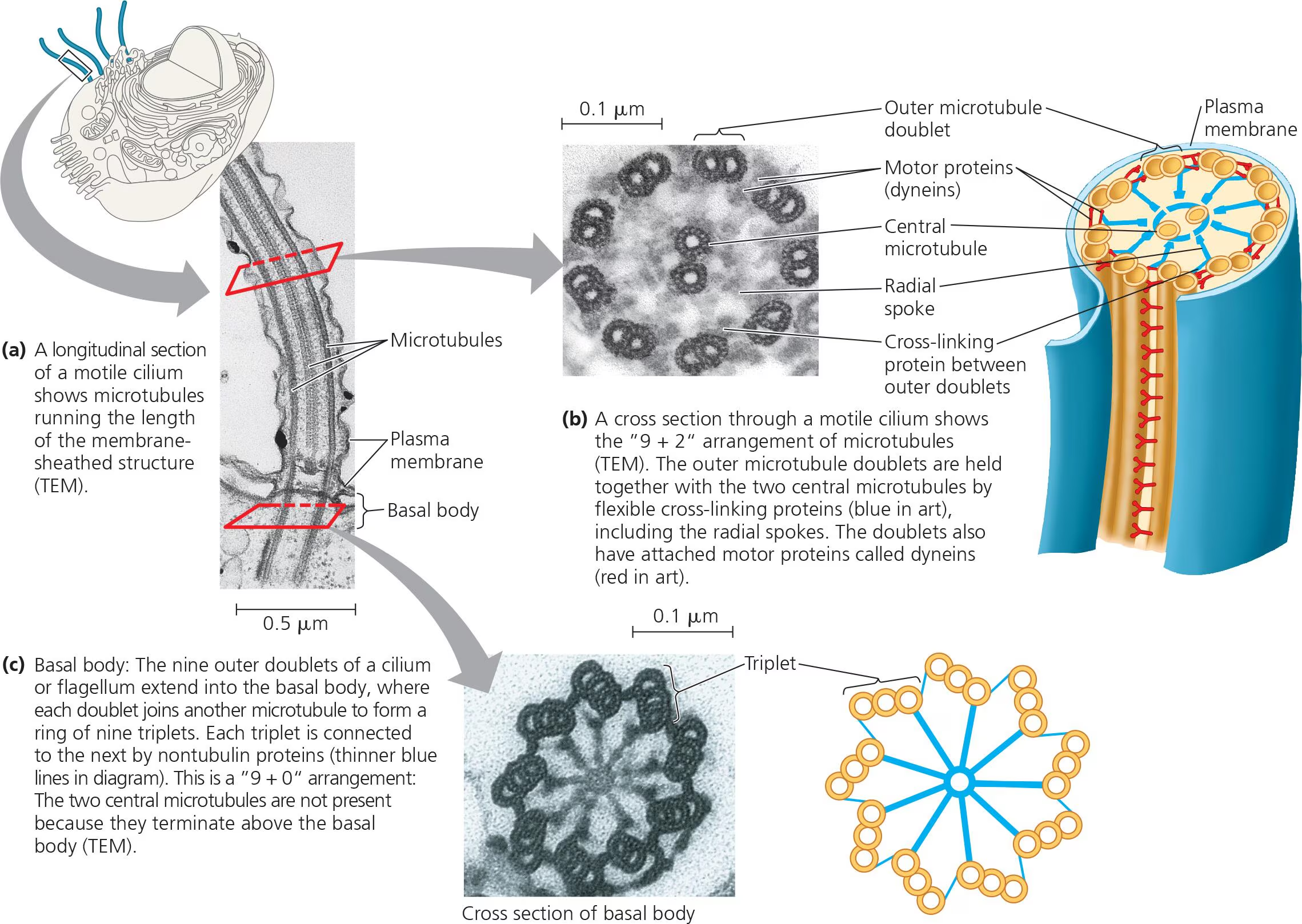
Flagella and Cilia contain microtubules that function in the motion of these structures
Flagella are long apendages that are specialized in locomotion, it has a core of nine outer doublet microtubules that allow it to move around
Cilia/Cilium are short apendages that specialized in locomotion or moving past the cell, these are usually nonmotile and play more of a sensory role

Microfilaments are thin solid rods that are built from molecules of actin
Actin is a globular protein that links into chains that twist helically forming microfilaments in muscle and other cells
They help form structural networks when proteins bind alongside the filament and creating a branch to form a new filament
Microfilaments function in cell motility including muscle contraction, amoeboid movement, and cytoplasmic streaming in plants.
Their function in cell motility through the protein myosin that interact causing muscle contractions
The structure is to help bear tension and support the cells shape, the outer layer known as the cortex is a semisolid gel consistency contrasting cytoplasms fluid state
Bundles of microfilaments can make up the core of microvilli that increase the cells surface area


Intermediate Filaments have larger diameters than microfilaments but are smaller than microtubules, these are only found in the cells of some animals
They bear tension and is constructed from diverse classes of cytoskeletal elements
They are more permanent fixtures of cells and don’t disassemble and reassemble
Even after cell death they filament networks continue to persist maintaining stability in the cell and the formation of nuclear lamina
Concept 6.7: Extracellular components and connections between cells help coordinate cellular activitiess
Cell Walls is an extracellular feature essential to plant cells
They help protect plant cells, maintain shape, and prevent excessive water uptake
Some prokaryotes also have cell walls
The inner structures of cell walls are the primary cell wall, middle lamella, and sometimes secondary cell wall
The Primary cell wall is a thin and flexible wall that young plants cells first secrete
Middle lamella is a thin layer composed of sticky polysaccharides known as pectins (thickening agent)
Secondary cell wall has strong and durable matrix offering cell support and protection
Ex. Wood is mainly secondary walls

The Extracellular Matrix is a structure surrounding animal cells made up of glycoproteins, polysaccharides, and proteoglycans that are synthesized and secreted by cells
ex. collagen is glycoprotein that is most abundant in animal cells
Glycoproteins are proteins that are covalently bonded carbohydrates
Proteoglycans are small core proteins consisting of many carbohydrate chains
Fibronectin binds to cell-surface receptor proteins known as integrins that are built into the plasma membrane
Fibronectin is a glycoprotein secreted by animal cells helping them attach to extracellular matrixes
Integrins are transmembrane receptor proteins that interconnect extracellullar matrix and cytoskeleton, they transmit singlas between the two regarding changes inside and outside the cell

Cell Junctions are the process of neighboring cells communicating via direct physcial contact to once another
Tight Junctions: are between animal cells and prevent the leak of material through spaces btw. cells
Desmosomes: in animal cells function as rivets that fasten cells together
Gap Junctions: consist of proteins surrounding a pore allowing the passing of materials btw. cells

Plasmodesmata: open channels through a cell wall connecting cytoplasms of adjacent plant cells allowing nutrients to pass between cells

Concept 6.8: A cell is greater than the sum of its parts
Cellular functions arise from cellular order= The cell is a living unit greater than the sum of it’s parts