Comprehensive Veterinary and Comparative Microanatomy of the Endocrine System
Cellular Surface Specializations: Microvilli vs. Cilia
- Microvilli:
- Non-motile cell surface extensions that do not possess active ciliary movement.
- Formed by specialized folding of the apical plasma membrane, supported internally by a core of longitudinal actin microfilaments anchored into the terminal web.
- Exhibit a uniform length of approximately .
- Primary physiological function is to maximize the apical surface area of epithelial cells to facilitate absorption, secretion, and membrane-bound enzymatic activity.

- Cilia:
- Motile, elongated surface projections capable of coordinated, whip-like rhythmic beating.
- Organized around a specialized internal axoneme exhibiting a characteristic microtubule arrangement, consisting of nine outer doublet microtubules surrounding a central pair of singlet microtubules.
- Length ranges between and .
- Primary physiological function is the directional movement of extracellular fluid, mucus, foreign particles, and cells across epithelial sheets.
Foundations of the Endocrine System & Cell Signaling Mechanisms
General Features of Endocrine Glands:
- Classic ductless glandular structures that synthesize and secrete chemical messengers (hormones) directly into the interstitial vascular network rather than onto an epithelial surface via ducts (in contrast to exocrine glands).
- Possess rich, extensive microvascular and capillary beds allowing immediate transport of secreted molecules into the systemic circulation.
- Hormones circulate throughout the systemic bloodstream to bind high-affinity receptors located at distant, specific target organs.
Modes of Chemical Signaling:
- Autocrine signaling: A cell secretes a hormone or chemical factor that binds to specific surface or intracellular receptors on the same cell, stimulating or inhibiting its own function.
- Paracrine signaling: A secretory cell releases bioactive molecules into local interstitial fluid to act on neighboring, adjacent cells without entering systemic circulation.
- Endocrine signaling: Specialized glandular cells release hormones directly into the vascular bloodstream, which conveys them systemically to distant effector organs.
Homeostatic Regulation and Feedback Loops:
- Endocrine glands typically function within closed feedback loops (predominantly negative feedback) that maintain internal physiologic equilibrium.
- The central axis coordinates three hierarchical tiers of signaling:
- Hypothalamic releasing hormones act on the anterior pituitary gland.
- Anterior pituitary stimulating (tropic) hormones act on peripheral endocrine organs.
- Peripheral target organ hormones execute terminal physiological effects and feed back to the hypothalamus and adenohypophysis to suppress further production of releasing and stimulating hormones.
Classification of Endocrine Organs:
- Primary Endocrine Organs: Glands whose exclusive or primary physiological function is hormone synthesis and systemic regulation:
- Pituitary gland (hypophysis cerebri).
- Pineal gland (epiphysis cerebri).
- Thyroid gland.
- Parathyroid glands.
- Adrenal glands (suprarenal glands).
- Secondary Endocrine Organs: Tissues and organs possessing specialized, non-endocrine primary tasks (e.g., digestion, excretion, circulation, gametogenesis) that concurrently contain endocrine cells producing hormones:
- Pancreas (islets of Langerhans).
- Kidneys.
- Heart.
- Gastrointestinal tract (stomach and intestines via enteroendocrine cells).
- Gonads (testes and ovaries).
- Adipose tissue.
- Placenta.
- Liver.
- Thymus.
The Hypothalamic-Pituitary Axis (HPA) & Hypophysis Cerebri
- Anatomical Relationships of the Hypothalamus and Pituitary:
- The pituitary gland is located ventral to the hypothalamus and caudal/inferior to the optic chiasm.
- It rests protected within the osseous hypophyseal fossa of the sella turcica of the sphenoid bone.
- The pituitary is anatomically suspended from the base of the brain by the infundibular stalk.
- The adenohypophysis physically envelops the ventral and lateral borders of the neurohypophysis.
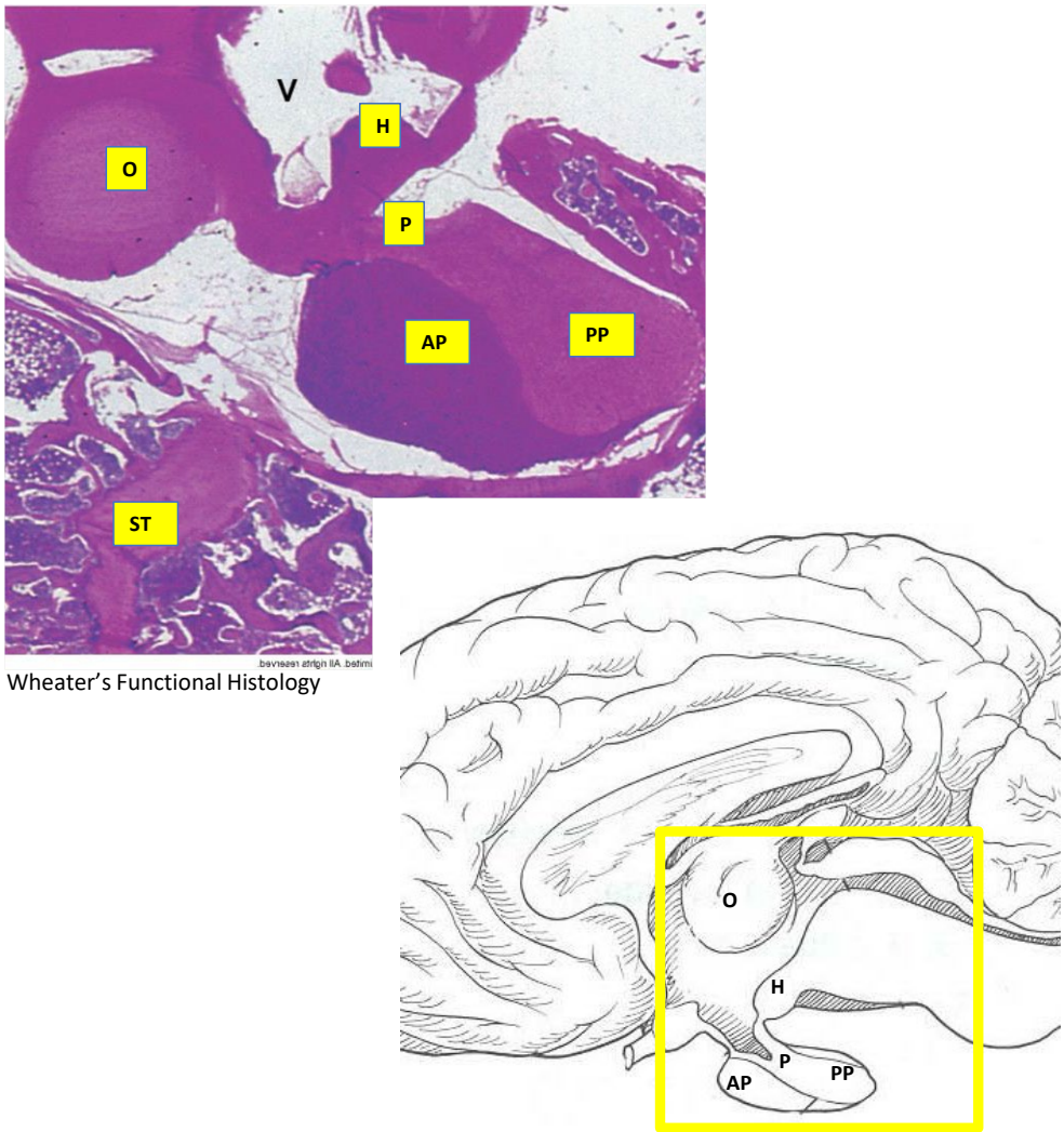
Hypothalamic Architecture and Neurosecretion:
- Integrates the central nervous system and endocrine control systems via neurosecretory neurons.
- Neurosecretion is defined as the synthesis and storage of neuropeptides within neuronal perikarya and their transport along axons to terminals, where they are released directly into the vascular circulation upon depolarization.
- Receives afferent input from all brain regions, the spinal cord, and special sensory pathways.
- The blood-brain barrier is absent at the median eminence and neurosecretory sites, allowing hypothalamic neurons to directly detect ionic, osmolar, nutritional, and circulating hormonal signals.
- Major neurosecretory cell clusters include:
- Paraventricular nuclei: Synthesize oxytocin, corticotropin-releasing hormone (CRH), and thyrotropin-releasing hormone (TRH).
- Supraoptic nuclei: Synthesize antidiuretic hormone (ADH / arginine vasopressin).
- Arcuate nucleus: Synthesizes growth hormone-releasing hormone (GHRH).
Dual Embryological Derivation of the Pituitary:
- Adenohypophysis (Anterior Pituitary): Derived from oral ectoderm via an upward invagination of the embryonic stomodeal epithelium of the pharyngeal roof, designated as Rathke\'s pouch.
- The lumen of Rathke\'s pouch persists histologically in many species as the hypophyseal cavity (hypophyseal cleft), which separates the pars distalis from the pars intermedia.
- Neurohypophysis (Posterior Pituitary): Derived from neuroectoderm as a downward projection of the diencephalon (ventral floor of the third ventricle).

- The Hypothalamic-Hypophyseal Portal System:
- Hypothalamic releasing hormones are secreted by neurosecretory terminals into the primary capillary plexus at the median eminence.
- Portal venules descend down the infundibular stalk to form a secondary capillary plexus within the sinusoids of the pars distalis.
- This specialized vascular arrangement delivers high local concentrations of releasing factors directly to anterior pituitary parenchymal cells without significant systemic dilution.
- Anterior pituitary stimulating hormones are subsequently exocytosed into the secondary plexus and carried through systemic collecting veins to reach target organs.
The Adenohypophysis: Pars Distalis, Pars Intermedia, and Pars Tuberalis
Anatomical Subdivisions of the Adenohypophysis:
- Pars distalis: The bulk of the anterior lobe, comprising glandular epithelial cells clustered in cords around thin-walled sinusoidal capillaries.
- Pars intermedia: An intermediate epithelial zone situated between the pars distalis and the pars nervosa, flanking the hypophyseal cleft.
- Pars tuberalis: A thin, vascularized collar of glandular tissue surrounding the infundibular stalk of the neurohypophysis.
Parenchymal Cytology of the Pars Distalis:
- Classified based on routine hematoxylin and eosin (H&E) staining into chromophils (cells whose cytoplasmic granules avidly bind dye) and chromophobes (poorly staining cells):
- Chromophils are categorized into acidophils and basophils, which represent distinct terminal differentiations producing specific tropins (-troph signifies a cell producing a tropin).
- Dyes such as Alcian Blue, Orange G, and Periodic Acid-Schiff (PAS) reagent, alongside immunohistochemistry (IHC), permit precise functional discrimination.
Acidophils:
- Somatotrophs:
- Specialized parenchymal cells concentrated laterally within the pars distalis.
- Synthesize and secrete growth hormone (GH / somatotropin).
- GHRH (somatotropin-releasing hormone [SRH]) from the hypothalamic arcuate nucleus binds somatotroph G-protein coupled receptors (GPCRs), signaling GH synthesis and exocytosis in response to neural stimuli (e.g., exercise, deep sleep).
- Physiological target tissues: Promotes broad anabolic responses in skeletal muscle, chondrocytes, and hepatocytes; triggers synthesis and systemic secretion of insulin-like growth factor I (IGF-I / somatomedin) in the liver and cartilage.
- IGF-I promotes chondroblast proliferation and epiphyseal plate elongation, resulting in linear skeletal growth.
- Downregulation occurs via negative feedback: circulating GH stimulates hypothalamic neurons to secrete somatostatin, which inhibits somatotroph secretion.
- Lactotrophs (Mammotrophs):
- Parenchymal cells producing prolactin (PRL).
- Individual cell size and cytoplasmic dye affinity increase during pregnancy and lactation.
- Primarily controlled by tonic hypothalamic neuroendocrine inhibition via dopamine; stimulated by prolactin-releasing factor (PRF).
- Physiological targets: Mammary alveolar and ductal epithelial cells, inducing proliferation and differentiation into active milk-synthesizing cells; contributes to luteotrophic maintenance of the corpus luteum in canines.
- Prolactin crosses the blood-brain barrier to bind receptors on dopaminergic neurons in the hypothalamus, enhancing dopamine release to execute negative feedback inhibition.
- Operates synergistically with GH to stimulate lymphocyte proliferation and immune system development.
Basophils:
- Thyrotrophs:
- Prominently clustered midventrally within the pars distalis.
- Synthesize thyroid-stimulating hormone (TSH / thyrotropin).
- Hypothalamic TRH from the paraventricular nucleus binds thyrotroph GPCRs, activating adenylyl cyclase to elevate intracellular cyclic adenosine monophosphate (), which triggers TSH exocytosis.
- Physiological target: Thyroid follicular epithelial cells, driving thyroglobulin iodination, endocytosis, and thyroid hormone release.
- Gonadotrophs:
- Relatively small cells that coexpress follicle-stimulating hormone (FSH) and luteinizing hormone (LH).
- Regulated by pulsatile secretion of gonadotropin-releasing hormone (GnRH) from diffusely scattered hypothalamic neurons.
- Target tissues and actions:
- Female: FSH induces proliferation of ovarian follicular granulosa cells and stimulates aromatase-dependent estrogen synthesis; LH drives theca interna cell synthesis of androgens (testosterone), triggers ovulation, and stimulates luteal cells of the corpus luteum to produce progesterone.
- Male: FSH binds receptors on testicular sustentacular (Sertoli) cells to support spermatogenesis and stimulate synthesis of inhibin, activin, and androgen-binding protein; LH (interstitial cell-stimulating hormone [ICSH]) binds interstitial (Leydig) cells to stimulate testosterone biosynthesis.
- Interstitial stroma cells in species such as pigs, dogs, cats, rabbits, and humans also produce testosterone under LH influence.
- Circulating gonadal steroids (estrogens, progesterone, testosterone) together with inhibin execute negative feedback on hypothalamic GnRH and adenohypophyseal gonadotrophs.
- Corticotrophs:
- Uniformly dispersed throughout the pars distalis, difficult to delineate via standard light microscopy due to weak basophilia.
- Cytoplasmic granules stain positively for adrenocorticotropic hormone (ACTH / corticotropin) and -lipotropin.
- Activated by hypothalamic corticotropin-releasing hormone (CRH) released from paraventricular neurons into the portal system.
- CRH binds GPCRs on corticotrophs, elevating intracellular and opening calcium channels to provoke exocytosis of ACTH.
- Physiological target: Cells of the adrenal cortex, activating adenylyl cyclase/ signaling to upregulate enzymes mediating steroidogenesis (principally glucocorticoids).
- Glucocorticoids execute negative feedback by inhibiting hypothalamic CRH release and suppressing POMC transcription and ACTH exocytosis in the anterior pituitary; acute severe stress triggers central neural pathways that override this glucocorticoid feedback.
Chromophobes:
- Small epithelial cells that show poor affinity for acidic and basic histologic dyes.
- Represent either quiescent, degranulated post-secretory acidophils/basophils or undifferentiated stellate stem cells capable of regenerating parenchymal populations.
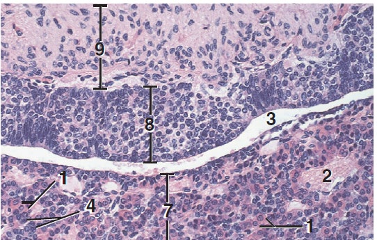
Pars Intermedia:
- Anatomically positioned between the neurohypophysis and the pars distalis; dimensions vary substantially across species.
- Innervated directly by hypothalamic axon terminals, including dopaminergic, serotoninergic, adrenergic, and GABAergic fibers that modulate parenchymal output.
- Predominant parenchymal cell is the melanotroph, characterized as a large, pale cell.
- Melanotrophs transcribe and translate proopiomelanocortin (POMC), a high-molecular-weight prohormone.
- POMC is enzymatically cleaved into bioactive peptides: melanotropins (specifically -melanocyte-stimulating hormone [-MSH]), lipotropins, and endogenous endorphins.
Pars Tuberalis:
- Forms a thin, folded collar composed of longitudinally oriented epithelial cell clusters and occasional small colloidal cysts around the infundibular stalk.
- Contains small numbers of interspersed gonadotrophs and thyrotrophs alongside ependymal lining cells.
- Parenchymal cells express high concentrations of surface melatonin receptors.
- Translates photoperiodic signals received from the pineal gland to regulate seasonal reproductive cycles and seasonal estrus transitions in domestic mammalian species.
The Neurohypophysis: Infundibulum and Pars Nervosa
Anatomical and Histological Organization:
- Composed of the infundibular stalk (which connects to the hypothalamus) and the pars nervosa (the posterior or neural lobe).
- Completely lacks neuronal perikarya (neuronal cell bodies);
- Composed predominantly of unmyelinated axons arising from hypothalamic neurons, specialized non-myelinating glial cells, and fenestrated capillaries.
Axonal Transport and Herring Bodies:
- Non-myelinated neurosecretory axons originate from magnocellular neurons residing in the supraoptic and paraventricular nuclei of the hypothalamus.
- Secretory vesicles containing neurohypophyseal peptide hormones complexed with neurophysin carrier proteins are actively transported down unmyelinated axons along longitudinal neurotubules (microtubules).
- Herring bodies represent focal, beaded axonal dilatations packed with membrane-bound neurosecretory granules along their trajectory and near capillary endings.
- Herring bodies stain selectively with aldehyde-fuchsin dyes, appearing as distinct, purplish, irregular eosinophilic expanses under light microscopy.
Pituicytes:
- Specialized glial cells (central gliocytes) distributed throughout the neurohypophyseal neuropil.
- Display irregular cytoplasmic contours and heterochromatic nuclei that are poorly demarcated on routine H&E sections.
- Function to provide structural, trophic, and metabolic support to unmyelinated neurosecretory axons and modulate hormone release into pericapillary spaces.
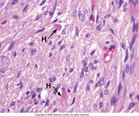
- Neurohypophyseal Hormones:
- Antidiuretic Hormone (ADH / Arginine Vasopressin [AVP]):
- Primarily synthesized by magnocellular neurons in the supraoptic nucleus (with minor contributions from the paraventricular nucleus).
- Transported to the pars nervosa and secreted directly into systemic fenestrated capillaries upon stimulation by plasma hyperosmolality or hypovolemia.
- Effector target: Epithelial cells of the renal collecting ducts and distal convoluted tubules, stimulating the insertion of aquaporin-2 water channels into the apical membrane to conserve water, concentrate urine, and restore circulating intravascular volume.
- Oxytocin (OTC):
- Primarily synthesized by neurons in the paraventricular nucleus (with minor contributions from the supraoptic nucleus).
- Released via neuroendocrine reflex arcs triggered by mechanical suckling at the teat/nipple or cervical dilation during parturition.
- Effector targets:
- Mammary gland: Binds oxytocin receptors on myoepithelial cells surrounding alveoli and alveolar ducts, stimulating cell contraction to drive alveolar milk ejection ("milk letdown").
- Uterus: Binds myometrial smooth muscle receptors during labor, eliciting powerful coordinated contractions to expel the fetus and fetal membranes.
The Pineal Gland (Epiphysis Cerebri)
Anatomical Position and Morphology:
- A small, conical, pinecone-shaped neuroendocrine evagination (epiphysis cerebri) arising from the posterior aspect of the roof of the diencephalon/third ventricle.
- Encapsulated by vascularized pia mater, with connective tissue septa penetrating inward to divide the parenchyma into indistinct lobules.
Pineal Parenchyma:
- Pinealocytes: The principal parenchymal endocrine cells, arranged in cords and clusters surrounded by extensive capillary networks.
- Possess round-to-oval euchromatic nuclei with prominent nucleoli and branched cytoplasmic processes terminating near perivascular spaces.
- Normally contain intracytoplasmic melanin pigment granules.
- Neuroglial Support Cells: Interstitial cells resembling astrocytes distributed among pinealocyte cords.
Corpora Arenacea ("Brain Sand"):
- Distinct, concentric, basophilic calcified concretions composed of calcium phosphate, calcium carbonate, and organic matrix proteins located within the pineal stroma.
- Accumulate progressively with advancing age; serve as a clear histological hallmark of pineal tissue.

- Functional Neuroendocrinology of Melatonin:
- Pinealocytes synthesize and secrete melatonin (derived from serotonin/tryptophan).
- Controlled by environmental photoperiods via a neural pathway: retina retinohypothalamic tract suprachiasmatic nucleus (SCN) of the hypothalamus thoracic spinal cord superior cervical ganglion sympathetic postganglionic noradrenergic fibers reaching pinealocytes.
- Melatonin synthesis and exocytosis are directly stimulated by darkness and inhibited by ambient light, establishing the continuous 24-hour circadian rhythm.
- Coordinates annual seasonal reproductive cycles in seasonal mammalian breeders by altering GnRH pulse frequency via melatonin receptors located in the adenohypophysis (pars tuberalis) and hypothalamus:
- Long-day breeders (e.g., horses): Reproductive estrus and breeding activity are stimulated by decreasing melatonin secretion during periods of extended day length (late spring/summer).
- Short-day breeders (e.g., sheep, goats): Reproductive estrus is stimulated by elevated melatonin levels occurring during periods of shorter daylight and prolonged darkness (autumn/winter).
The Thyroid Gland: Follicular Architecture, Biosynthesis, and Parafollicular Cells
- Gross and Microscopic Morphology:
- Composed of two lateral lobes situated adjacent to the proximal trachea and cricoid cartilage; connected across the tracheal ventral midline by an isthmus in some species.
- Lobes are subdivided by thin fibrovascular connective tissue septa into functional glandular lobules.
- The structural and functional units are spherical, closed thyroid follicles:
- The wall of each follicle consists of a simple continuous layer of follicular epithelial cells (thyrocytes) resting on an intact basal lamina.
- The follicular lumen is filled with homogeneous, viscous, eosinophilic colloid, which functions as an extracellular storage reservoir for hormone precursors.
- Cell morphology reflects functional state:
- Active (stimulated) follicles: Follicular cells become cuboidal or low-columnar, and the colloid exhibits peripheral scalloping (resorption lacunae) representing active pinocytotic uptake.
- Inactive (resting) follicles: Follicular cells flatten into a simple squamous epithelium, and follicles become distended with dense, un-resorbed colloid.

Biosynthesis and Release of Thyroid Hormones ( and ):
- Thyroglobulin Synthesis: Cuboidal follicular cells synthesize thyroglobulin, a large glycosylated protein containing numerous tyrosine residues, and exocytose it into the follicular lumen.
- Iodide Uptake and Oxidation: Circulating inorganic iodide () is actively transported into follicular cells from interstitial blood via basal sodium-iodide symporters (NIS), transferred across the apical membrane, and oxidized to active iodine () by thyroid peroxidase (TPO).
- Organification and Coupling: Iodine atoms are covalently linked to tyrosine residues of thyroglobulin, forming monoiodotyrosine (MIT) and diiodotyrosine (DIT). TPO subsequently catalyzes oxidative coupling:
- ( / thyroxine).
- ().
- Endocytosis and Proteolysis: Under TSH stimulation (acting via thyrocyte GPCRs and ), apical cytoplasmic pseudopodia internalize colloid droplets via endocytosis. Endosomes fuse with lysosomes, where lysosomal proteases hydrolyze thyroglobulin, cleaving and releasing free and .
- Systemic Transport: Active and diffuse across the basolateral membrane into sinusoidal capillaries. They circulate systemically bound to plasma carrier proteins (such as thyroxine-binding globulin [TBG], transthyretin, and albumin) or as unbound free hormones, while uncoupled MIT and DIT are deiodinated and their iodine recycled.
Intracellular Method of Action and Systemic Physiological Effects of and :
- Intracellular Mode of Action: Free and enter peripheral target cells by carrier-mediated transport and passive diffusion. Target tissues express intracellular deiodinase enzymes that convert circulating prohormone into biologically potent . Free enters the cell nucleus and binds with high affinity to thyroid hormone receptors (TRs) bound to specific Thyroid Hormone Response Elements (HREs) on promoter DNA, regulating transcription of target genes.
- Physiological Roles:
- Basal Metabolic Rate and Thermogenesis: Elevates cellular oxygen consumption, ATP turnover, and basal metabolic rate across tissues; drives non-shivering thermogenesis.
- Carbohydrate and Lipid Metabolism: Accelerates glycogenolysis, stimulates hepatic gluconeogenesis, increases intestinal hexose absorption, stimulates lipolysis in adipose tissue, and upregulates hepatic low-density lipoprotein (LDL) receptors to lower serum cholesterol.
- Protein Metabolism: Enhances structural protein synthesis at physiological levels.
- Cardiovascular Dynamics: Upregulates myocardial -adrenergic receptor density and myosin heavy chain expression, elevating cardiac contractility (inotropy), heart rate (chronotropy), and cardiac output.
- Neurological and Skeletal Growth: Essential for early postnatal development of the central nervous system; promotes oligodendrocyte myelin synthesis, axonal arborization, and neurotransmitter biosynthesis. Potentiates growth hormone and somatomedins to drive epiphyseal maturation and linear skeletal growth.
Parafollicular Cells (C Cells):
- Large, pale-staining, oval or polyhedral cells situated singly or in small clusters within the basal lamina of follicles without direct exposure to the follicular lumen, or interspersed within interfollicular stroma.
- Calcitonin Secretion: Synthesize and store calcitonin in cytoplasmic dense-core granules, exocytosing it in direct response to elevated plasma calcium () levels.
- Physiological Actions: Acts as a physiological antagonist to parathyroid hormone to lower circulating blood calcium:
- Direct osteoclast inhibition: Suppresses osteoclast motility and enzymatic bone resorption, promoting calcium deposition and mineral retention within bone.
- Renal excretion: Inhibits tubular calcium and phosphate reabsorption, increasing urinary calcium clearance.
The Parathyroid Glands: Calcium Homeostasis and Counter-Regulation
- Anatomical Arrangement Across Domestic Species:
- Mammals possess two distinct pairs of parathyroid glands, categorized anatomically based on their relationship to the thyroid:
- Internal Parathyroid Glands (Parathyroid IV): Embedded within the deeper parenchyma of the thyroid lobes.
- External Parathyroid Glands (Parathyroid III): Situated externally, outside or adjacent to the peripheral fibrous capsule of the thyroid lobes.
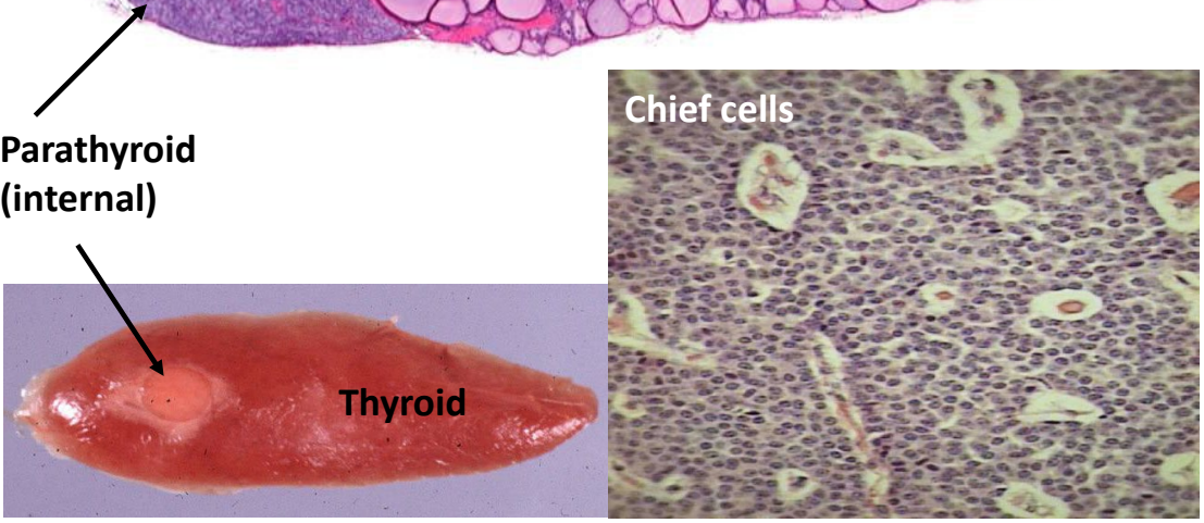
Parenchymal Cytology:
- Chief Cells (Principal Cells):
- The predominant functional parenchymal cells of the parathyroid gland, organized in dense cords, anastomosing plates, or small clusters surrounded by delicate reticular stroma and capillary plexuses.
- Display small, round, centrally placed vesicular nuclei and pale, amphophilic-to-lightly eosinophilic cytoplasm containing glycogen and secretory granules.
- Express cell-surface calcium-sensing receptors (CaSRs), which continuously monitor extracellular ionized calcium levels.
Biosynthesis and Actions of Parathormone (PTH):
- Trigger: Declining concentrations of extracellular ionized calcium () relieve CaSR inhibition, triggering immediate exocytosis of parathormone (PTH / parathyroid hormone) from chief cell granules.
- Physiological Target Actions: Acts systemically to elevate plasma calcium concentration:
- Bone Resorption: Binds surface PTH receptors on osteoblasts, upregulating receptor activator of nuclear factor- ligand (RANKL), which binds RANK on osteoclast precursors to stimulate mature osteoclast differentiation and osteolysis, mobilizing calcium and phosphate into circulation.
- Renal Reabsorption: Stimulates active calcium reabsorption in the distal convoluted tubules of the nephron while simultaneously promoting phosphate excretion (phosphaturia) in the proximal tubule.
- Intestinal Uptake: In the kidney, PTH stimulates renal mitochondrial -hydroxylase, catalyzing the conversion of -hydroxyvitamin D into active -dihydroxyvitamin D (), which enters circulation to induce calbindin and transport proteins in enterocytes, accelerating dietary calcium and phosphate absorption.
Counter-Regulatory Dual-Hormone Loop:
- Blood calcium equilibrium is preserved through the coordinated antagonism between calcitonin (from thyroid C cells) and PTH (from parathyroid chief cells):
- Elevated plasma triggers thyroid C-cell calcitonin exocytosis, suppressing bone resorption and increasing renal calcium excretion to bring levels down.
- Depressed plasma triggers parathyroid chief-cell PTH release, driving osteoclast bone resorption, renal calcium conservation, and calcitriol-mediated intestinal uptake to elevate levels back to the homeostatic baseline.
The Adrenal (Suprarenal) Glands: Cortex and Medulla
- Gross Anatomy, Vascular Supply, and Dual Embryological Origins:
- Paired retroperitoneal organs positioned craniomedial to each kidney, enclosed within an external connective tissue capsule.
- Vascular architecture: Arterial branches arising from the suprarenal and phrenic arteries form a subcapsular plexus; blood flows centripetally through cortical sinusoids into a central medullary sinusoidal network, which empties into a central medullary vein.
- Dual Embryological Origins:
- Adrenal Cortex: Derives from the coelomic intermediate mesoderm (mesodermal origin).
- Adrenal Medulla: Derives from neuroectodermal neural crest cells that migrate ventrally into the developing cortical blastema (ectodermal origin).
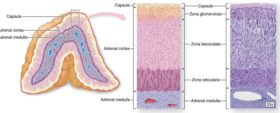
Adrenal Cortex Zonation and Steroidogenesis:
- Zona Glomerulosa (Outer Cortex):
- Formed by compact epithelial cells arranged in rounded clusters, nests, or tufts (in humans and ruminants) or in curved arcade-like arches beneath the capsule (termed the zona arcuata in equids, suids, and carnivores).
- Primary site of mineralocorticoid synthesis, specifically aldosterone and corticosterone.
- Regulated primarily by the renin-angiotensin-aldosterone system (RAAS) and extracellular potassium () levels, with minor sensitivity to ACTH.
- Aldosterone binds mineralocorticoid receptors in the distal convoluted tubules and collecting ducts of the kidneys, upregulating basolateral ATPase pumps and apical epithelial sodium channels (ENaC) to promote sodium and water reabsorption while enhancing potassium and hydrogen ion excretion, elevating intravascular blood volume and systemic arterial pressure.
- Zona Intermedia:
- A thin, demarcated transition layer composed of small, undifferentiated blastemic stem cells situated at the interface between the outer cortex (zona glomerulosa) and inner cortex (zona fasciculata).
- Functions as a mitotic zone that generates replacement parenchymal cells for both the outer and inner cortical zones.
- Zona Fasciculata (Inner Cortex):
- The thickest cortical layer, composed of spherical epithelial cells (spongiocytes) arranged in long, straight, parallel cords (fascicles) oriented perpendicular to the capsule, bordered by fenestrated sinusoids.
- Spongiocytes contain abundant cytoplasmic lipid droplets filled with cholesterol esters, rendering the cytoplasm pale, foamy, and highly vacuolated on routine H&E sections.
- Primary site of glucocorticoid synthesis, producing cortisol and cortisone.
- Regulated directly by adenohypophyseal ACTH via GPCRs, which stimulate adenylyl cyclase/ signaling to activate steroidogenic enzymes (cholesterol desmolase cascade).
- Cortisol actions: Induces hepatic gluconeogenesis, reduces peripheral glucose uptake in skeletal muscle and adipose tissue, promotes skeletal muscle protein catabolism, stimulates lipolysis, reduces gastrointestinal calcium absorption, downregulates collagen synthesis, and suppresses cell-mediated and humoral immunity by suppressing inflammatory cytokines.
- Zona Reticularis (Innermost Cortex):
- Composed of small, polyhedral cells organized in an irregular, anastomosing network of cords and plates separated by wide, irregular vascular sinusoids.
- Cells feature dense, darkly staining eosinophilic cytoplasm containing lipofuscin granules.
- Synthesizes weak androgens (sex steroids), including dehydroepiandrosterone (DHEA) and androstenedione, which can be peripherally converted to active testosterone and estrogens.
- Stimulates the development of secondary sexual characteristics, regulates auxiliary hair and body growth, and supports reproductive fertility.
Adrenal Medulla:
- Cytological Structure: Essentially a modified sympathetic ganglion lacking postganglionic axonal processes, innervated directly by preganglionic sympathetic fibers descending via splanchnic nerves.
- Chromaffin Cells (Phaeochromocytes):
- Large, ovoid, pale-staining epithelioid cells arranged in clusters and cords surrounding wide venous sinusoids.
- Contain fine cytoplasmic neurosecretory granules that oxidize upon exposure to chromium salts, forming brown dichromate precipitates (the chromaffin reaction).
- Synthesize, store, and secrete the catecholamines epinephrine (adrenaline) and norepinephrine (noradrenaline).
- Functional Differentiation:
- Norepinephrine-producing cells: Rounded epithelioid cells situated deeper within the medulla that store unmodified norepinephrine.
- Epinephrine-producing cells: Columnar cells located peripherally along medullary sinusoids (especially prominent in horses, ruminants, and pigs); contain the enzyme phenylethanolamine N-methyltransferase (PNMT), which catalyzes the methylation of norepinephrine into epinephrine.
- High local glucocorticoid concentrations reaching the medulla via cortical sinusoids upregulate PNMT transcription.
- Catecholamine Actions:
- Acute physical stress, fear, pain, or exercise triggers sympathetic preganglionic release of acetylcholine, which depolarizes chromaffin cells and elicits calcium-dependent catecholamine exocytosis.
- Catecholamines mediate the systemic "fight-or-flight" autonomic response: activate -adrenergic receptors to elevate heart rate and myocardial contractility; activate -adrenergic receptors to induce bronchodilation and muscular vasodilation; activate -adrenergic receptors to induce peripheral splanchnic vasoconstriction and elevate blood pressure; stimulate hepatic and muscular glycogenolysis and adipose lipolysis to rapidly supply circulating metabolic fuels.
The Endocrine Pancreas: Islets of Langerhans and Glucose Regulation
- Morphological Organization:
- The pancreas is a secondary endocrine organ consisting of two functionally distinct parenchymal compartments:
- Exocrine Pancreas: Constitutes approximately of the total organ mass, organized into tubuloacinar units that synthesize and secrete digestive enzymes into pancreatic ducts.
- Endocrine Pancreas: Constitutes approximately of the total organ mass, consisting of discrete, spherical or oval neurovascular aggregates termed the islets of Langerhans scattered throughout the acinar tissue.
- Islets are surrounded by a fine reticular connective tissue capsule and contain an extensive, fenestrated capillary network to facilitate immediate exchange of nutrients and hormones.

- Cell Types and Secretory Products:
- Specific islet cell lineages are differentiated using specialized immunohistochemical (IHC) antibody staining:
- Beta () Cells:
- Constitute the most abundant cell population ( of islet tissue).
- Synthesize, store, and secrete insulin.
- Senses elevations in extracellular plasma glucose through facilitated GLUT2 transporter uptake and glucokinase phosphorylation, triggering ATP generation, closure of ATP-sensitive potassium channels (), membrane depolarization, calcium influx, and insulin exocytosis.
- Physiological target actions: Lowers blood glucose by binding receptor tyrosine kinases on target tissues:
- Liver: Stimulates glycogen synthesis, accelerates glycolysis, enhances lipid accumulation, suppresses gluconeogenesis and glycogenolysis.
- Skeletal Muscle: Drives GLUT4 transporter translocation to the membrane to enhance glucose uptake, accelerates glycogen synthesis, promotes amino acid uptake and protein synthesis (increasing muscle mass).
- Adipose Tissue: Drives glucose uptake, stimulates lipogenesis, inhibits hormone-sensitive lipase (suppressing lipolysis), reduces systemic inflammation, and promotes an anti-inflammatory macrophage phenotypic shift.
- Alpha () Cells:
- Constitute approximately of islet tissue, generally located along the periphery of the islet.
- Synthesize, store, and secrete glucagon.
- Exocytosis is triggered by hypoglycemia, gastric inhibitory polypeptide (GIP), and elevated circulating amino acids; exocytosis is inhibited by insulin and hyperglycemia.
- Physiological target actions: Elevates blood glucose and energy availability:
- Liver: Promotes hepatic glycogenolysis, drives gluconeogenesis, stimulates amino acid catabolism, stimulates lipolysis and mitochondrial ketone body synthesis.
- Adipose Tissue: Enhances lipolysis and resting energy expenditure (including in brown adipose tissue).
- Brain: Interacts with central satiety networks to decrease food intake and appetite while increasing satiety.
- Heart: Increases myocardial contractility (inotropy) and elevates heart rate (chronotropy).
- Pancreas: Provides local paracrine stimulation for insulin secretion.
- Delta () Cells:
- Constitute approximately of islet tissue.
- Synthesize and secrete somatostatin.
- Functions as a broad paracrine inhibitor that suppresses neighboring and cell secretion, while also inhibiting exocrine pancreatic enzyme output, gastrointestinal motility, and gallbladder contraction.
- F Cells (PP Cells):
- Minor islet cell population synthesizing pancreatic polypeptide.
- Plays modulatory roles in gastrointestinal motility and pancreatic exocrine secretion; specific functions remain largely obscure.
Secondary Endocrine Organs and Tissues
Gastrointestinal Tract (Enteroendocrine System):
- Composed of single, enteroendocrine cells dispersed throughout the gastric and intestinal mucosal epithelium, formerly known as diffuse neuroendocrine system (DNES) cells, enterochromaffin (EC) cells, enterochromaffin-like (ECL) cells, or amine precursor uptake and decarboxylation (APUD) cells.
- Gastrin:
- Synthesized by G cells in the pyloric antrum of the stomach and duodenum.
- Stimulated by luminal amino acids, gastric distention, and vagal gastrin-releasing peptide (GRP).
- Directly stimulates gastric parietal cells and promotes histamine release from ECL cells to drive hydrochloric acid () and gastric juice secretion.
- Ghrelin:
- Known as the "hunger hormone"; synthesized primarily by neuroendocrine P/D1 cells in the stomach fundus.
- Secretion peaks during fasting and states of negative energy balance; acts centrally on hypothalamic arcuate neurons to stimulate appetite, trigger hunger sensations, and promote growth hormone secretion.
- Cholecystokinin (CCK):
- Secreted by I cells in the duodenum and jejunum in response to luminal free fatty acids and amino acids.
- Triggers gallbladder contraction, stimulates exocrine pancreatic enzyme secretion, delays gastric emptying, and induces postprandial satiety.
Adipose Tissue:
- Unilocular white adipocytes function as an active endocrine organ in addition to storing neutral triacylglycerols.
- Synthesize and secrete leptin into systemic circulation in direct proportion to total body lipid mass.
- Leptin crosses the blood-brain barrier and binds cytokine-family leptin receptors on neurons of the hypothalamic "lipostat" in the arcuate nucleus.
- Suppresses appetite and inhibits food intake by downregulating orexigenic neuropeptides (neuropeptide Y [NPY] and agouti-related peptide [AgRP]) while upregulating anorexigenic signaling (-MSH), maintaining long-term energy homeostasis.
The Heart as an Endocrine Organ:
- Atrial myocardial cells synthesize, store, and secrete Atrial Natriuretic Peptide (ANP) in response to atrial wall stretch caused by hypervolemia, elevated venous return, or increased arterial blood pressure.
- Effector target: Epithelial cells of the distal convoluted tubules and collecting ducts of the kidneys.
- Physiological actions: Directly promotes natriuresis (sodium excretion) and diuresis (water loss) while relaxing vascular smooth muscle, counteracting ADH and the RAAS to decrease effective circulating volume and lower arterial blood pressure.
The Kidneys as an Endocrine Organ:
- Renin:
- Synthesized, stored, and secreted by specialized myoepithelial juxtaglomerular (JG) cells situated in the wall of the afferent glomerular arteriole.
- Released in response to renal hypoperfusion, decreased transmural stretch, reduced sodium chloride () delivery to the macula densa, or sympathetic -adrenergic stimulation.
- Acts enzymatically as an aspartyl protease, cleaving circulating hepatic angiotensinogen to yield the inactive decapeptide angiotensin I, the initiating rate-limiting step of the Renin-Angiotensin-Aldosterone System (RAAS).
- Erythropoietin (EPO):
- Synthesized and secreted by specialized peritubular interstitial fibroblastic cells situated in the deep renal cortex and outer medulla.
- Exocytosis is triggered by tissue hypoxia detected via oxygen-dependent prolyl hydroxylase and hypoxia-inducible factor () pathways.
- Functions as an essential hematopoietic cytokine, circulating to the red bone marrow to bind erythropoietin receptors on colony-forming unit-erythroid (CFU-E) progenitors, rescuing them from apoptosis and accelerating erythropoiesis to expand systemic red blood cell mass.
Endocrine Pathologies and Clinical Syndromes
Hyperadrenocorticism (Cushing\'s Disease):
- Results from prolonged systemic overproduction of glucocorticoids (cortisol excess).
- Pathogenesis involves either ACTH-secreting adenomas of the anterior pituitary (pituitary-dependent Cushing\'s) or autonomous functional adrenocortical adenomas/carcinomas of the zona fasciculata.
- Manifests with bilateral symmetric alopecia, muscle wasting, abdominal pendulous enlargement (potbelly), polyuria, polydipsia, and secondary diabetes mellitus.
Hypoadrenocorticism (Addison\'s Disease):
- Characterized by idiopathic immune-mediated atrophy, destruction, or necrosis of the adrenal cortex, resulting in deficient secretion of both glucocorticoids (cortisol) and mineralocorticoids (aldosterone).
- Mineralocorticoid deficiency disrupts renal electrolyte handling, precipitating severe hyponatremia, hyperkalemia, progressive hypovolemia, dehydration, hypotension, and circulatory collapse.
Adrenal-Associated Endocrinopathy (AAE):
- Arises from functional nodular hyperplasia or neoplasia involving the zona reticularis, resulting in uncontrolled overproduction of circulating androgens (sex steroids).
- Leads to alopecia, vulvar or prostatic enlargement, and aberrant sexual behavior (frequently recognized in domestic ferrets).
Hyperaldosteronism (Conn\'s Syndrome):
- Caused by autonomous, excessive secretion of aldosterone resulting from functional adenomas or bilateral hyperplasia of the zona glomerulosa; commonly diagnosed in cats.
- Manifests with marked renal potassium wasting, causing profound hypokalemia, muscle weakness, systemic hypertension, and cervical ventroflexion.

Hyperthyroidism:
- Functional autonomy or multinodular adenomatous hyperplasia of thyroid follicular cells leading to excessive secretion of active and .
- Highly prevalent in aging domestic felines; causes progressive weight loss despite voracious polyphagia, tachycardia, hypertrophic cardiomyopathy, hyperactivity, and poor coat condition.
Hypothyroidism:
- Inadequate circulating levels of active thyroid hormones ( and ) resulting from immune-mediated lymphocytic thyroiditis or idiopathic follicular atrophy; most frequently diagnosed in domestic canines.
- Clinically characterized by a depressed basal metabolic rate, lethargy, mental dullness, unexplained weight gain without increased caloric intake, bilaterally symmetrical non-pruritic endocrine alopecia, and hyperkeratosis.
Endocrine Pancreatic Pathology & Diabetes Mellitus:
- Hypoinsulinemia and Type 1 Diabetes Mellitus:
- Results from progressive immune-mediated destruction or extensive loss of insulin-producing pancreatic cells in the islets of Langerhans.
- The resulting absolute insulin deficiency prevents cellular glucose uptake, precipitating marked persistent hyperglycemia, glycosuria, osmotic diuresis, polyuria, polydipsia, weight loss, and life-threatening diabetic ketoacidosis.
Pituitary Pathology & Diabetes Insipidus:
- Central Diabetes Insipidus:
- Results from trauma, compression, or neoplastic destruction of the supraoptic/paraventricular hypothalamic nuclei, the infundibular stalk, or the pars nervosa of the hypophysis.
- Impairs synthesis or secretion of antidiuretic hormone (ADH), preventing aquaporin-2 translocation in the collecting ducts and causing profound polyuria with hyposthenuric urine and secondary compensatory polydipsia.
Endocrine Gland Neoplasia:
- Autonomous neoplasms can arise in any primary or secondary endocrine tissue (e.g., pituitary adenomas, thyroid adenomas and carcinomas, parathyroid adenomas, adrenocortical adenomas/carcinomas, medullary pheochromocytomas, pancreatic insulinomas, gastrinomas).
- Neoplasms are classified clinically as functional (manifesting as clinical syndromes of hormone excess) or non-functional (manifesting through local tissue compression and destruction of adjacent parenchymal structures).
Questions and Discussion
Question 1:
- Prompt: Which of the following hormones is stored by the neurohypophysis?
- A) CRH
- B) PRF
- C) TRH
- D) OTC
- Response: The correct answer is D) OTC (Oxytocin).
- Explanation: Oxytocin (OTC) and antidiuretic hormone (ADH) are synthesized within the neuronal perikarya of the hypothalamus, transported down unmyelinated axons, and stored within Herring bodies in the neurohypophysis (pars nervosa) for direct vascular release. In contrast, corticotropin-releasing hormone (CRH), prolactin-releasing factor (PRF), and thyrotropin-releasing hormone (TRH) are releasing hormones produced by the hypothalamus that are transported through the hypophyseal portal system to act on parenchymal cells of the adenohypophysis.
Question 2:
- Prompt: Which -troph will synthesize GH (Growth Hormone)?
- A) Somatotrophs
- B) Lactotrophs
- C) Gonadotrophs
- D) Corticotrophs
- Response: The correct answer is A) Somatotrophs.
- Explanation: Somatotrophs are acidophilic parenchymal cells concentrated laterally within the pars distalis that synthesize and secrete growth hormone (GH). Lactotrophs produce prolactin (PRL), gonadotrophs synthesize follicle-stimulating hormone (FSH) and luteinizing hormone (LH), and corticotrophs synthesize adrenocorticotropic hormone (ACTH).
Question 3:
- Prompt: Which layer of the pituitary gland produces POMC?
- A) Pars distalis
- B) Pars intermedia
- C) Pars tuberalis
- D) Pars nervosa
- Response: The correct answer is B) Pars intermedia.
- Explanation: The pars intermedia contains melanotrophs that synthesize proopiomelanocortin (POMC), which is post-translationally cleaved into -melanocyte-stimulating hormone (-MSH), lipotropin, and endorphins. The pars distalis contains standard acidophils and basophils, the pars tuberalis surrounds the infundibular stalk and expresses melatonin receptors, and the pars nervosa functions as a storage site for ADH and oxytocin.
Question 4:
- Prompt: Where is this structure located? (Showing a high-power histological field containing a concentric calcified body surrounded by parenchymal cells)
- A) Thyroid gland
- B) Parathyroid gland
- C) Pituitary gland
- D) Pineal gland
- Response: The correct answer is D) Pineal gland.
- Explanation: The depicted structure is a corpus arenaceum ("brain sand"), a lamellated calcification pathognomonic for the pineal gland that accumulates progressively with age amidst pinealocytes and neuroglial fibers.
Question 5:
- Prompt: Which hormone can be made in the absence of iodine?
- A) TSH
- B) Thyroxine
- C) Triiodothyronine
- D)
- Response: The correct answer is A) TSH.
- Explanation: Thyroxine () and triiodothyronine () are iodinated tyrosine derivatives that require covalent bonding of oxidized iodine atoms to thyroglobulin within the thyroid follicle. Thyroid-stimulating hormone (TSH) is an adenohypophyseal glycoprotein hormone whose peptide synthesis by basophilic thyrotrophs does not incorporate iodine.
Question 6:
- Prompt: Which cells will respond to increased plasma calcium?
- A) Pinealocytes
- B) Parafollicular cells
- C) Chief cells
- D) Phaeochromocytes
- Response: The correct answer is B) Parafollicular cells.
- Explanation: Parafollicular cells (C cells) of the thyroid gland detect hypercalcemia and respond by secreting calcitonin, which lowers circulating calcium by suppressing osteoclastic bone resorption and enhancing renal calcium excretion. Pinealocytes secrete melatonin in response to darkness; parathyroid chief cells secrete PTH in response to decreased plasma calcium; and medullary phaeochromocytes (chromaffin cells) release catecholamines in response to sympathetic neural stimulation.
Question 7:
- Prompt: If there is an issue producing mineralocorticoids, which layer of the adrenal gland is affected?
- A) Zona glomerulosa
- B) Zona fasciculata
- C) Zona reticularis
- D) Adrenal medulla
- Response: The correct answer is A) Zona glomerulosa.
- Explanation: The zona glomerulosa (or zona arcuata in certain domestic species) forms the outer cortex and specifically synthesizes mineralocorticoids, principally aldosterone. The zona fasciculata produces glucocorticoids (cortisol), the zona reticularis synthesizes androgens, and the adrenal medulla secretes catecholamines.
Question 8:
- Prompt: Which layer of the adrenal gland will produce cortisol?
- A) Zona glomerulosa
- B) Zona fasciculata
- C) Zona reticularis
- D) Medulla
- Response: The correct answer is B) Zona fasciculata.
- Explanation: Cortisol is a glucocorticoid synthesized by the lipid-rich spongiocytes that form the parallel cords of the zona fasciculata under the regulatory influence of ACTH.
Question 9:
- Prompt: Which hormone directly counteracts the effects of ADH?
- A) Erythropoietin
- B) ACTH
- C) Renin
- D) ANP
- Response: The correct answer is D) ANP.
- Explanation: Antidiuretic hormone (ADH) promotes water retention and renal vascular fluid conservation via collecting duct aquaporin-2 channel insertion. Atrial natriuretic peptide (ANP), released by atrial myocytes during hypervolemia, directly counteracts these fluid-retaining effects by promoting renal sodium excretion (natriuresis) and water loss (diuresis) to decrease circulating blood pressure. Erythropoietin stimulates erythropoiesis; ACTH stimulates glucocorticoid production; and renin activates the RAAS to raise blood pressure and conserve fluid.
Question 10:
- Prompt: If you have damage to the zona fasciculata, which disease is likely to occur?
- A) Cushing\'s disease
- B) Conn\'s disease
- C) Adrenal-associated endocrinopathy
- D) Addison\'s disease
- Response: The correct answer is D) Addison\'s disease.
- Explanation: Primary hypoadrenocorticism (Addison\'s disease) results from destructive lesions or atrophy of the adrenal cortex, specifically compromising the zona fasciculata and zona glomerulosa, leading to glucocorticoid (cortisol) and mineralocorticoid deficiencies. Cushing\'s disease involves cortisol excess; Conn\'s syndrome involves aldosterone excess stemming from zona glomerulosa lesions; and adrenal-associated endocrinopathy involves androgen excess driven by pathology of the zona reticularis.