PROTOCOLS
THYROID:
Transverse Thyroid (comparison)
Grayscale
Color
Transverse Isthmus
Grayscale (decrease depth)
Measure AP
Transverse Thyroid Right Lobe Superior in Grayscale
Transverse Thyroid Right Lobe Mid
Grayscale
Measure width and AP
Color
Transverse Thyroid Right Lobe Inferior in Grayscale
Long Thyroid Right Lobe Lateral in Grayscale
Long Thyroid Right Lobe Mid
Grayscale
Measure length and AP
Color
Long Thyroid Right Lobe Medial in Grayscale
Long Right Lateral Neck in Grayscale
Transverse Thyroid Left Lobe Superior in Grayscale
Transverse Thyroid Left Lobe Mid
Grayscale
Measure width and AP
Color
Transverse Thyroid Left Lobe Inferior in Grayscale
Long Thyroid Left Lobe Lateral in Grayscale
Long Thyroid Left Lobe Mid
Grayscale
Measure length and AP
Color
Long Thyroid Left Lobe Medial in Grayscale
Long Left Lateral Neck in Grayscale
THYROID NOTES:
RUSH radiologist prefers for mid to be split screen with long mid and transverse mid
Split screen should include measurements for BOTH planes
SCROTUM:
Transverse Scrotum (comparison)
Grayscale
Color
Transverse Right Testicle Superior in Grayscale
Transverse Right Testicle Mid
Grayscale
Measure width
Color
Transverse Right Testicle Inferior in Grayscale
Long Right Sac
Long Right Testicle Lateral in Grayscale
Long Right Testicle Mid
Grayscale
Measure length and AP
Color
PW
Long Right Testicle Medial in Grayscale
Transverse Right Epi Head
Grayscale
Measure width
Long Right Epi Head
Grayscale
Measure length and AP
Color
Transverse Left Testicle Superior in Grayscale
Transverse Left Testicle Mid
Grayscale
Measure width
Color
Transverse Left Testicle Inferior in Grayscale
Long Left Sac
Long Left Testicle Lateral in Grayscale
Long Left Testicle Mid
Grayscale
Measure length and AP
Color
PW
Long Left Testicle Medial in Grayscale
Transverse Left Epi Head
Grayscale
Measure width
Long Left Epi Head
Grayscale
Measure length and AP
Color
SCROTUM NOTES:
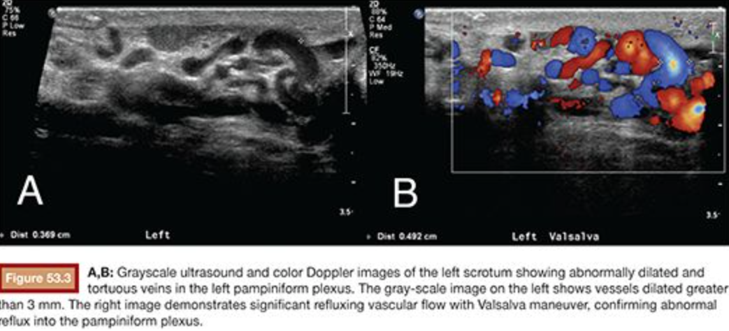
Valsalva maneuver is used to identify enlarged veins or a varicocele and is performed by asking a patient to
Take a breath
Pinch nose and close mouth
Forcefully exhale while bearing down
Hold
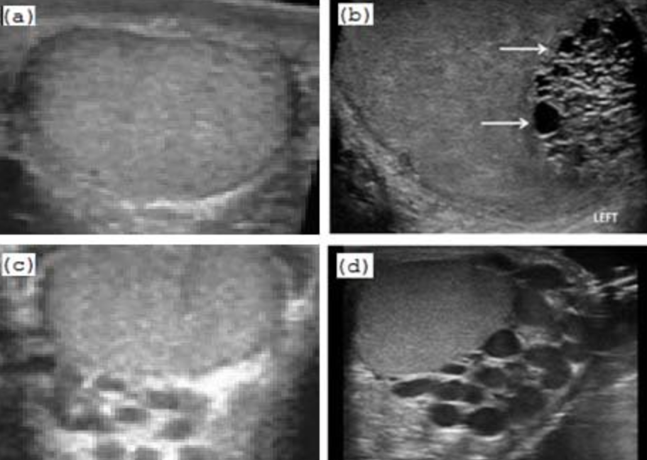
Varicocele is identified if vessel measures larger than 3 mm and is a normal finding
BREAST:
Anti-Radial
Grayscale with chest wall
Grayscale with decreased depth focused on mass
Measure width of mass
Color
Radial
Grayscale
Measure length and height of mass
Color
RUQ:
Transverse Pancreas (1-2 images) showing as much pancreas as possible
Grayscale
Color
Long Left Liver in Grayscale (3-4 images) sweeping through including landmarks
Dorito
Aorta
IVC
Caudate
Transverse Left Liver in Grayscale (3-4 images) sweeping through stopping at pancreas
Long Right Liver in Grayscale (6-8 images)
Liver and Right Kidney Interface
Measure length
Color and PW of MPV
Transverse Right Liver in Grayscale (6-8 images) starting at dome and ending at kidney
Color of Hepatic Veins
CBD
Grayscale
Measure inner to inner
Color
Long GB in Grayscale (3-4 images)
Transverse GB in Grayscale (3-4 images
Long GB LLD in Grayscale (3-4 images)
Transverse GB LLD in Grayscale (3-4 images)
Measure GB wall
Long Right Kidney Lateral in Grayscale
Long Right Kidney Mid
Grayscale
Measure length
Color
Long Right Kidney Medial in Grayscale
Transverse Right Kidney Superior in Grayscale
Transverse Right Kidney Mid
Grayscale
Measure height and width
Color
Transverse Right Kidney Inferior in Grayscale
COMPLETE ABDOMEN:
Long Aorta Proximal
Grayscale
Measure outer to outer
Long Aorta Mid in Grayscale
Grayscale
Measure outer to outer
Color
PW
Long Aorta Distal in Grayscale
Grayscale
Measure outer to outer
Long IVC
Grayscale
Color
Transverse Pancreas (1-2 images)
Grayscale
Color
Long Left Liver in Grayscale (3-4 images) sweeping through including landmarks
Dorito
Aorta
IVC
Caudate
Transverse Left Liver in Grayscale (3-4 images) sweeping through stopping at pancreas
Long Right Liver in Grayscale (6-8 images)
Liver and Right Kidney Interface
Measure length
Color and PW of MPV
Transverse Right Liver in Grayscale (6-8 images) starting at dome and ending at kidney
Color of Hepatic Veins
Make sure to include and label each hepatic vein in color
RHV
MHV
LHV
Patients with HCC on indication: SWITCH TO LINEAR TRANSDUCER Right Liver Surface
Patients with HCC on indication: SWITCH TO LINEAR TRANSDUCER Left Liver Surface
Long Right Kidney Lateral in Grayscale
Long Right Kidney Mid
Grayscale
Measure length
Color
Long Right Kidney Medial in Grayscale
Transverse Right Kidney Superior in Grayscale
Transverse Right Kidney Mid
Grayscale
Measure height and width
Color
Transverse Right Kidney Inferior in Grayscale
Long GB in Grayscale (3-4 images)
Transverse GB in Grayscale (3-4 images)
Measure GB wall
Long GB LLD in Grayscale (3-4 images)
Transverse GB LLD in Grayscale (3-4 images)
CBD
Grayscale
Measure inner to inner
Color
Long Left Kidney Lateral in Grayscale
Long Left Kidney Mid
Grayscale
Measure length
Color
Long Left Kidney Medial in Grayscale
Transverse Left Kidney Superior in Grayscale
Transverse Left Kidney Mid
Grayscale
Measure height and width
Color
Transverse Left Kidney Inferior in Grayscale
Long Spleen
Spleen and Left Kidney Interface
Grayscale
Measure
Color
Transverse Spleen
Grayscale
Color
COMPLETE ABDOMEN NOTES:
RUSH does not PW on MPV unless specified by order
RUSH does not measure kidneys in transverse
ABDOMINAL DOPPLER:
Aorta
Grayscale
Measure
Color
PW
Celiac Artery
Grayscale
Color
PW
Superior Mesenteric Artery (SMA)
Grayscale
Color
PW
Hepatic Artery
Grayscale
Color
PW
Splenic Artery
Grayscale
Color
PW
Hepatic Veins including Right, Middle, and Left
Grayscale
Color
PW
Portal Veins including Right, Left, and Main
Grayscale
Color
PW
RENAL:
Long Right Kidney L-M (sweep) in Grayscale
Long Right Kidney Mid
Grayscale
Measure length
Color
Transverse Right Kidney S-I (sweep) in Grayscale
Transverse Right Kidney Mid
Grayscale
Measure height and width
Color
Long Left Kidney L-M (sweep) in Grayscale
Long Left Kidney Mid
Grayscale
Measure length
Color
Transverse Left Kidney S-I (sweep) in Grayscale
Transverse Left Kidney Mid
Grayscale
Measure height and width
Color
Transverse Bladder S-I (sweep) in Grayscale
Transverse Bladder Mid
Grayscale
Measure
Transverse Bladder Jets in Color
Long Bladder ML-RT (sweep) in Grayscale
Long Bladder ML-LT (sweep) in Grayscale
Pre-Void Bladder SPLIT SCREEN
Transverse bladder: Measure width
Long bladder: Measure length and height
Post-Void Bladder SPLIT SCREEN
Transverse bladder: Measure width
Long bladder: Measure length and height
RENAL DOPPLER (RUSH PROTOCOL):
Aorta
Grayscale
Color
PW
Renal Artery Origin
Grayscale
Color
PW
Renal Artery Proximal
Grayscale
Color
PW
Renal Artery Mid
Grayscale
Color
PW
Renal Artery Distal/Hilum
Grayscale
Color
PW
Renal Vein at Kidney Hilum or Origin
Grayscale
Color
PW
Arcuate Artery Superior
Grayscale
Color
PW
Arcuate Artery Mid
Grayscale
Color
PW
Arcuate Artery Inferior
Grayscale
Color
PW
Repeat on other kidney
RENAL DOPPLER NOTES:
Native kidneys: More concerned with peak systole (PS)
PS > 180 cm/sec = STENOSIS
Transplant kidneys: More concerned with resistive index (RI)
RI > 0.7 = STENOSIS
APPENDIX (RUSH PROTOCOL):
Linear probe: SPLIT SCREEN of Transverse RLQ & W/CMP (with compression)
Physically move transducer from iliacs (lateral) to belly-button or bladder (medial)
Image at each new location in Grayscale
Color on iliacs
Measure appendix width IF visible
Color appendix IF visible
Linear probe: SPLIT SCREEN of Long RLQ & W/CMP (with compression)
Physically move transducer from iliacs (lateral) to belly-button or bladder (medial)
Image at each new location in Grayscale
Color on iliacs
Measure appendix length and AP IF visible
Color appendix IF visible
Curved probe: Long RUQ to check for fluid near liver
Curved probe: Transverse RUQ to check for fluid near kidneys
Image kidneys in grayscale to check for hydronephrosis
Image kidneys in color to check for stones
APPENDIX NOTES:
Those with appendicitis will have REBOUND PAIN
Normal patients: Pain upon compression or pressing down
Appendicitis: Pain after relieving compression or stopping pressure
Report: MUST STATE THAT APPENDIX IS NOT VISUALIZED, NOT THAT PATIENT IS NEGATIVE FOR APPENDICITIS
Appendix not visualized due to overlying bowel gas
No free fluid in abdomen
AORTA/IVC:
Long Aorta Proximal (Supraclavicular)
Grayscale
Measure (outer to outer)
Color
PW (calipers on PS and ED)
Transverse Aorta Proximal (Supraclavicular)
Grayscale
Measure (outer to outer)
Color
Long Aorta Mid (Juxtarenal)
Grayscale
Measure (outer to outer)
Color
PW (calipers on PS)
Transverse Aorta Mid (Juxtarenal)
Grayscale
Measure (outer to outer)
Color
Long Aorta Distal (Infrarenal)
Grayscale
Measure (outer to outer)
Color
PW (calipers on PS)
Transverse Aorta Distal (Infrarenal)
Grayscale
Measure (outer to outer)
Color
Bifurcation
Grayscale
Color
PW
Long Right Common Iliac
Grayscale
Measure (outer to outer)
Color
PW (calipers on PS)
Transverse Right Common Iliac
Grayscale
Measure (outer to outer)
Color
Long Left Common Iliac
Grayscale
Measure (outer to outer)
Color
PW (calipers on PS)
Transverse Left Common Iliac
Grayscale
Measure (outer to outer)
Color
FEMALE ABDOMINAL PELVIS:
Long Midline (ML) Uterus that includes endometrium, uterus, cervix
Grayscale
Measure length and AP
Color
Long Midline Endometrium
Grayscale
Measure
Long Right Uterus that includes uterus, cervix in Grayscale
Right Midline (ML) Sweep or Right Lateral Pelvis in Grayscale
Long Left Uterus that includes uterus, cervix in Grayscale
Left Midline (ML) Sweep or Left Lateral Pelvis in Grayscale
Transverse vagina (inferior to cervix) in Grayscale
Transverse cervix in Grayscale
Transverse Inferior Uterus in Grayscale
Transverse Mid Uterus
Grayscale
Measure width
Transverse Superior Uterus in Grayscale
Transverse Superior Pelvis that includes bladder in Grayscale
Transverse Right Ovary
Grayscale
Measure length and width
Color
Arterial PW
Venous PW
Long Right Ovary
Grayscale
Measure AP
Color
Arterial PW
Venous PW
Transverse Right Adnexa that includes right side of uterus, ovary, pelvis, in Grayscale
Long Right Adnexa that DOES NOT include right side of uterus, ovary in Grayscale
Transverse Left Ovary
Grayscale
Measure length and width
Color
Arterial PW
Venous PW
Long Left Ovary
Grayscale
Measure AP
Color
Arterial PW
Venous PW
Transverse Left Adnexa that includes left side of uterus, ovary, pelvis in Grayscale
Long Left Adnexa that DOES NOT include left side of uterus, ovary in Grayscale
PARACENTESIS:
1-2 images in grayscale RUQ
1-2 images in grayscale RLQ
1-2 images in grayscale LUQ
1-2 images in grayscale LLQ
PARACENTESIS NOTES:
Normal lab values include
Platelet count > 50
INR/PT (prothrombin time) < 1.5