Abdomen VI
At which vertebral level does the abdominal aorta split into the left and right common iliac arteries?
A) T7
B) T12
C) L4
D) S1
E) S4
2) Which of the following is a paired branch of the abdominal aorta?
A) celiac artery
B) renal artery
C) superior mesenteric artery
D) inferior mesenteric artery
E) median sacral artery
3) An unpaired artery that branches from the abdominal aorta includes the ________.
A) suprarenal artery
B) superior mesenteric artery
C) phrenic artery
D) gonadal artery
E) lumbar artery
4) Which of the following is the right branch of the celiac trunk?
A) cystic artery
B) splenic artery
C) superior mesenteric artery
D) common hepatic artery
E) gastroduodenal artery
5) Which gastric secretory cells are known as "zymogen cells," and are most abundant near the back of gastric glands?
A) mucous neck cells
B) parietal cells
C) oxyntic cells
D) chief cells
E) enteroendocrine cells
6) Parietal cells, chief cells, and G cells dominate which structure?
A) gastric pits
B) serosa
C) rugae
D) villi
E) gastric glands
7) Which portion of the stomach is connected to the duodenum?
A) cardia
B) body
C) fundus
D) cardiac orifice
E) pyloric canal
The stomach mucosa contains numerous shallow depressions called ________.
A) gastric cardia
B) gastric pits
C) gastric glands
D) intestinal glands
E) pyloric glands
9) The inferior third of the esophageal muscularis externa consists of which tissue?
A) stratified nonkeratinized squamous epithelial tissue
B) skeletal muscle and adipose connective tissue
C) an even mixture of skeletal and smooth muscle cells
D) only smooth muscle
E) only skeletal muscle
10) The muscularis externa of the large intestine differs from that of other intestinal regions because the longitudinal layer has been reduced to the muscular bands of the ________.
A) omental appendices
B) haustra
C) rugae
D) taenia coli
E) plicae circulares
11) The mucosa of the esophagus contains an abrasion-resistant ________.
A) inner circular and outer longitudinal smooth muscle layers
B) simple, branched tubular glands
C) nonkeratinized stratified squamous epithelium
D) pseudostratified ciliated columnar epithelium
E) connective tissue
12) The ________ is the region of the stomach, which extends to the entrance to the duodenum and whose muscular sphincter regulates the passage of materials into the duodenum.
A) pylorus
B) body
C) cardia
D) antrum
E) ileum
13) The ________ muscle is divided longitudinally by a median collagenous partition called the linea alba.
A) rectus abdominis
B) internal oblique
C) transversus abdominis
D) diaphragm
E) external oblique
14) Of the following muscles, which can compress the abdomen?
A) internal oblique
B) external oblique
C) transversus abdominis
D) rectus abdominis
E) All of the answers are correct.
The deepest lateral layer of the abdominal muscles is the ________.
A) transversus abdominis
B) rectus abdominis
C) internal oblique
D) external oblique
E) psoas major
16) The ascending limb travels in the medulla toward the renal pelvis, and the descending limb returns toward the cortex. FALSE
17) Which of the following are cells found between the endothelial cells of adjacent capillaries that play a role in regulating glomerular blood flow and filtration?
A) filtration slits
B) mesangial cells
C) podocytes
D) fenestrated cells
E) pyramidal cells
18) Which of the following are described as the gaps between the pedicles of the podocytes?
A) urachus
B) glomeruli
C) filtration slits
D) convoluted tubules
E) renal papillae
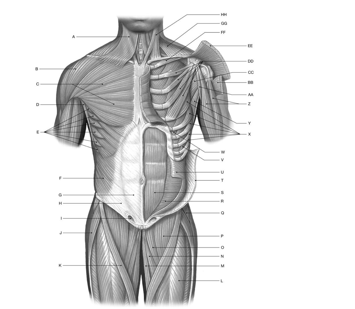
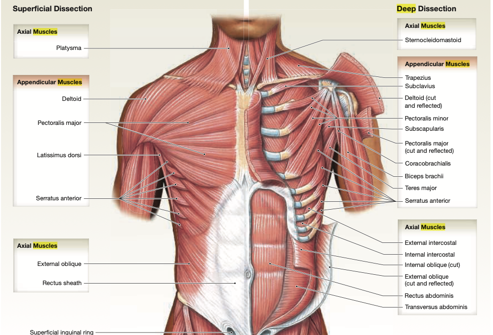
Identify the structure(s) indicated by Label F.
A) Rectus abdominis
B) Serratus anterior
C) External oblique
D) Transversus abdominis
E) Internal oblique
20) Identify the structure(s) indicated by Label G.
A) Serratus anterior
B) External oblique
C) Platysma
D) Rectus sheath
E) Internal oblique
21) Identify the structure(s) indicated by Label S.
A) External oblique
B) Transversus abdominis
C) Rectus abdominis
D) Serratus anterior
E) Internal oblique
22) Identify the structure(s) indicated by Label U.
A) Serratus anterior
B) Rectus abdominis
C) External oblique
D) Internal oblique
E) Iliopsoas
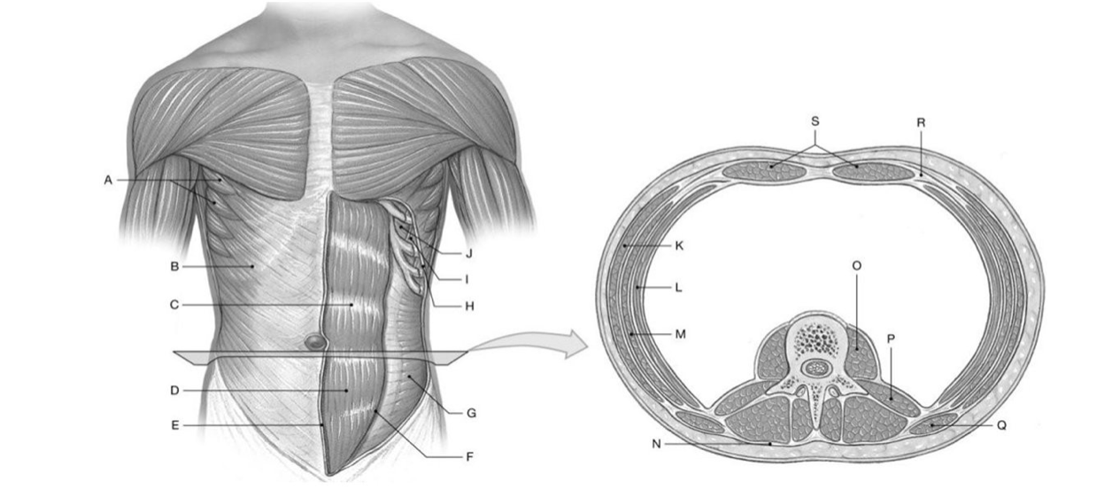
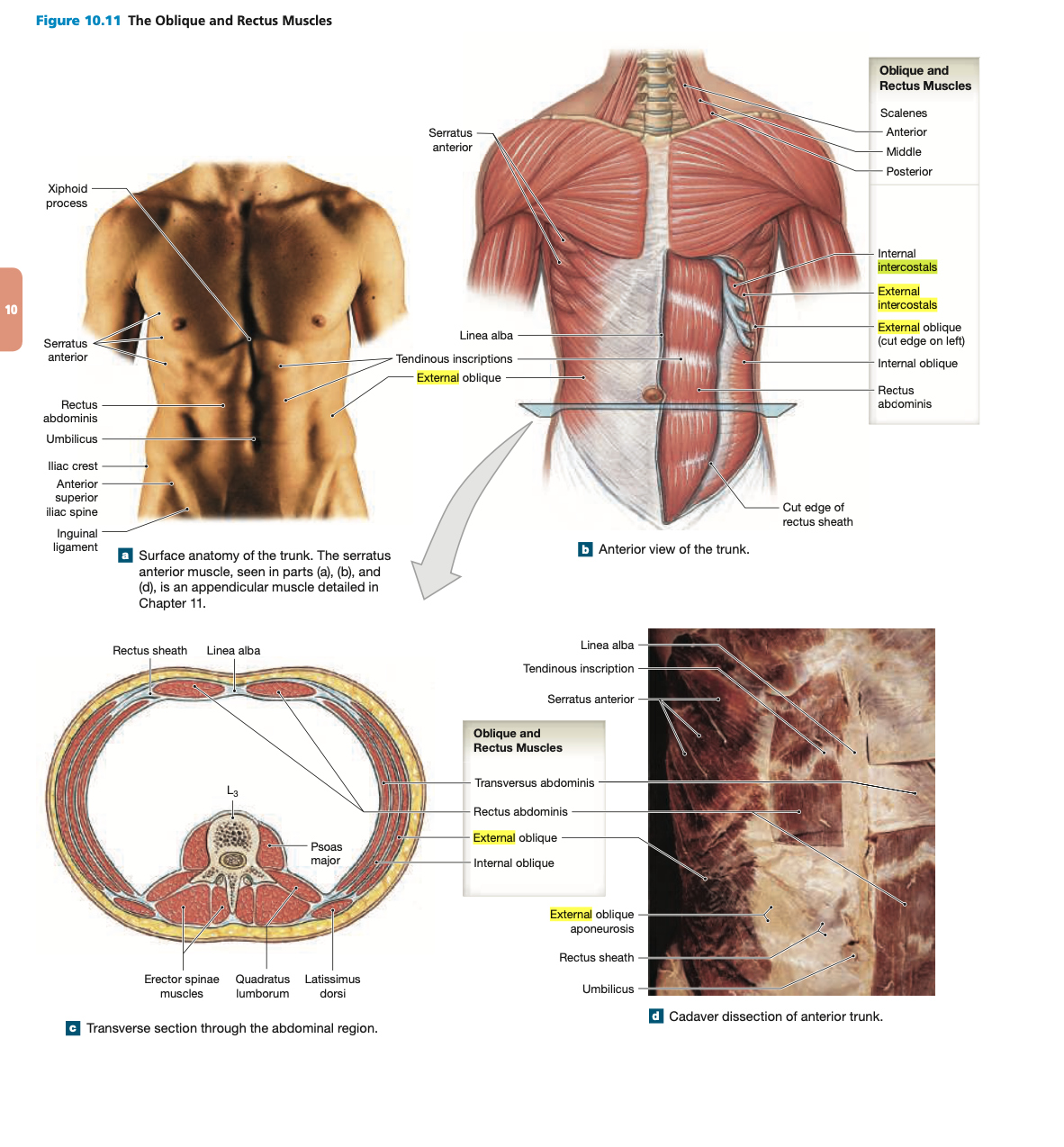
Identify the structure(s) indicated by Label B.
A) Internal oblique
B) External intercostals
C) External oblique
D) Rectus abdominis
E) Internal intercostals
24) Identify the structure(s) indicated by Label D.
A) Rectus abdominis
B) Internal oblique
C) Transversus abdominis
D) External oblique
E) Linea alba
25) Identify the structure(s) indicated by Label E.
A) Serratus anterior
B) Tendinous inscriptions
C) Umbilicus
D) External oblique aponeurosis
E) Linea alba
26) Identify the structure(s) indicated by Label G.
A) Rectus abdominis
B) External oblique
C) Transversus abdominis
D) Internal intercostals
E) Internal oblique
27) Identify the structure(s) indicated by Label H.
A) External intercostals
B) Rectus abdominis
C) Internal intercostals
D) External oblique
E) Internal oblique
28) Identify the structure(s) indicated by Label K.
A) External oblique
B) Rectus abdominis
C) Internal oblique
D) Transversus abdominis
E) Internal intercostals
Identify the structure(s) indicated by Label L.
A) Internal oblique
B) External oblique
C) Transversus abdominis
D) Rectus abdominis
E) Quadratus lumborum
30) Identify the structure(s) indicated by Label M.
A) External oblique
B) Rectus sheath
C) Internal oblique
D) Transversus abdominis
E) Serratus anterior
31) Identify the structure(s) indicated by Label R.
A) Latissimus dorsi
B) Rectus abdominis
C) Linea alba
D) Erector spinae muscles
E) Rectus sheath
32) Identify the structure(s) indicated by Label S.
A) Internal intercostals
B) Rectus abdominis
C) External oblique
D) Transversus abdominis
E) Internal oblique
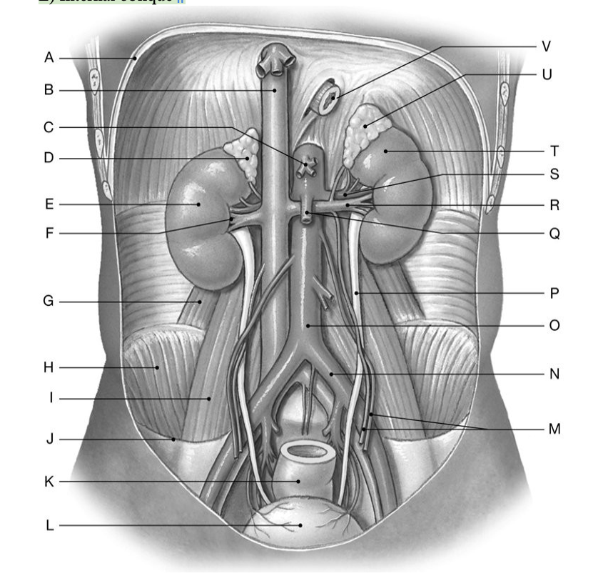
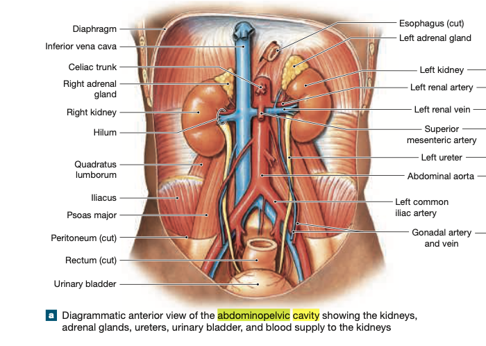
Identify the structure(s) indicated by Label B.
A) Aorta
B) Hepatic portal vein
C) Superior vena cava
D) Renal vein
E) Inferior vena cava
34) Identify the structure(s) indicated by Label D.
A) Pancreas
B) Spleen
C) Right kidney
D) Stomach
E) Right suprarenal gland
35) Identify the structure(s) indicated by Label E.
A) Right kidney
B) Liver
C) Right suprarenal gland
D) Spleen
E) Stomach
36) Identify the structure(s) indicated by Label F.
A) Medulla
B) Hilum
C) Greater omentum
D) Hepatoduodenal ligament
E) Lesser omentum
37) Identify the structure(s) indicated by Label L.
A) Uterus
B) Fallopian tube
C) Suprarenal gland
D) Urinary bladder
E) Spleen
38) Identify the structure(s) indicated by Label P.
A) Inferior vena cava
B) Left ureter
C) Aorta
D) Left renal vein
E) Left urethra
39) Identify the structure(s) indicated by Label R.
A) Left urethra
B) Aorta
C) Left renal vein
D) Inferior vena cava
E) Left ureter
40) Identify the structure(s) indicated by Label S.
A) Left renal vein
B) Inferior vena cava
C) Left ureter
D) Left renal artery
E) Aorta