Chapter 16 Spinal Cord and Spinal Cord
The spinal cord and its attached spinal nerves are a pathway for sensory and motor impulses
The diameter of a typical adult spinal cord is about ¾ of an inch or 19 mm
The thoracic part of the spinal cord lies inferior to the cervical part, it contains neurons for the thoracic spinal nerves
The cervical part of the spinal cord is the superior most region of the spinal cord
In superior segments, the anterior horns are relatively small and the posterior horns are relatively large, while in the inferior segments, the anterior horns are larger. This describes the cervical region of the spinal cord
The spinal cord and spinal nerves are responsible for reflexes, which are our quickest reactions to a stimulus
The lumbar part of the spinal cord contains the neurons for the lumbar spinal nerves
The spinal cord extends inferiorly from the brain through the foramen magnum and then through the vertebral canal and ends at the inferior level of the L1 vertebra.
The cervical part of the spinal cord contains cell bodies of motor neurons whose axons contribute to the cervical nerves. These neurons receive input from sensory neurons through these same spinal nerve
The spinal cord in an adult is shorter than the vertebral canal that houses it
Inferior to conus medullaris, nerve roots collectively called the cauda equina project inferiorly from the spinal cord

The sacral part of the spinal cord lies inferior to the lumbar part and contains the neurons for the sacral spinal nerves.
In areas of the spinal cord that control the limbs, the cross-sectional diameter is larger compared to other areas.
The tapering inferior end of the spinal cord is called the conus equina
The spinal cord has two longitudinal depressions. These are the posterior median sulcus and the anterior median fissure
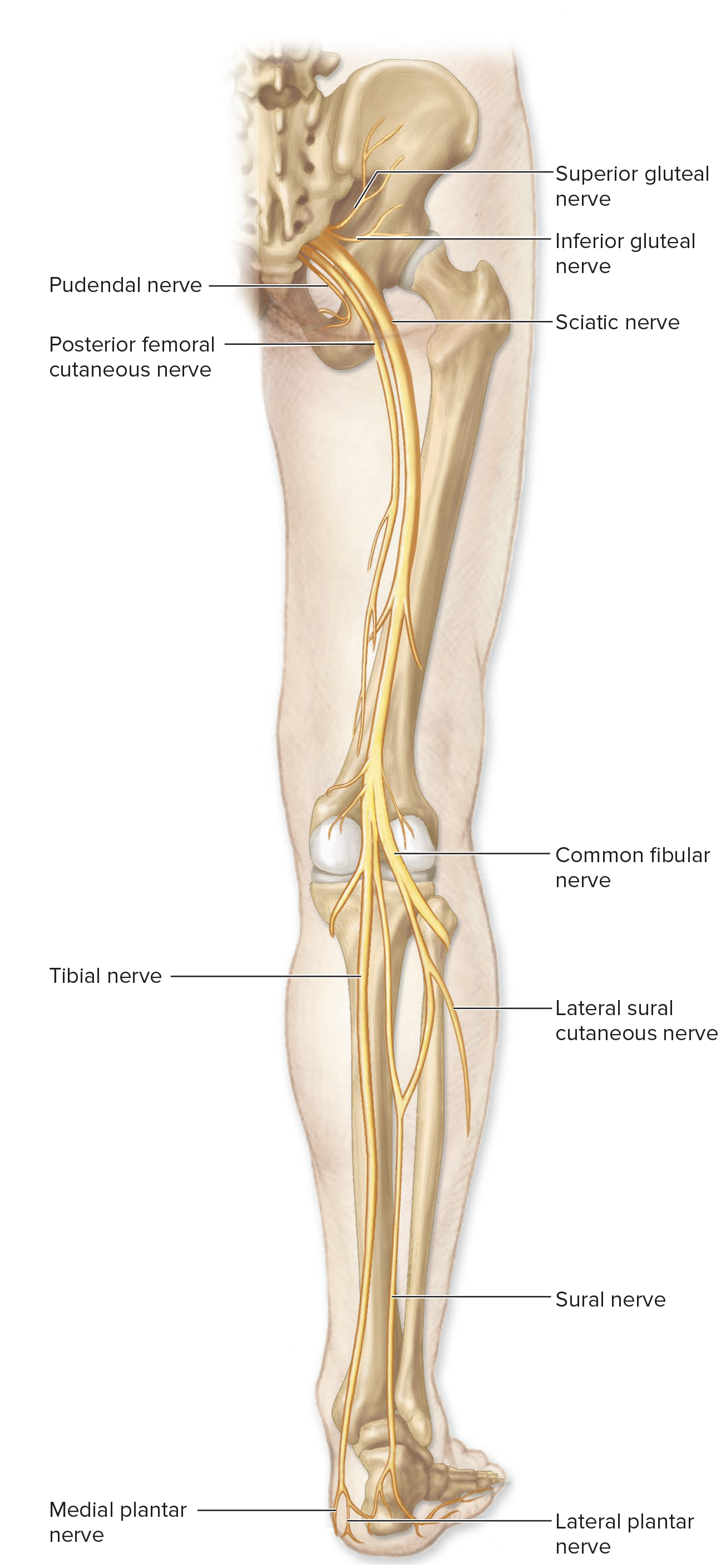
The spinal nerves connect the central nervous system to muscles, glands, and receptors
The enlargement of the spinal cord that contains the neurons that innervate the upper limbs is the cervical enlargement
The epidural space contains areolar connective tissue, blood vessels, and adipose connective tissue.
Spinal nerves are considered mixed nerves because they contain both motor and sensory axons.
The space that lies between the dura mater and the periosteum that covers the inner walls of a vertebra is the epidural space.
A filum terminale is a thin strand of pia mater that helps anchor the conus medullaris to the coccyx
Immediately deep to the epidural space is the most external of the meninges, the dura mater.
There are 7 cervical vertebrae and 8 pairs of cervical spinal nerves.
The spinal cord is protected and encapsulated by spinal cord meninges, which are continuous with the cranial meninges
A narrow space separates the dura mater from the arachnoid mater. This space is called the subdural space
The gray matter of the spinal cord is dominated by the dendrites and cell bodies of neurons.
The spinal cord contains 8 pairs of cervical nerves, 12 pairs of thoracic nerves, 5 pairs of lumbar nerves, 5 pairs sacral nerves and 1 pair of coccygeal nerves.
Which ligaments are paired, lateral triangular extensions of the spinal pia mater that attach to the dura mater?
Denticulate ligaments
The cell bodies of somatic motor neurons are primarily housed by the anterior horns
anterior horns are left & right anterior masses of gray matter
cell bodies of somatic motor neurons: innervate skeletal muscle
Sequence the structures and spaces surrounding the spinal cord from superficial to deep. Please put the most superficial structure at the top of the list.
Vertebra
Epidural space
Dura mater
Subdural space
Arachnoid mater
Subarachnoid space
Pia mater
The spinal cord is partitioned into a inner gray matter region and an outer white matter region.
The gray matter region is dominated by dendrites neuron cell bodies, glial cells, unmyelinated axons
White matter is composed primarily of myelinated axons
The cell bodies of interneurons are found in the posterior horns of the spinal cord
The white matter region on each lateral side of the spinal cord is the lateral funiculus

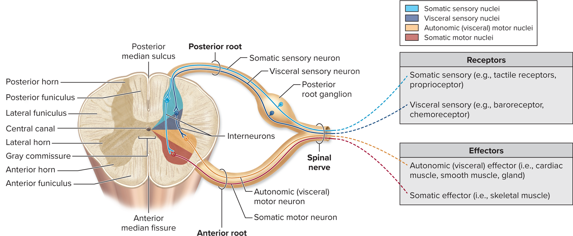
The gray commissure is a horizontal bar of gray matter that surrounds a narrow central canal in the spinal cord.
The lateral horns contain the cell bodies of autonomic motor neurons.
Which sensory nuclei receive information from sensory receptors, such as the stretch receptors in the smooth muscle walls of small intestine?
Visceral
The axons of sensory neurons and the cell bodies of interneurons are located in the posterior horn
The gray commissure primarily contain Unmyelinated axons and serves as a communication route between the right and left sides of the gray matter of the spinal cord
Sensory nuclei in the posterior horns contain interneuron cell bodies.
Motor nuclei in the anterior and lateral horns contain motor neuron cell bodies that send nerve impulses to muscles and glands.
A posterior funiculus lies between the posterior gray horns on the posterior side of the cord and the posterior median sulcus.
The anterior funiculi are interconnected by the white commissure
The gray commissure is a horizontal bar of gray matter that surrounds a narrow central canal in the spinal cord.
The anterior and lateral funiculi contain both ascending and descending tracts, so they contain both motor and sensory axons
The posterior funiculi contain only ascending (sensory) tracts
Some individual fasciculi conduct motor impulses as descending tracts from the brain to the spinal cord.
The anterior funiculus is composed of tracts of white matter that occupy the space on each anterior side of the cord between the anterior gray horns and the anterior median fissure
Some of the individual tracts conduct sensory impulses as ascending tracts from the spinal cord to the brain
Which type of neuron processes information in the central nervous system?
Interneuron
The Golgi tendon reflex is a Blank______ reflex that prevents skeletal muscles from tensing excessively
polysynaptic
As a result of the Golgi tendon reflex, the motor neurons that cause the muscle contraction are Blank
Inhibited