Brain Behavior
Definition (#f7aeae)
Important (#edcae9)
Extra (#fffe9d)
KEY CONCEPTS:
Branches of the nervous system.
Neurons: Building blocks of the nervous system & how it works.
Action potential & Synaptic transmissions.
Brain structure.
Exploring brain structure and function.
BRANCHES OF THE NERVOUS SYSTEM:
2 Main branches:
Central Nervous System (CNS):
Consisting of the brain, spinal cord and majority of the neurons.
Peripheral Nervous System (PNS):
The spinal cord transmits information between the brain and PNS.
Includes all parts of the nervous system beyond the CNS.
Further divided into Somatic Nervous System (SNS) and Autonomic Nervous System (ANS).
Somatic Nervous System (SNS):
Network linking the spinal cord with the upper body and sense organs.
Controls voluntary behavior.
Autonomic Nervous System (ANS):
Collection of neurons that carry information to and from internal organs.
Divided into parasympathetic- to converse energy & sympathetic- to arouse.
NEURONS:
Neurons are not nerves.
A neuron is a single cell in the nervous system that transmits information through the nervous system.
Comes in different shapes and sizes but has 4 basic parts.
Dendrites: Tree, roots, 1000s of branches, receiving incoming information.
Cell-body (Soma): Receives information from dendrites, stores genetic information.
Axon: Carries information away from soma.
Axon terminals: bulb shaped at the end of axon to form synapses with other dendrites & release chemicals.
How Neurons Works:
Primarily electric, the charged molecules are called ions - found inside and outside neurons.
Some ions positively charged, some are negative.
Neurons at rest, where more positive ions accumulate outside as more negative charges exist within- resting potential.
As messages are received, the resting potential becomes slightly raised while overall electric charges rise.
When this charge breaches a threshold, it turns into an action potential; change in the neuron’s overall charge.
Myelin sheath: an insulating material, made up of fatty substances coating the axon, to allow electrical impulses transmit more quickly and effectively.
Under the microscope, these sheaths appear white and neurons appear grey.
Areas of the brain containing mainly neuron cell bodies are referred to as grey matter, areas containing myelinated axons are called white matter.
Damaged myelin may result in slowing down the transmission of charges leading to sensations numbness, weakness or paralysis.
Action Potential:
Ion channels: Tiny tunnels in the axon membrane function as doors.
Ion channels are blocked by molecules that prevent sodium (Na+) ions from entering the axon.
During an action potential, the gates open and Na+ ions rush into the axon.
The channels first open near the soma, then down the length of the axon as the action potential travels down.
In the end, a negative after potential occurs where it dips slightly below a resting potential.
Synaptic Transmissions:
The microscopic space over which messages pass between 2 neurons.
Synaptic transmissions occur when an action potential reaches the tips of an axon terminal and releases a neurotransmitter.
Transmissions: Dendrites → Soma → Axons → Axon terminals
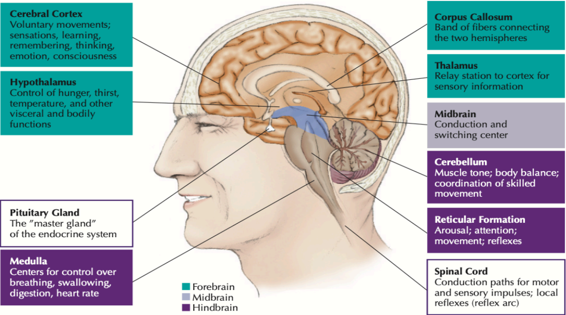
BRAIN STRUCTURE:
Hemispheric lateralisation: Specialization of abilities of the left vs right hemisphere.
Left brain: Language, speech, time sense, writing, rhythm, calculation, complex movements.
Right brain: nonverbal, perceptual skills, visualization, recognition, expression of emotions.
Lobes of the cerebral cortex:
Each hemisphere can be divided into smaller lobes:
Frontal: Sense of self, motor control and higher mental abilities; reasoning.
Occipital: Vision.
Parietal: Sensations such as touch, temperature and pressure.
Temporal: Hearing and language.
Cerebellum: Posture, coordination, muscle tone and memory.
Brain structure & function:
Structure:
Computed Tomography (CT) scans:
Multiple X-ray scans of a single location taken from different angles and forms an image.
Magnetic Resonance Imaging (MRI) scan:
Placing body into strong magnetic fields, as computers processing creates a 3D model of the brain or body, more detailed than CT scans.
Function:
Electroencephalography (EEG):
Measures electrical activity near the surface of the brain through small electrodes.
Functional Magnetic Resonance Imaging (fMRI) scan:
Uses MRI technology to record activity level in various areas of the brain.
Positron Emission Tomography (PET) scan:
High resolution imaging technique that captures brain activity by attaching radioactive brain particles to glucose molecules.